Abstract
In patients with muscle injury or muscle disease, assessment of muscle damage is typically limited to clinical signs, such as tenderness, strength, range of motion, and more recently, imaging studies. Biological markers can also be used in measuring muscle injury, such as increased creatine kinase levels in the blood, but these are not always correlated with loss in muscle function (i.e. loss of force production). This is even true of histological findings from animals, which provide a “direct measure” of damage, but do not account for loss of function. The most comprehensive measure of the overall health of the muscle is contractile force. To date, animal models testing contractile force have been limited to the muscle groups moving the ankle. Here we describe an in vivo animal model for the quadriceps, with abilities to measure torque, produce a reliable muscle injury, and follow muscle recovery within the same animal over time. We also describe a second model used for direct measurement of force from an isolated quadriceps muscle in situ.
Keywords: skeletal muscle, lengthening contraction, injury, contractile function, torque, recovery, quadriceps, specific force
INTRODUCTION
Several animal models exist to measure muscle contractility and to induce muscle injury via eccentric contractions. Until now, these have been limited to testing small, thin muscles in vitro or testing the dorsiflexor and plantarflexor muscles of the ankle in vivo. One difficulty in using either “muscle group” is that it is not always clear what the relative contribution of each muscle is to overall dorsiflexion or plantarflexion contractile forces. In addition, it is potentially imprecise to compare the histology of one muscle to contractility data from a group of muscles. We have successfully adapted our procedures to test contractility and susceptibility to injury for the quadriceps, the sole mover of extension at the knee.
Testing the quadriceps muscle provides several advantages. First, it is a very large muscle and therefore differences in contractility between experimental groups are often more readily apparent. The minute forces generated by the slight ankle muscles of small animals can make comparisons between animals difficult. Second, because of its large size, there is a copious amount of muscle for histological and biochemical analyses. Third, the quadriceps are the only knee extensors, leaving no doubt regarding which muscles contributed to the force generated and making comparisons of histological analysis to contractile data potentially more accurate. Finally, the quadriceps is arguably more crucial to gait than the plantarflexors and dorsiflexors. It is easier to compensate gait for foot drop than knee weakness and such differences could have important ramifications for comparing muscle function and/or injury to gait analysis. In addition to current methods for measuring contractility, we propose the use of two quadriceps models that, collectively, can measure torque and direct contractile force, induce muscle injury, as well as follow muscle function (e.g. recovery from injury) over time in a single animal.
MATERIALS AND PROCEDURE
Animal preparation
-
1
All experimental procedures were approved by the University of Maryland Institutional Animal Care & Use Committee. These procedures can be used for rats or mice [1–3]. To begin, place the animal supine under inhalation anesthesia (~4–5% isoflurane for induction in an induction chamber, then ~2% isoflurane via a nosecone for maintenance) using a precision vaporizer (cat # 911103, Vet Equip, Inc., Pleasanton, CA). If the animal is to be anesthetized for a long period of time, apply sterile ophthalmic cream (Paralube Vet Ointment, PharmaDerm, Floham Park, NJ) to each eye to protect the corneas from drying. During the procedure, the animal is kept warm by use of a heat lamp.
CAUTION: Animals lose a lot of body heat while under inhalation anesthesia, so need to be kept warm. However, the heat lamp should be kept at a safe distance, at least 12 in ches from the animal.
-
2
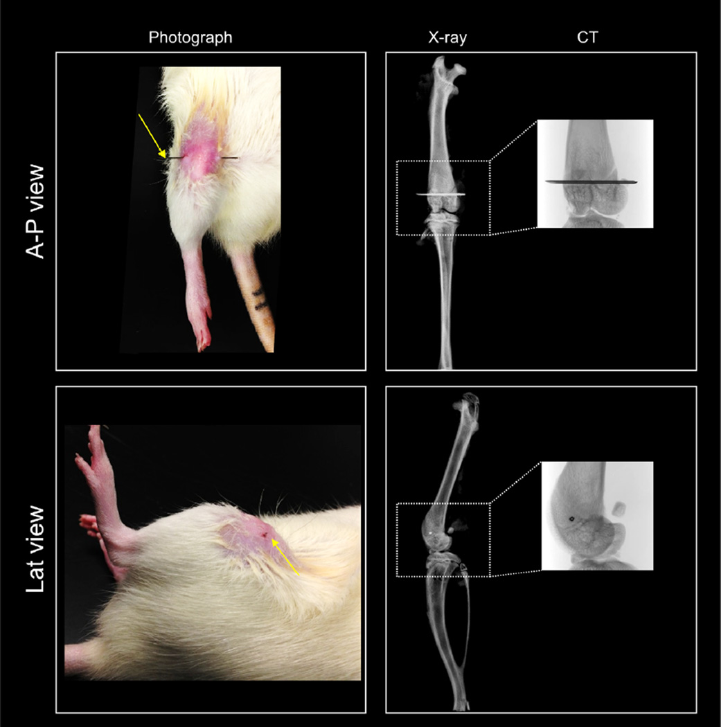
Confirm proper anesthesia by lack of a deep tendon reflex (no foot withdrawal in response to pinching the foot). Prep the skin around the knee by removing hair and by cleaning with alternating scrubs of betadine and 70% alcohol to prevent seeding skin bacteria into the soft tissue or bone. A needle (25 G for rat or 27 G for mouse) is manually placed through the distal femur (Fig. 1, femoral condyles) by hand (see Movie S1). The needle will be used to stabilize the femur onto the rig (see step 3).
TIP: The widest aspect of the knee is formed by the femoral epicondyles, which sit just superficial to the condyles and are good landmarks to palpate for needle placement. Stabilizing the thigh and moving the knee passively to visually identify the femur (not moving) from the tibia (moving) is also helpful for those new to this type of procedure.
CAUTION: The needle should not enter the anterior compartment of the thigh, the quadriceps, or knee joint. Needle placement is easy, but can take practice. Correct needle placement can only be confirmed by opening the skin, which can be done during muscle harvesting, whenever the investigator decides to sacrifice the animal.
-
3
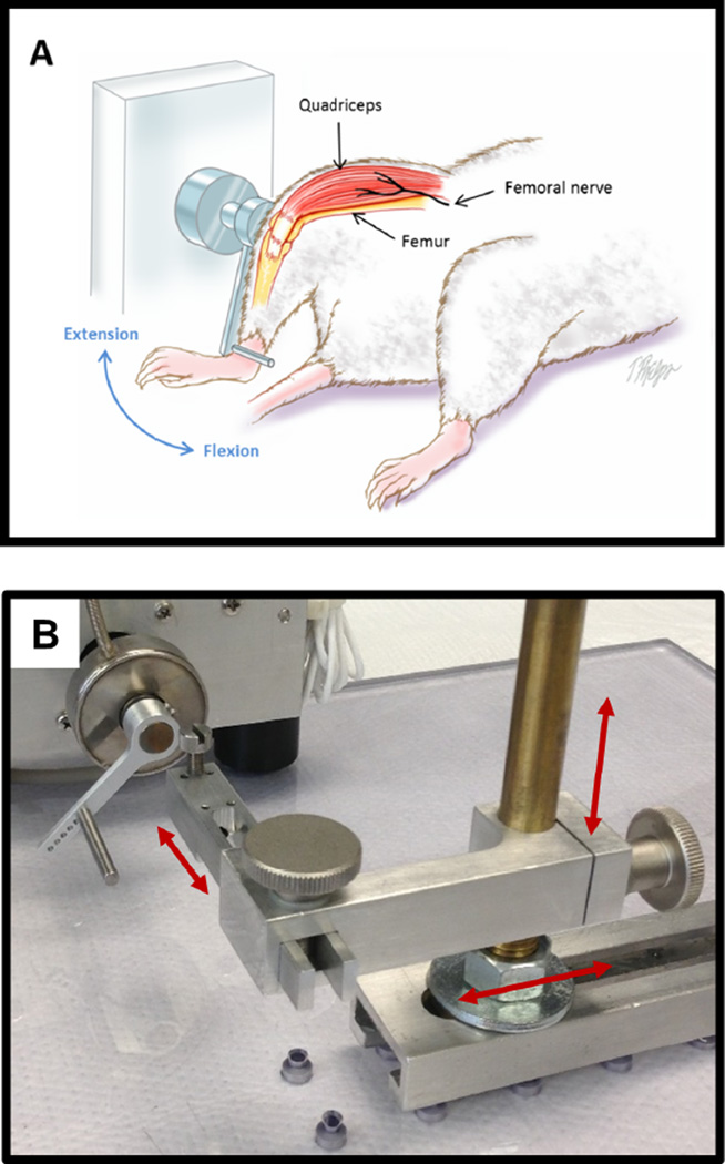
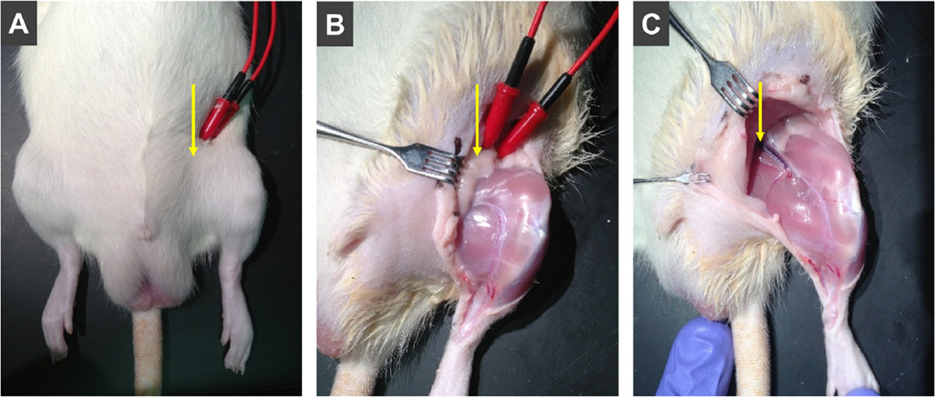
Lock the needle (and consequently, the femur) into a fixed position with the animal supine, and on a raised bed such that the thigh lies flat on the bed and the knee is free to rotate through a full range of motion. We use a custom-made device to secure the needle and thereby stabilize the thigh (see Fig. 3B and Movie S1). Subcutaneous electrodes (J05 Needle Electrode Needles, 36BTP, Jari Electrode Supply, Gilroy, CA) are used to stimulate the femoral nerve anterior to the hip joint, where the nerve lies in a superficial position within the femoral triangle (Fig. 2). Visually confirm isolated knee extension by performing a series of twitches (0.1 ms pulse for the mouse and 1 ms pulse for the rat) before the ankle is secured. Knee extension should occur in a sagittal plane, such that the leg moves only forward and backward.
CAUTION: The knee is essentially a hinge joint, so if the femoral condyles are not aligned parallel to the floor, extension could occur in an oblique plane, which could alter torque readings.
TIP: The device we use to stabilize the needle allows three degrees of freedom for positioning, but this is for convenience; the method used to stabilize the needle is not important, as long as the femur is immobilized and the position is accurate (for positioning, see Step 4). Our prototype was simply a vise that was positioned correctly.
TIP: One can obtain knee extension and a decent trace recording with the electrodes in the approximately correct position. Be sure to try several positions, from superficial to deep, in order to determine which electrode placement results in maximal torque. This type of trial and error is not necessary after you have an idea of what values animals of a specific gender/age/species generate on your device. Although the femoral nerve is “superficial” to the hip joint and surrounding musculature, it is still ~1cm deep to the skin.
-
4
Secure the ankle to a custom-machined adjustable lever arm [4] (Fig. 3A) with adhesive tape. The ankle rod should be adjusted so that it lies anteriorly on the distal leg just above the foot. The position of the ankle rod should not be altered between age-matched animals of the same species so that the lever is constant between tests. The lever arm is attached to a stepper motor (model T8904, NMB Technologies, Chatsworth, CA) and a torque sensor (model QWFK-8M, Sensotec, Columbus, OH). Align the axis of knee rotation (i.e. needle) with the axis of motor rotation.
TIP: If the femoral condyles are parallel to the floor, the foot will be aligned so the toes are pointing anteriorly, as in Figure 3, and not to the left or to the right.
Figure 1. Location of pin placement for thigh stabilization.
The images show the anterior-posterior (A-P) view and lateral (Lat) view for pin placement. Corresponding X-rays and computed scans (CT, inset) are shown to illustrate in detail proper orientation of the pin through the femoral condyles. The pin should be placed to result in a medial-lateral axis as closely as possible, without any obliquity in any of the anatomical planes. A thin needle is placed at the distal end of the femur through the femoral condyles. Once secured by the apparatus, the distal thigh will then be immobilized, which is paramount for obtaining accurate recordings of torque or force.
Figure 3. In vivo apparatus.
A. Drawing illustrates the general setup used to assess quadriceps strength, the thigh is stabilized and the leg attached to a motor-driven lever arm. The quadriceps muscle is stimulated via the femoral nerve while the lever arm records torque. To produce a contraction-induced injury, the quadriceps muscle is stimulated via the femoral nerve to produce extension while the lever arm forces the knee into flexion (arrow). Image modified from Pratt et al., J Physiology, 2013, used with permission. B. The apparatus is shown without the animal. The custom-made device we use to lock the needle into a fixed position is adjustable in all three anatomical planes (red arrows). Any device would work, as long as the needle is immobilized and, assuming it is placed through the axis of the knee, in line with the axis of the stepper motor.
Figure 2. Location of stimulation.
The femoral nerve is used to stimulate the quadriceps muscles. A. Placement of electrodes: The femoral nerve passes over the anterior aspect of the hip (yellow arrow) before innervating the quadriceps. B. Subcutaneous view: The femoral nerve is subcutaneous, but covered in a layer of peritoneal fat. C. Depth of neurovascular bundle: Despite being in a “superficial” location (relative to the musculoskeletal anatomy), due to the fat the subcutaneous needle electrodes must be pushed ~1 cm into the groin for optimal stimulation.
In vivo measurement of isometric torque and induction of muscle injury
A visual presentation of the procedure can be found in Movie S1.
-
5
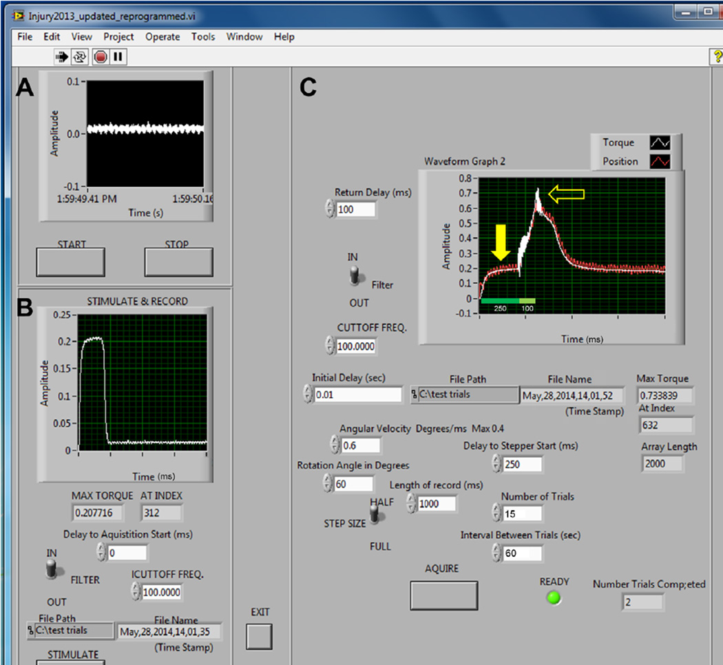
Before injury, and at selected time points after an injury, the maximal force producing capacity of the quadriceps is recorded as the “maximal isometric torque” (Fig. 4B). Torque measurements are performed on the same rig that is used to induce injury. In order to obtain maximal isometric torque, the pulse amplitude is adjusted to optimize twitch tension and the optimal position of the leg is determined by measuring twitches at different lengths of the quadriceps. This is done by sequentially rotating the lever arm at 5° increments from start position at 0° (full knee extension) through full range of flexion at about 120°. After obtaining a torque-angle curve to determine the optimal length (Lo) of the quadriceps (Lo, aka “resting length”, is where peak twitch amplitude occurs), a torque frequency plot is obtained by progressively increasing the frequency of pulses during a 200 ms pulse train. Finally, an increase in fused amplitude in response to an increase in voltage confirms that opposing muscles (hamstrings) are not being simultaneously stimulated by excessive current and that optimal voltage is reached (Fig. 4B). Three separate twitches and tetanic contractions are recorded and saved for further analysis.
TIP: Lo, or resting length, is typically at ~40° for us. Similar to humans, peak torque occurs midway through the available range of motion.
TIP: There are a variety of stimulation parameters in the literature that are difficult to make sense of. For example, some studies suggest that maximal tension of a muscle is obtained at a stimulation frequency of 250–300 Hz. This very high frequency is typical for in vitro procedures (removing a muscle from the animal and ‘hanging it’ in a bath for field stimulation). However, for in vivo procedures (stimulating through the motor nerve), maximal contractile tension occurs at a much lower frequency. In our hands, a maximal fused tetanic contraction for the quadriceps in vivo is obtained at ~100–150 Hz. The frequency to obtain maximal stimulation can be even lower in other hindlimb muscles (e.g., dorsiflexors and plantarflexors) during in vivo testing [5;6].
-
6
We use commercial software (LabView version 2013, National Instruments, Austin, TX) to synchronize contractile activation, onset of knee rotation, and torque data collection (Fig. 4). For lengthening contractions, stimulation of the quadriceps muscles occurs while the computer-controlled motor simultaneously moves the lever arm against the direction of knee extension, thus leading to a lengthening contraction (also called “eccentric” contraction which, if maximal and repeated, causes injury of the muscle). The specific protocol depends on the desired magnitude of injury and is designed by the investigator. The magnitude of injury, or tissue damage, can be regulated by manipulation of variables such as angular velocity, timing of muscle activation, range of motion, and the number of lengthening contractions.
-
7
To induce injury, superimpose a lengthening contraction onto a maximal isometric contraction (a contraction during which muscle length is held constant), varying the range of motion, velocity of lengthening, and timing of stimulation as needed. For example, a maximal isometric contraction is obtained in the quadriceps (250 ms train, Fig. 4C, closed arrow) before the onset of motor movement and the muscle is immediately lengthened (100 ms train, Fig. 4C, open arrow) at a selected velocity to approximate normal movement (900°/sec) resulting in a high force peak. We have shown that activation before and during motor movement, and the degree of lengthening are important factors in obtaining an injury [7]. As reported previously, this model results in injury to muscle [7–10]. The quadriceps remains stimulated throughout lengthening.
TIP: Many variables affect the magnitude of injury (operationally defined as a loss in maximal isometric torque). Assuming the velocity is above 100°/sec, faster velocity of lengthening is not likely to matter. Obtaining a full isometric contraction before (at least 200 ms long) lengthening occurs, as well as using a large range of motion (but within physiological limits), will assist in producing a reliable injury that does not require an excessive number of contractions.
-
8
After injury, the animal is removed from the apparatus. The pin is removed, the leg is cleaned again with betadine, and over-the-counter antibiotic cream is applied to the needle placement site. The animal is returned to the cage (placed on a temperature-controlled heating block at 37°C) and monitored until recovery. If follow-up measurements are desired, a new sterile needle is placed into the same location.
TIP: In our experience, it is relatively easy to place the tip of the needle under the skin and slide along the condyle to find the same location used for earlier procedures.
CAUTION: If excessive pin re-insertions are needed for extended periods of time (e.g., daily for weeks), this could lead to joint inflammation, which could result in joint scarring and limit knee extension range of motion. Before follow-up testing, be sure to check that the knee freely moves through a full range of motion. We find daily insertion acceptable, but for only up to 3–4 days; we have seen no complications re-inserting every 3 days for 1–2 weeks.
Figure 4. Torque data from in vivo apparatus.
A. Before experiments, the torque sensor can be tested for proper sensitivity/calibration and the baseline adjusted to zero. B. Once the animal is secured into the apparatus, maximal isometric torque is recorded from a fused tetanic contraction in the quadriceps before and after lengthening contractions. Maximal isometric torque is achieved using optimal muscle length, stimulation rate (Hz), and voltage (see step 5). C. Recordings from lengthening contractions show peak amplitude from a 250 ms train stimulation before onset of motor movement (closed arrow). Stimulation of the femoral nerve continues (an additional 100 ms) throughout forced flexion of the quadriceps via the motor driven lever arm. This produces a high force peak (closed arrow) during the lengthening contraction.
In situ measurement of whole muscle tension
A visual presentation of the procedure can be found in Movie S2.
-
9
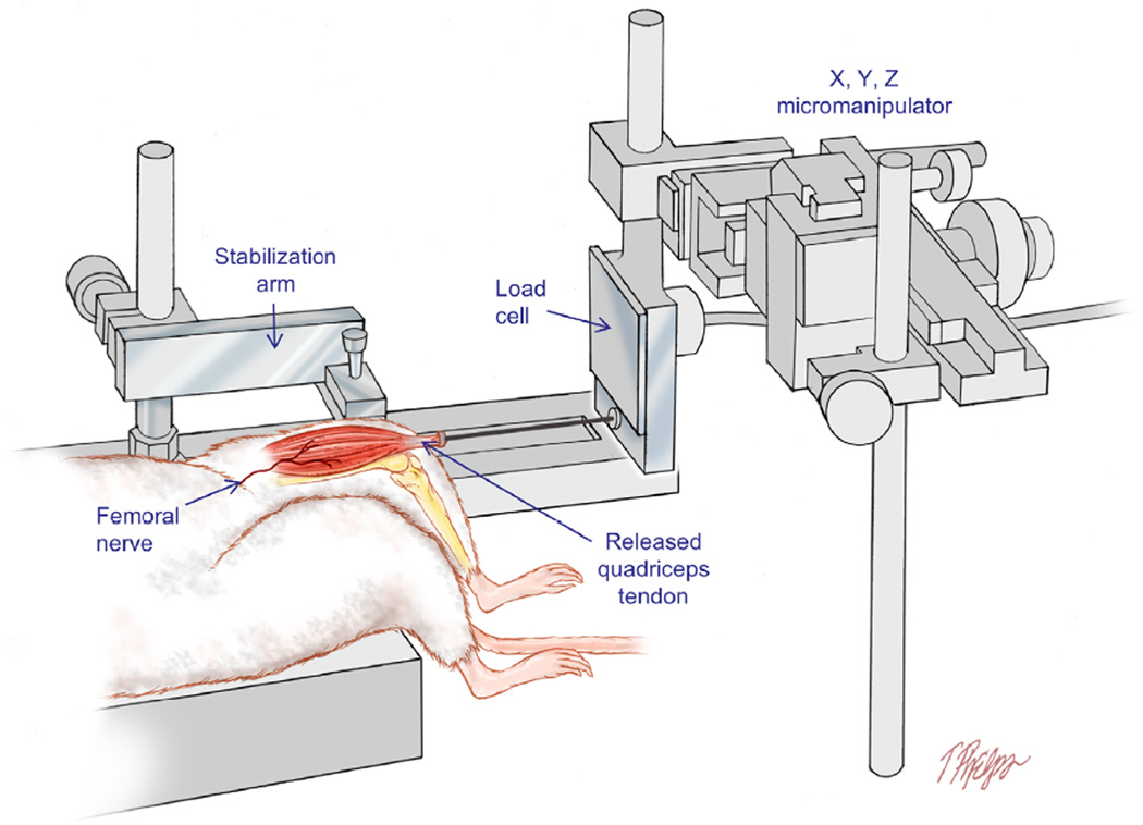
The animal is prepared, the femur is stabilized (Fig. 5), and the femoral nerve is stimulated as described above (the device used to stabilize the needle, thus immobilizing the femur, is the same one as shown in Fig 3B). All instrumentation is turned on at least 30 min prior to testing for proper calibration and to minimize thermal drift of the force transducer. In contrast to the in vivo torque measurements method, this technique involves a surgical procedure (releasing the muscle insertion) and is therefore terminal.
NOTE: Both the in vivo (torque) and in situ (force) systems are calibrated by using precision weights (10 g – 200 g) and a known moment arm (for in vivo torque system), which allows us to determine accuracy and linearity. Our systems record torque, or force, in volts and a conversion factor is then used to convert to N·mm, or N, respectively.
-
10
Incise the skin anterior to the knee and sever the tendon of the quadriceps muscle. Carefully tie 4.0 Ethicon silk non-absorbable suture to the tendon and attach the vicryl suture to the load cell (Fig. 5) via the provided S-hook (weight = 0.1 g), (model FT03, Grass Instruments, Warwick, RI). Alternatively, a custom clamp (weight = 0.5 g) can be used to attach the tendon to the vicryl suture.
TIP: After incising the skin anterior to the knee, it is helpful to make a small vertical incision on either side of the patella tendon so as to release it from the lateral fascial attachments. After this, attaching suture or a clamp is typically easier to do before making a horizontal cut to release the entire tendon.
-
11
The load cell is mounted to a micromanipulator (Fig. 5, Kite Manipulator, World Precision Instruments Inc., Sarasota, FL) so that the quadriceps can be adjusted to resting length and aligned properly (a straight line of pull between the origin and insertion of the muscle and the load cell). The quadriceps muscle is protected from cooling by a heat lamp. If the user wishes to expose more than just the tendon, dehydration of the exposed muscle can be minimized by application of mineral oil, as needed (to which extent the quadriceps is released is up to the user. For more on muscle release, see Step 12 TIP). Alternatively, a continuous drip of 37°C culture medium (DMEM) over the muscle can be used. The signals from the load cell (calibrated before each test) are fed via a DC amplifier (model P122, Grass Instruments, Warwick, RI) to an A/D board to be collected and stored by acquisition software (PolyVIEW™ 16, Grass Instruments, Warwick, RI).
-
12
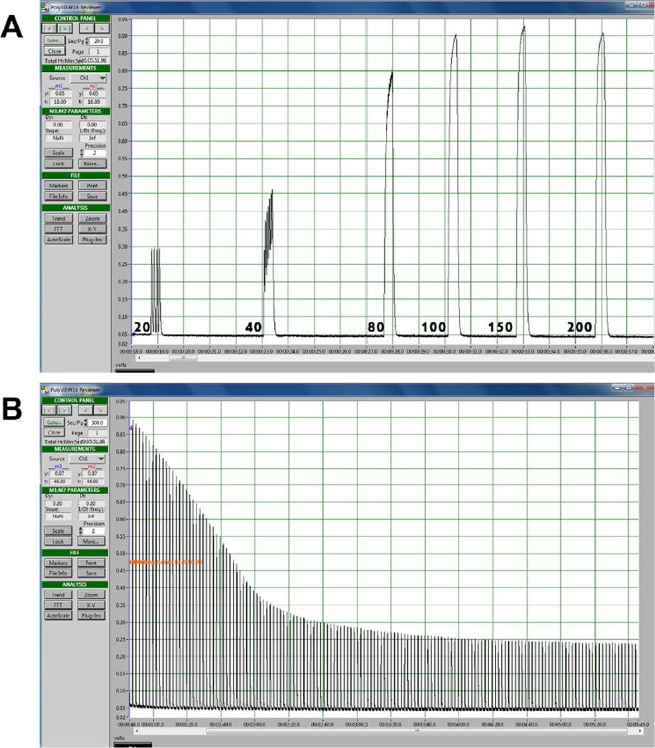
Attach the quadriceps to the load cell and measure single twitches (rectangular pulse of 1 ms) at different muscle lengths in order to determine resting length (Lo). Muscle resting length is defined as the length at which maximal contractile force is produced. At this length, gradually increase the pulse amplitude and then the pulse frequency to establish a force-frequency relationship. A maximally fused tetanic contraction is obtained at approximately 100–150 Hz (300 ms train duration comprised of 0.1 ms or 1 ms pulses). Use 150% of the maximum stimulation intensity to activate the quadriceps in order to induce maximal contractile tension (Po). Three separate twitches and tetanic contractions are recorded and saved for further analysis. Maximal tetanic contractions can be performed repeatedly (typically every 0.5 s – 1 s) with the final tension expressed as percentage of Po, providing an index of fatigue at any desired point in time.
TIP: Muscles can transmit some of their contractile force laterally, such that muscles in a group (e.g., dorsiflexors) can contribute to force measurements even when only one of the muscles (e.g., tibialis anterior) is tied to the load cell for assessment. Thus, for muscles of the leg (dorsi- and plantar-flexors), it can be important to release the muscle entirely to its origin to avoid lateral transmission of force from nearby muscles. For the quadriceps, since there are no additional knee extensor muscles, one can release just the tendon. Releasing the quadriceps completely up to its origin is more difficult than with other muscles due to its more complex attachments.
CAUTION: Once the quadriceps is attached to the load cell there is a small amount of passive force present when the muscle is stretched to resting length. This passive tension can ‘drift’ after the first few contractions. Having moist suture and performing a series of ‘warm up’ twitches can minimize this drift, but the investigator should adjust resting length as needed.
Figure 5. In situ apparatus.
The load cell is mounted to a micromanipulator so that the quadriceps can be adjusted to resting length and aligned properly (a straight line of pull between the origin and insertion of the muscle and the load cell) in the X, Y, and Z directions. The distal tendon of the quadriceps is attached to the load cell and single twitches (1 ms duration) are induced at different muscle lengths (thus a length-tension, or L-T, curve) in order to determine Lo. A maximal tetanic contraction is obtained to determine maximal contractile tension (Po). Maximal tetanic contractions can be performed repeatedly over time with the final tension expressed as percentage of Po, providing an index of fatigue at a desired point in time.
Harvesting and storing muscles
-
13
At the end of experiments, animals are euthanized per institutional animal care guidelines and the quadriceps muscles are harvested, weighed, snap frozen in liquid nitrogen, and then stored at −80°C. This can be performed at any point in time after the in vivo experiments. Muscles are always harvested immediately after the in situ experiments, as this is a terminal procedure. For detailed morphological studies, the animal is fixed with 4% paraformaldehyde via perfusion through the left ventricle.
TIP: Care should be taken to remove the entire quadriceps. It attaches along the length of the femur, including the postero-lateral aspect, as well as to the surrounding fascia
REPRESENTATIVE RESULTS
Figure 4C shows representative curves from a mouse on the in vivo apparatus during an injury protocol of 15 repetitions with an arc of motion from 40° – 100°. Torque is recorded before and after injury (Fig. 4B) and converted from volts into units of N•mm, but the absolute value depends on the size of the animal and its condition (e.g., injured muscle, fatigued muscle, or muscle lacking a certain protein due to genetic modification). Such values are used to calculate torque deficits after lengthening contractions and are commonly reported as percent loss from pre-injury torque.
A cartoon depiction of the in situ apparatus is shown in Figure 5. Our in situ apparatus does not involve lengthening contractions; rather, it allows us to isolate, properly align, and measure maximal tension produced by an individual muscle at a known length. Figure 6 shows a representative force frequency plot and fatigue curves from a mouse quadriceps. Tension (force) is typically recorded in Newtons (or grams), but like torque, the absolute value depends on the size and condition of the animal. Because muscle weight is obtained immediately after this procedure, the force can be normalized (called “specific force”) to muscle cross sectional area.
Figure 6. Sample tension data from in situ apparatus.
A. A screen shot display of typical data showing how a force-frequency curve is generated. Curves shown were produced from maximal contractions (200 ms duration) at incrementing higher frequencies in the quadriceps muscle in a rat. Note that maximal force is generated at ~150 Hz and diminishes with higher frequency. This optimal frequency can vary depending on the muscle group tested and the conditions (e.g., direct nerve stimulation as here versus field stimulation when muscles are removed for in vitro experiments, not shown here). B. One can also measure the decline in maximal isometric tetanic tension during repeated stimulation to assess fatigue. In this example, the quadriceps was isolated, adjusted to optimal length (Lo), and then stimulated with a 200 ms tetanic contraction once every second for 5 minutes.
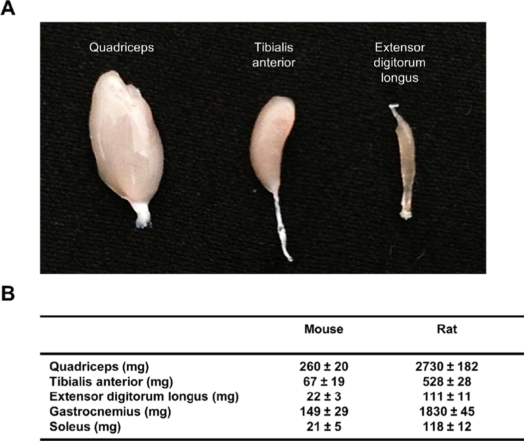
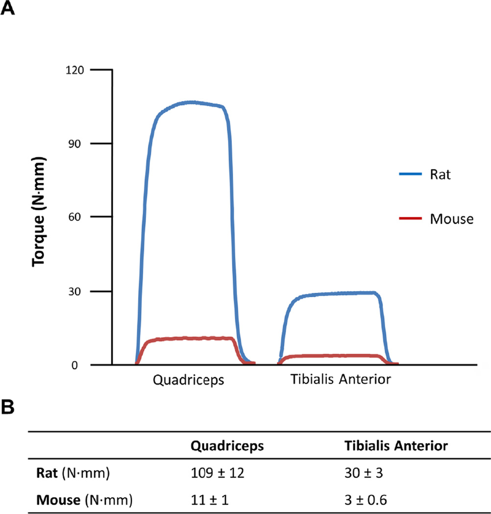
The benefits of using a larger muscle were discussed earlier. Figure 7 shows a representative image of the quadriceps and dorsiflexor muscles from a mouse for comparison with a corresponding table indicating the weight of commonly researched hindlimb muscles from mouse and rat. Figure 8 illustrates how the differences in muscle sizes are manifested in the functional changes. In addition, Figure 8 also provides a reference to compare mouse and rat functional outcomes.
Figure 7. Comparison of quadriceps to other commonly tested hindlimb muscles.
A. The quadriceps and the dorsiflexor muscles (tibialis anterior and extensor digitorum longus) from a mouse hindlimb are shown for size comparison. B. The table shows weights of commonly assessed muscles from the rodent hindlimb for comparison of muscle mass (from animals 2–3 months old, N = 3 each species).
Figure 8. Comparison of mouse/rat quadriceps and tibialis anterior muscles.
A. Representative in vivo trace recordings from a rat (blue) and mouse (red) are shown for the quadriceps and tibialis anterior muscles. In this example, muscles were stimulated for 200 ms at optimal muscle length to induce a maximal isometric contraction (torque). Contractile forces are much higher in the quadriceps compared to the ankle dorsiflexors (i.e., tibialis anterior), which have, in our experience, made it easier to discern differences in function. B. For reference, the table shows torque data derived from 12 Sprague-Dawley rats and 12 C57BL/10 mice; as noted in the text, the absolute values shown can be normalized to muscle weight. For experiments (or comparing different studies), care should be taken to evaluate animals that are age-matched, gender-matched, and of the same strain and species. Ideally, muscle measurements are obtained on the same rig.
CAUTION: In addition to species, it is important to realize that “typical” functional data are dependent on the animal strain, the muscle being tested, age, gender, muscle quality (e.g. disease states), and likely many other variables. Calibration of equipment is also important, therefore caution is recommended if values are not obtained from the identical apparatus.
DISCUSSION
“Muscle damage” has been defined and measured in many ways. Structural damage is evident in histological findings [11] after muscle injury, but one problem with many of the biological markers used to assess injury, including those used in animal studies, is that they usually do not correlate with the loss of force. Muscle damage is often defined within the context of the assay used to examine it and no one finding can account for the changes in contractility after injury. Since full contractile function can persist despite the presence of injury markers, loss of force is likely the most valid measure of injury [13].
It is difficult to study muscle injuries in humans, as the incidence is a random event that is difficult to predict and the clinical presentation varies greatly. Therefore much of the data regarding muscle injuries have been ascertained from studies on animals, which provides control over many variables and the ability to study mechanisms of injury and recovery. Several devices are currently available to test the dorsiflexor and plantarflexor muscles in animals such as mice and rats. Interestingly, there have been no published descriptions of testing the quadriceps muscles, as is used in human dynamometers such as the Biodex and KinCom devices. The in vivo injury apparatus we have described provides a method for assessing quadriceps contractile function. Our quadriceps model has previously been briefly described [4,14], but our main goal here is to share the procedural methods in step-by-step detail.
The apparatus used for in vivo torque measurements has several advantages. It does not involve any dissection, so there is no need to euthanize the animal under study. The result is that one can measure contractility in the same animal over time, comparing results to animal growth, or additional assays such as gait analysis or in vivo MRI [15–16]. Other advantages are that normal anatomy is not altered and the nerve is not bypassed for stimulation (such as for in vitro preparations). Furthermore, the muscle remains in its normal environment, so the effects of inflammation, hormones, or other factors can be studied. Because it requires the use of fewer animals, whose muscles are subjected to fewer manipulations (e.g., dissection prior to assay of function), we prefer to use the torque measurements whenever possible.
The in situ apparatus we describe is used to determine the “specific force” (force per unit of cross sectional area) of an individual muscle. This setup is more typical of classic muscle physiology whereby the muscle is isolated and positioned properly; this also avoids force transmission from nearby muscles [17], although this is less of a concern in this protocol as there are no additional knee extensors. The in situ apparatus provides an alternative for measuring contractility of only one muscle with a known length and mass. Although the in situ apparatus provides more experimental control when measuring the force of an individual muscle, the trade-off is that the experiment becomes less physiologic. It requires a surgical release of the quadriceps muscle, which can alter the architecture (muscle fiber orientation) and affect force transmission. The experiment is also terminal, so the muscle cannot be monitored over time.
In summary, as a primary mover of the leg and a mixed fiber muscle with an important role in gait, future studies in the quadriceps will provide interesting comparisons to other muscles in injury and recovery.
Supplementary Material
Acknowledgements
This work was supported by a grant to RML from the National Institutes of Health (1R01AR059179).
Footnotes
Competing Interests: The authors have declared that no competing interests exist.
Supplementary information
Movie S1. Movie showing how to obtain measurements of maximal torque and induce injury in the quadriceps muscle
Movie S2. Movie showing the procedures for the in situ setup.
Supplementary information of this article can be found online at http://www/jbmethods.org.
References
- 1.Hammond JW, Hinton RY, Curl LA, Muriel JM, Lovering RM. Use of Autologous Platelet-rich Plasma to Treat Muscle Strain Injuries. Am J Sports Med. 2009;37:1135–1142. doi: 10.1177/0363546508330974. (2009) [DOI] [PMC free article] [PubMed] [Google Scholar]
- 2.Roche JA, Lovering RM, Bloch RJ. Impaired recovery of dysferlin-null skeletal muscle after contraction-induced injury in vivo. Neuroreport. 2008;19:1579–1584. doi: 10.1097/WNR.0b013e328311ca35. [DOI] [PMC free article] [PubMed] [Google Scholar]
- 3.Stone MR, Neill A, O’Lovering RM, Strong J, Resneck WG, et al. Toivola DM, Ursitti JA, Omary MB, Bloch RJ. Absence of keratin 19 in mice causes skeletal myopathy with mitochondrial and sarcolemmal reorganization. J Cell Sci. 2007;120:3999–4008. doi: 10.1242/jcs.009241. (2007) [DOI] [PMC free article] [PubMed] [Google Scholar]
- 4.Pratt SJP, Lawlor MW, Shah SB, Lovering RM. An in vivo rodent model of contraction-induced injury in the quadriceps muscle. Injury. 2011;43:788–793. doi: 10.1016/j.injury.2011.09.015. [DOI] [PMC free article] [PubMed] [Google Scholar]
- 5.Lovering RM, O’Neill A, Muriel JM, Prosser BL, Strong J, et al. Physiology, structure, and susceptibility to injury of skeletal muscle in mice lacking keratin 19-based and desmin-based intermediate filaments. Am J Physiol Cell Physiol. 2011;300:C803–C813. doi: 10.1152/ajpcell.00394.2010. [DOI] [PMC free article] [PubMed] [Google Scholar]
- 6.Pratt SJP, Xu S, Mullins RJ, Lovering RM. Temporal changes in magnetic resonance imaging in the mdx mouse. BMC Res Notes. 2013;6:262. doi: 10.1186/1756-0500-6-262. [DOI] [PMC free article] [PubMed] [Google Scholar]
- 7.Lovering RM, Hakim M, Moorman CT, 3rd, De Deyne PG. The contribution of contractile pre-activation to loss of function after a single lengthening contraction. J Biomech. 2005;38:1501–1507. doi: 10.1016/j.jbiomech.2004.07.008. [DOI] [PMC free article] [PubMed] [Google Scholar]
- 8.Hakim M, Hage W, Lovering RM, Moorman CT, 3rd, Curl LA, et al. Dexamethasone and recovery of contractile tension after a muscle injury. Clin Orthop Relat Res. 2005;439:235–242. doi: 10.1097/01.blo.0000177716.70404.f9. [DOI] [PubMed] [Google Scholar]
- 9.Lovering RM, De Deyne PG. Contractile function, sarcolemma integrity, and the loss of dystrophin after skeletal muscle eccentric contraction-induced injury. Am J Physiol Cell Physiol. 2004;286:230–238. doi: 10.1152/ajpcell.00199.2003. (2003) [DOI] [PMC free article] [PubMed] [Google Scholar]
- 10.Lovering RM, Roche JA, Bloch RJ, De Deyne PG. Recovery of function in skeletal muscle following 2 different contraction-induced injuries. Arch Phys Med Rehabil. 2007;88:617–625. doi: 10.1016/j.apmr.2007.02.010. [DOI] [PubMed] [Google Scholar]
- 11.Hamer PW, McGeachie JM, Davies MJ, Grounds MD. Evans Blue Dye as an in vivo marker of myofibre damage: optimising parameters for detecting initial myofibre membrane permeability. J Anat. 2002;200:69–79. doi: 10.1046/j.0021-8782.2001.00008.x. [DOI] [PMC free article] [PubMed] [Google Scholar]
- 12.Brooks SV, Zerba E, Faulkner JA. Injury to muscle fibres after single stretches of passive and maximally stimulated muscles in mice. J Physiol. 1995;488(Pt 2):459–469. doi: 10.1113/jphysiol.1995.sp020980. [DOI] [PMC free article] [PubMed] [Google Scholar]
- 13.Pratt SJ, Shah SB, Ward CW, Inacio MP, Stains JP, Lovering RM. Effects of in vivo injury on the neuromuscular junction in healthy and dystrophic muscles. J Physiol. 2013;591:559–570. doi: 10.1113/jphysiol.2012.241679. (2012) [DOI] [PMC free article] [PubMed] [Google Scholar]
- 14.Lovering RM, Roche JA, Goodall MH, Clark BB, McMillan A. An in vivo Rodent Model of Contraction-induced Injury and Non-invasive Monitoring of Recovery. J Vis Exp. 2011;51:2782. doi: 10.3791/2782. [DOI] [PMC free article] [PubMed] [Google Scholar]
- 15.McMillan AB, Shi D, Pratt SJP, Lovering RM. Diffusion tensor MRI to assess damage in healthy and dystrophic skeletal muscle after lengthening contractions. J Biomed Biotechnol. 2011;2011:970726. doi: 10.1155/2011/970726. [DOI] [PMC free article] [PubMed] [Google Scholar]
- 16.Lovering RM, McMillan AB, Gullapalli RP. Location of myofiber damage in skeletal muscle after lengthening contractions. Muscle Nerve. 2009;40:589–594. doi: 10.1002/mus.21389. [DOI] [PMC free article] [PubMed] [Google Scholar]
- 17.Huijing PA, Baan GC. Myofascial force transmission causes interaction between adjacent muscles and connective tissue: effects of blunt dissection and compartmental fasciotomy on length force characteristics of rat extensor digitorum longus muscle. Arch Physiol Biochem. 2001;109:97–109. doi: 10.1076/apab.109.2.97.4269. [DOI] [PubMed] [Google Scholar]
Associated Data
This section collects any data citations, data availability statements, or supplementary materials included in this article.


