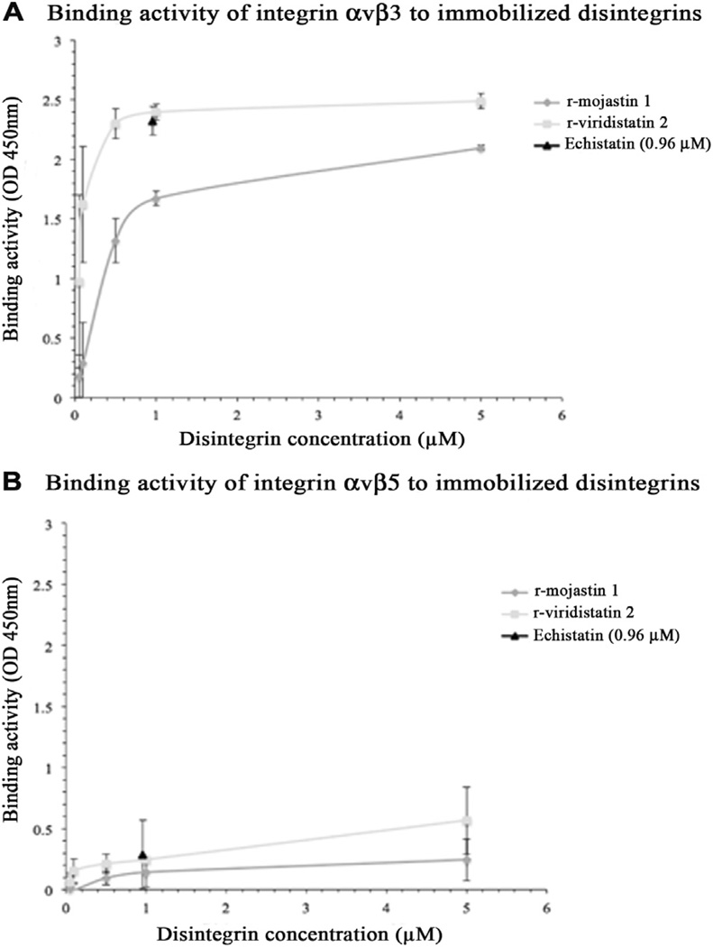
Fig. 6.
Interaction of immobilized r-disintegrins with A) integrin αvβ3 and B) integrin αvβ5. A 96-well plate was coated with various concentrations of rmojastin 1 or r-viridistatin 2. After blocking with 1% BSA in PBS-T for 1 h, soluble integrins αvβ3 or αvβ5 (2 µg) were added to each well and the plates were incubated for additional 2 h at room temperature. After washing with PBS-T, anti-αvβ3 or anti-αvβ5 was used as the primary antibody. After 1 h incubation at room temperature and subsequent washing, an HRP conjugated-goat anti-mouse IgG was used as the secondary antibody. The color was developed by TMB substrate solution. Absorbance at 450 nm of the individual well was measured to determine the binding activity. The disintegrin echistatin was used as a positive control and prepared by coating the plate with 0.96 µM echistatin instead of r-mojastin 1 and r-viridistatin 2. The error bars represent the standard deviation from two independent experiments.

