

Abstract
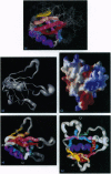
The pleckstrin homology (PH) domain is a recognition motif thought to be involved in signal-transduction pathways controlled by a variety of cytoplasmic proteins. Assignments of nearly all 1H, 13C, and 15N resonances of the PH domain from dynamin have been obtained from homonuclear and heteronuclear NMR experiments. The secondary structure has been elucidated from the pattern of nuclear Overhauser enhancements, from 13C chemical shift deviations, and from observation of slowly exchanging amide hydrogens. The secondary structure contains one alpha-helix and eight beta-strands, seven of which are arranged in two contiguous, antiparallel beta-sheets. The structure is monomeric, in contrast to the well-defined intimate dimerization of the crystal structure of this molecule. Residues possibly involved in ligand binding are in apparently flexible loops. Steady-state 15N(1H) nuclear Overhauser effect measurements indicate unequivocally the boundaries of this PH domain, and the structured portion of the domain appears to be more extended to the C terminus than previously suggested for other PH domains.
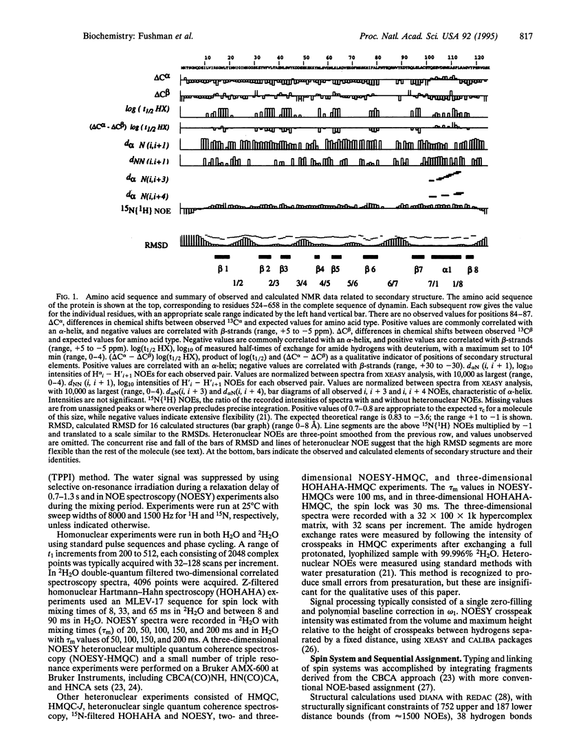
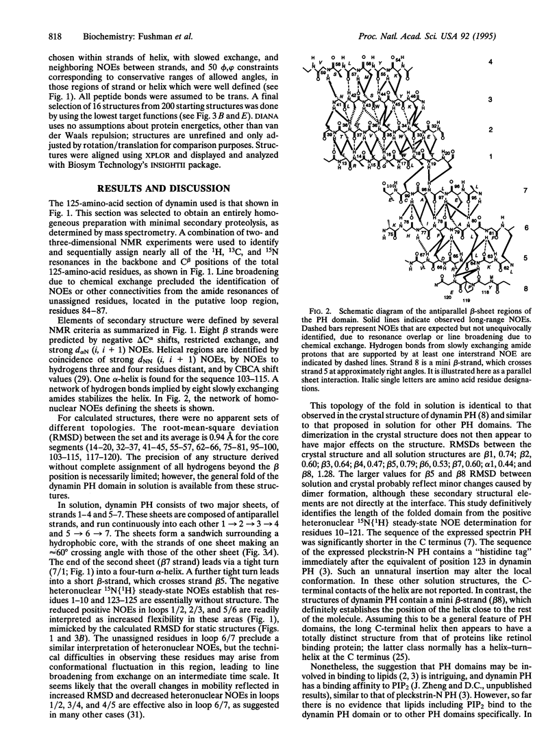
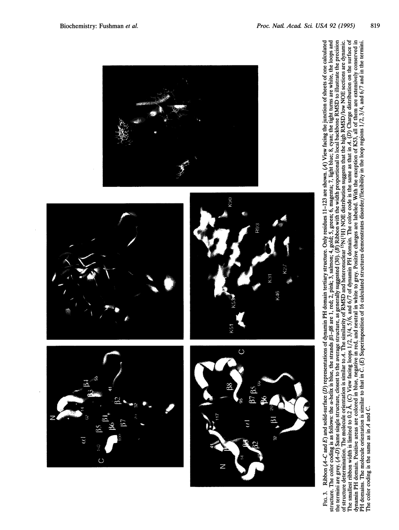
Full text
PDF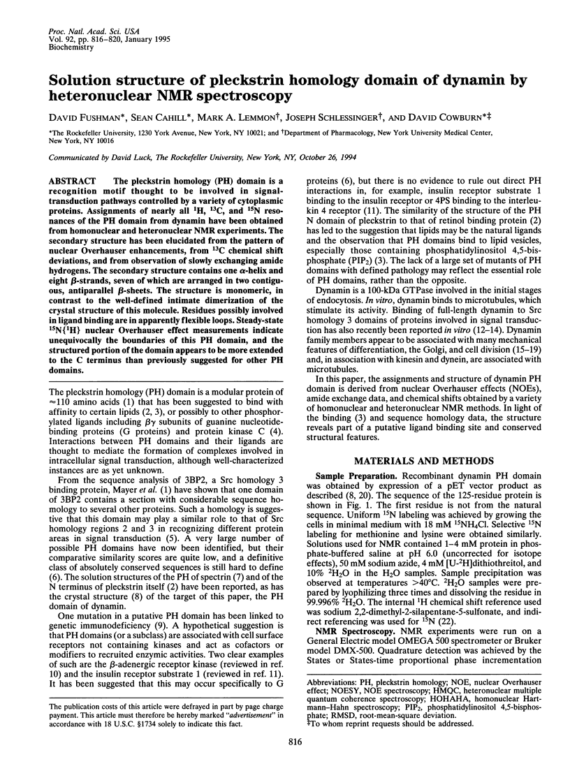
Images in this article
Selected References
These references are in PubMed. This may not be the complete list of references from this article.
- Bloom G. S. Motor proteins for cytoplasmic microtubules. Curr Opin Cell Biol. 1992 Feb;4(1):66–73. doi: 10.1016/0955-0674(92)90060-p. [DOI] [PubMed] [Google Scholar]
- Booker G. W., Gout I., Downing A. K., Driscoll P. C., Boyd J., Waterfield M. D., Campbell I. D. Solution structure and ligand-binding site of the SH3 domain of the p85 alpha subunit of phosphatidylinositol 3-kinase. Cell. 1993 May 21;73(4):813–822. doi: 10.1016/0092-8674(93)90259-s. [DOI] [PubMed] [Google Scholar]
- Chen M. S., Burgess C. C., Vallee R. B., Wadsworth S. C. Developmental stage- and tissue-specific expression of shibire, a Drosophila gene involved in endocytosis. J Cell Sci. 1992 Nov;103(Pt 3):619–628. doi: 10.1242/jcs.103.3.619. [DOI] [PubMed] [Google Scholar]
- Downing A. K., Driscoll P. C., Gout I., Salim K., Zvelebil M. J., Waterfield M. D. Three-dimensional solution structure of the pleckstrin homology domain from dynamin. Curr Biol. 1994 Oct 1;4(10):884–891. doi: 10.1016/s0960-9822(00)00197-4. [DOI] [PubMed] [Google Scholar]
- Ferguson K. M., Lemmon M. A., Schlessinger J., Sigler P. B. Crystal structure at 2.2 A resolution of the pleckstrin homology domain from human dynamin. Cell. 1994 Oct 21;79(2):199–209. doi: 10.1016/0092-8674(94)90190-2. [DOI] [PubMed] [Google Scholar]
- Flower D. R., North A. C., Attwood T. K. Structure and sequence relationships in the lipocalins and related proteins. Protein Sci. 1993 May;2(5):753–761. doi: 10.1002/pro.5560020507. [DOI] [PMC free article] [PubMed] [Google Scholar]
- Gibson T. J., Hyvönen M., Musacchio A., Saraste M., Birney E. PH domain: the first anniversary. Trends Biochem Sci. 1994 Sep;19(9):349–353. doi: 10.1016/0968-0004(94)90108-2. [DOI] [PubMed] [Google Scholar]
- Gout I., Dhand R., Hiles I. D., Fry M. J., Panayotou G., Das P., Truong O., Totty N. F., Hsuan J., Booker G. W. The GTPase dynamin binds to and is activated by a subset of SH3 domains. Cell. 1993 Oct 8;75(1):25–36. [PubMed] [Google Scholar]
- Grzesiek S., Bax A. Amino acid type determination in the sequential assignment procedure of uniformly 13C/15N-enriched proteins. J Biomol NMR. 1993 Mar;3(2):185–204. doi: 10.1007/BF00178261. [DOI] [PubMed] [Google Scholar]
- Güntert P., Braun W., Wüthrich K. Efficient computation of three-dimensional protein structures in solution from nuclear magnetic resonance data using the program DIANA and the supporting programs CALIBA, HABAS and GLOMSA. J Mol Biol. 1991 Feb 5;217(3):517–530. doi: 10.1016/0022-2836(91)90754-t. [DOI] [PubMed] [Google Scholar]
- Güntert P., Wüthrich K. Improved efficiency of protein structure calculations from NMR data using the program DIANA with redundant dihedral angle constraints. J Biomol NMR. 1991 Nov;1(4):447–456. doi: 10.1007/BF02192866. [DOI] [PubMed] [Google Scholar]
- Harlan J. E., Hajduk P. J., Yoon H. S., Fesik S. W. Pleckstrin homology domains bind to phosphatidylinositol-4,5-bisphosphate. Nature. 1994 Sep 8;371(6493):168–170. doi: 10.1038/371168a0. [DOI] [PubMed] [Google Scholar]
- Herskovits J. S., Shpetner H. S., Burgess C. C., Vallee R. B. Microtubules and Src homology 3 domains stimulate the dynamin GTPase via its C-terminal domain. Proc Natl Acad Sci U S A. 1993 Dec 15;90(24):11468–11472. doi: 10.1073/pnas.90.24.11468. [DOI] [PMC free article] [PubMed] [Google Scholar]
- Hollenbeck P. J. Cell motility. Dynamin joins the family. Nature. 1990 Sep 20;347(6290):229–229. doi: 10.1038/347229a0. [DOI] [PubMed] [Google Scholar]
- Izutsu K. [Cell division and the microtubular cytoskeleton]. Hum Cell. 1991 Jun;4(2):100–108. [PubMed] [Google Scholar]
- Kay L. E., Torchia D. A., Bax A. Backbone dynamics of proteins as studied by 15N inverse detected heteronuclear NMR spectroscopy: application to staphylococcal nuclease. Biochemistry. 1989 Nov 14;28(23):8972–8979. doi: 10.1021/bi00449a003. [DOI] [PubMed] [Google Scholar]
- Keegan A. D., Nelms K., White M., Wang L. M., Pierce J. H., Paul W. E. An IL-4 receptor region containing an insulin receptor motif is important for IL-4-mediated IRS-1 phosphorylation and cell growth. Cell. 1994 Mar 11;76(5):811–820. doi: 10.1016/0092-8674(94)90356-5. [DOI] [PubMed] [Google Scholar]
- Lefkowitz R. J. G protein-coupled receptor kinases. Cell. 1993 Aug 13;74(3):409–412. doi: 10.1016/0092-8674(93)80042-d. [DOI] [PubMed] [Google Scholar]
- Lemmon M. A., Ladbury J. E. Thermodynamic studies of tyrosyl-phosphopeptide binding to the SH2 domain of p56lck. Biochemistry. 1994 May 3;33(17):5070–5076. doi: 10.1021/bi00183a010. [DOI] [PubMed] [Google Scholar]
- Macias M. J., Musacchio A., Ponstingl H., Nilges M., Saraste M., Oschkinat H. Structure of the pleckstrin homology domain from beta-spectrin. Nature. 1994 Jun 23;369(6482):675–677. doi: 10.1038/369675a0. [DOI] [PubMed] [Google Scholar]
- Mayer B. J., Ren R., Clark K. L., Baltimore D. A putative modular domain present in diverse signaling proteins. Cell. 1993 May 21;73(4):629–630. doi: 10.1016/0092-8674(93)90244-k. [DOI] [PubMed] [Google Scholar]
- Musacchio A., Gibson T., Rice P., Thompson J., Saraste M. The PH domain: a common piece in the structural patchwork of signalling proteins. Trends Biochem Sci. 1993 Sep;18(9):343–348. doi: 10.1016/0968-0004(93)90071-t. [DOI] [PubMed] [Google Scholar]
- Rawlings D. J., Saffran D. C., Tsukada S., Largaespada D. A., Grimaldi J. C., Cohen L., Mohr R. N., Bazan J. F., Howard M., Copeland N. G. Mutation of unique region of Bruton's tyrosine kinase in immunodeficient XID mice. Science. 1993 Jul 16;261(5119):358–361. doi: 10.1126/science.8332901. [DOI] [PubMed] [Google Scholar]
- Shaw G. Identification of novel pleckstrin homology (PH) domains provides a hypothesis for PH domain function. Biochem Biophys Res Commun. 1993 Sep 15;195(2):1145–1151. doi: 10.1006/bbrc.1993.2164. [DOI] [PubMed] [Google Scholar]
- Sutcliffe M. J. Representing an ensemble of NMR-derived protein structures by a single structure. Protein Sci. 1993 Jun;2(6):936–944. doi: 10.1002/pro.5560020607. [DOI] [PMC free article] [PubMed] [Google Scholar]
- Timm D., Salim K., Gout I., Guruprasad L., Waterfield M., Blundell T. Crystal structure of the pleckstrin homology domain from dynamin. Nat Struct Biol. 1994 Nov;1(11):782–788. doi: 10.1038/nsb1194-782. [DOI] [PubMed] [Google Scholar]
- Vuister G. W., Kim S. J., Wu C., Bax A. NMR evidence for similarities between the DNA-binding regions of Drosophila melanogaster heat shock factor and the helix-turn-helix and HNF-3/forkhead families of transcription factors. Biochemistry. 1994 Jan 11;33(1):10–16. doi: 10.1021/bi00167a002. [DOI] [PubMed] [Google Scholar]
- Yeh E., Driscoll R., Coltrera M., Olins A., Bloom K. A dynamin-like protein encoded by the yeast sporulation gene SPO15. Nature. 1991 Feb 21;349(6311):713–715. doi: 10.1038/349713a0. [DOI] [PubMed] [Google Scholar]
- Yoon H. S., Hajduk P. J., Petros A. M., Olejniczak E. T., Meadows R. P., Fesik S. W. Solution structure of a pleckstrin-homology domain. Nature. 1994 Jun 23;369(6482):672–675. doi: 10.1038/369672a0. [DOI] [PubMed] [Google Scholar]