

Abstract
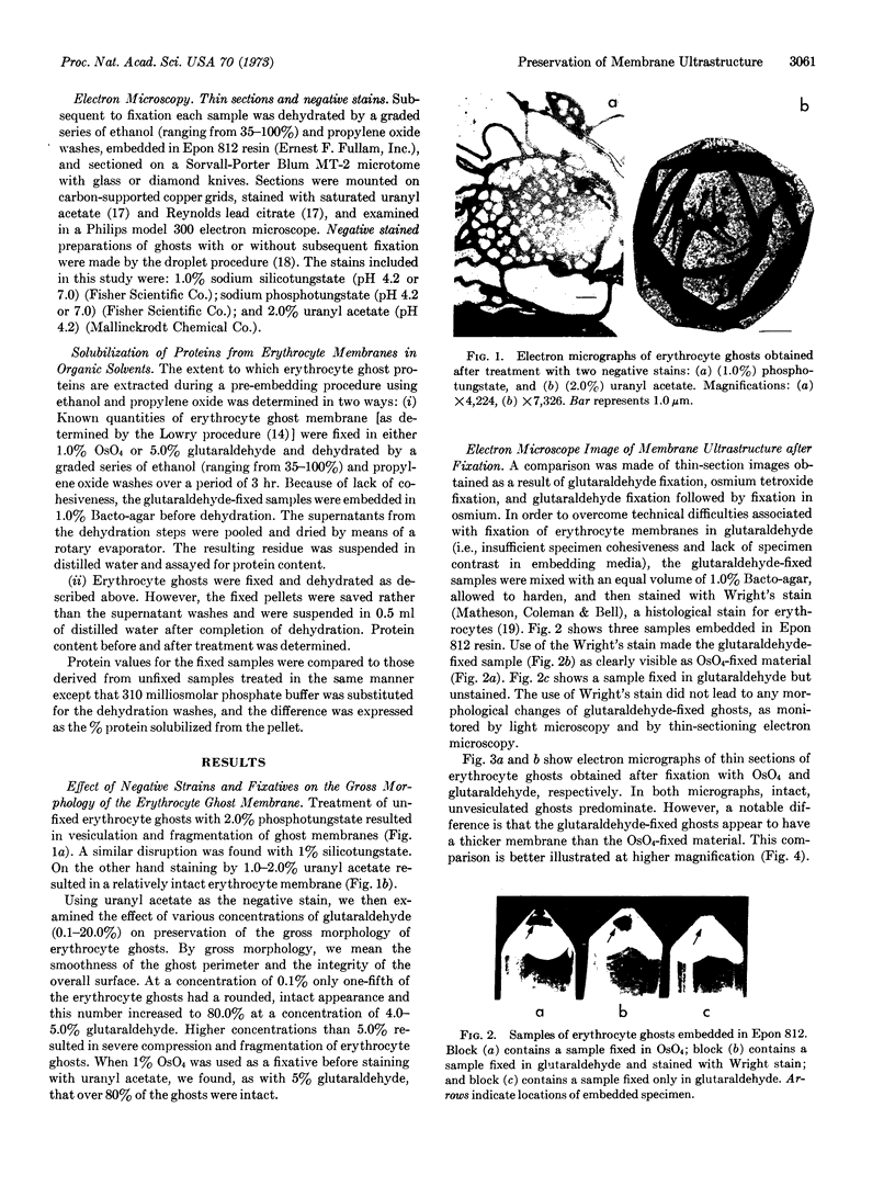
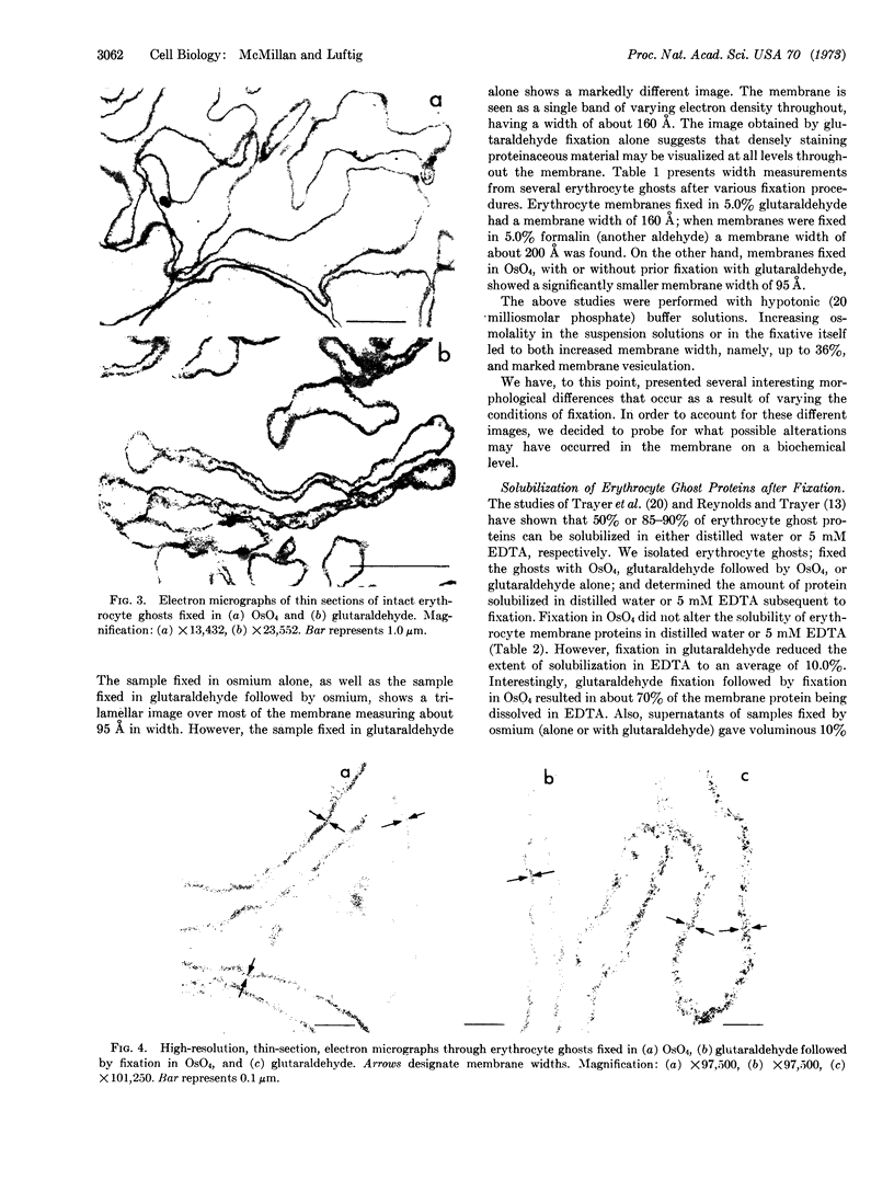
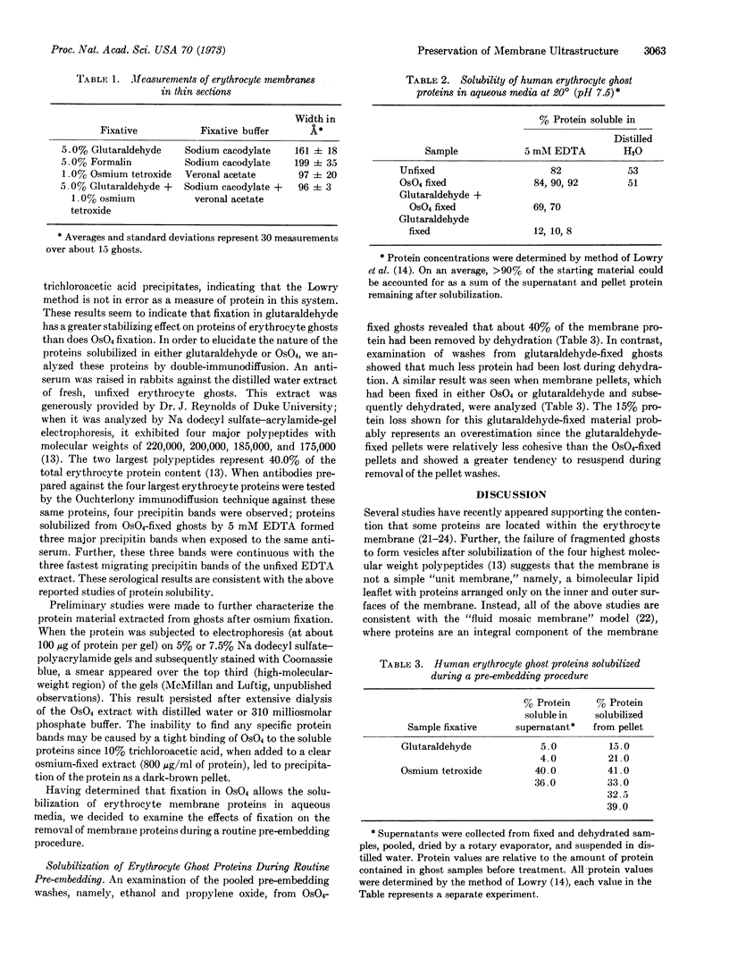
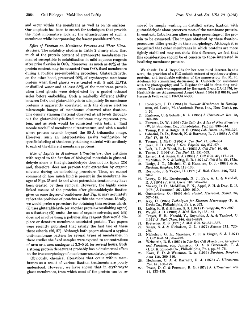
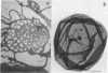
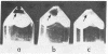
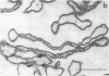
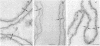
We have evaluated the quality of ultrastructural preservation of erythrocyte ghosts achieved under various electron microscopic preparative conditions. Initially, negative staining was used to monitor gross morphology of the ghosts. Of several negative stains used (phosphotungstate, silicotungstate, ammonium molybdate, and uranyl acetate), all but uranyl acetate resulted in fragmentation of membranes and the appearance of small vesicular structures. Further, preservation of gross membrane ultrastructure was greatly enhanced when samples were fixed with either 4.0-5.0% glutaraldehyde or 1% osmium tetroxide (OsO4) before they were stained with uranyl acetate. We then examined the ability of these fixatives to preserve membrane fine structure, as monitored by thin-sectioning procedures. In these studies, fixation with 1% osmium tetroxide (alone or in conjunction with 5% glutaraldehyde) resulted in a trilamellar image about 95 Å in width. Fixation with 5% glutaraldehyde alone provided a markedly different result. The membrane now appeared as a single line about 160 Å wide with regions of varying electron density throughout. This result suggests that glutaraldehyde used alone may reveal the location of membrane proteins that are obscured or removed by OsO4 fixation. This point would seem to be supported by the results obtained when erythrocyte membranes were extracted with 5 mM EDTA after fixation in either 5% glutaraldehyde or 1% OsO4. While only 10% of the detectable protein was solubilized from glutaraldehyde-treated erythrocyte membranes, 85% was solubilized from OsO4-treated ghosts. Among these latter proteins are three that migrated on Ouchterlony double-diffusion agar plates at the same position as three known proteins with molecular weights of about 200,000. Additional studies indicated that, even during a routine pre-embedding procedure, OsO4 led to solubilization of as much as 8 times the amount of protein as glutaraldehyde alone. Although the erythrocyte membrane has a notoriously weak association with its proteins, we feel that our studies provide a cautionary note with regard to the use of OsO4 as a fixative in other membrane systems.
Keywords: plasma membranes, electron microscopy, fixation procedures, membrane proteins
Full text
PDF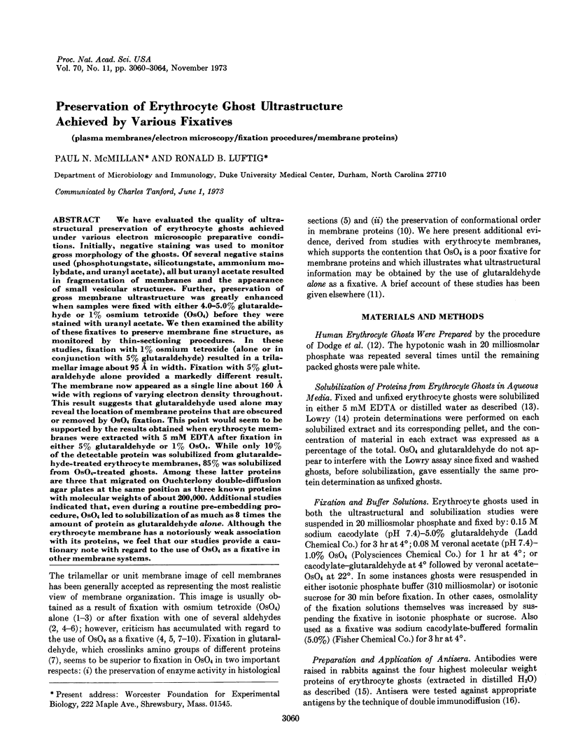
Images in this article
Selected References
These references are in PubMed. This may not be the complete list of references from this article.
- Bretscher M. S. A major protein which spans the human erythrocyte membrane. J Mol Biol. 1971 Jul 28;59(2):351–357. doi: 10.1016/0022-2836(71)90055-6. [DOI] [PubMed] [Google Scholar]
- DODGE J. T., MITCHELL C., HANAHAN D. J. The preparation and chemical characteristics of hemoglobin-free ghosts of human erythrocytes. Arch Biochem Biophys. 1963 Jan;100:119–130. doi: 10.1016/0003-9861(63)90042-0. [DOI] [PubMed] [Google Scholar]
- Heckman C. A., Barrnett R. J. GACH: a water-miscible, lipid-retaining embedding polymer for electron microscopy. J Ultrastruct Res. 1973 Jan;42(1):156–179. doi: 10.1016/s0022-5320(73)80013-9. [DOI] [PubMed] [Google Scholar]
- KARLSSON U., SCHULTZ R. L. FIXATION OF THE CENTRAL NERVOUS SYSTEM FROM ELECTRON MICROSCOPY BY ALDEHYDE PERFUSION. I. PRESERVATION WITH ALDEHYDE PERFUSATES VERSUS DIRECT PERFUSION WITH OSMIUM TETROXIDE WITH SPECIAL REFERENCE TO MEMBRANES AND THE EXTRACELLULAR SPACE. J Ultrastruct Res. 1965 Feb;12:160–186. doi: 10.1016/s0022-5320(65)80014-4. [DOI] [PubMed] [Google Scholar]
- Korn E. D., Weisman R. A. I. Loss of lipids during preparation of amoebae for electron microscopy. Biochim Biophys Acta. 1966 Apr 4;116(2):309–316. doi: 10.1016/0005-2760(66)90013-0. [DOI] [PubMed] [Google Scholar]
- LOWRY O. H., ROSEBROUGH N. J., FARR A. L., RANDALL R. J. Protein measurement with the Folin phenol reagent. J Biol Chem. 1951 Nov;193(1):265–275. [PubMed] [Google Scholar]
- Lenard J., Singer S. J. Alteration of the conformation of proteins in red blood cell membranes and in solution by fixatives used in electron microscopy. J Cell Biol. 1968 Apr;37(1):117–121. doi: 10.1083/jcb.37.1.117. [DOI] [PMC free article] [PubMed] [Google Scholar]
- Luftig R. B., Kilham S. S. An electron microscope study of Rauscher leukemia virus. Virology. 1971 Nov;46(2):277–297. doi: 10.1016/0042-6822(71)90030-4. [DOI] [PubMed] [Google Scholar]
- Mickey D. D., McMillan P. N., Appel S. H., Day E. D. The specificity and cross-reactivity of antisynaptosome antibodies as determined by sequential adsorption analysis. J Immunol. 1971 Dec;107(6):1599–1610. [PubMed] [Google Scholar]
- Nicolson G. L., Marchesi V. T., Singer S. J. The localization of spectrin on the inner surface of human red blood cell membranes by ferritin-conjugated antibodies. J Cell Biol. 1971 Oct;51(1):265–272. doi: 10.1083/jcb.51.1.265. [DOI] [PMC free article] [PubMed] [Google Scholar]
- Pease D. C., Peterson R. G. Polymerizable glutaraldehyde-urea mixtures as polar, water-containing embedding media. J Ultrastruct Res. 1972 Oct;41(1):133–159. doi: 10.1016/s0022-5320(72)90043-3. [DOI] [PubMed] [Google Scholar]
- Reynolds J. A., Trayer H. Solubility of membrane proteins in aqueous media. J Biol Chem. 1971 Dec 10;246(23):7337–7342. [PubMed] [Google Scholar]
- SABATINI D. D., BENSCH K., BARRNETT R. J. Cytochemistry and electron microscopy. The preservation of cellular ultrastructure and enzymatic activity by aldehyde fixation. J Cell Biol. 1963 Apr;17:19–58. doi: 10.1083/jcb.17.1.19. [DOI] [PMC free article] [PubMed] [Google Scholar]
- Singer S. J., Nicolson G. L. The fluid mosaic model of the structure of cell membranes. Science. 1972 Feb 18;175(4023):720–731. doi: 10.1126/science.175.4023.720. [DOI] [PubMed] [Google Scholar]
- TOOZE J. MEASUREMENTS OF SOME CELLULAR CHANGES DURING THE FIXATION OF AMPHIBIAN ERYTHROCYTES WITH OSMIUM TETROXIDE SOLUTIONS. J Cell Biol. 1964 Sep;22:551–563. doi: 10.1083/jcb.22.3.551. [DOI] [PMC free article] [PubMed] [Google Scholar]
- TORMEY J. M. DIFFERENCES IN MEMBRANE CONFIGURATION BETWEEN OSMIUM TETROXIDE-FIXED AND GLUTARALDEHYDE-FIXED CILIARY EPITHELIUM. J Cell Biol. 1964 Dec;23:658–664. doi: 10.1083/jcb.23.3.658. [DOI] [PMC free article] [PubMed] [Google Scholar]
- Trayer H. R., Nozaki Y., Reynolds J. A., Tanford C. Polypeptide chains from human red blood cell membranes. J Biol Chem. 1971 Jul 25;246(14):4485–4488. [PubMed] [Google Scholar]
- Trump B. F., Bulger R. E. New ultrastructural characteristics of cells fixed in a glutaraldehyde-osmium tetroxide mixture. Lab Invest. 1966 Jan;15(1 Pt 2):368–379. [PubMed] [Google Scholar]