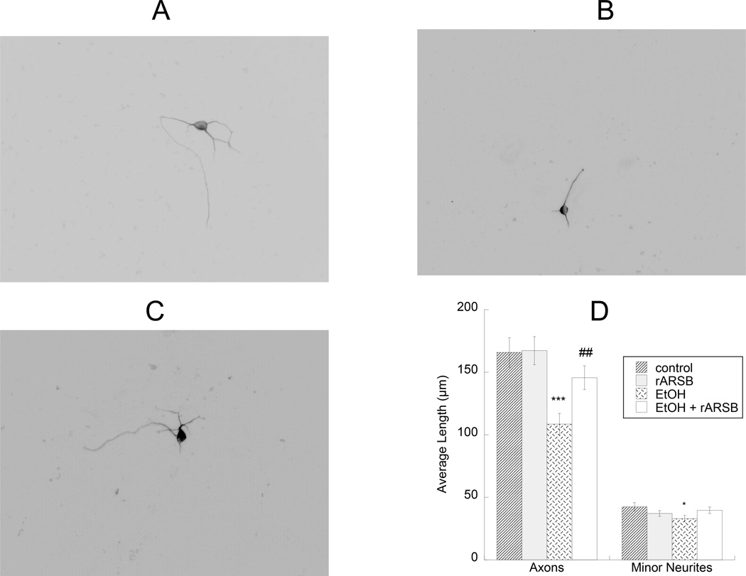
Figure 4. Effect of ethanol- and rARSB-treated astrocytes on hippocampal neuron neurite outgrowth.
Astrocytes were treated for 24 h with 75 mM ethanol, 1 ng/ml rARSB, or ethanol plus rARSB. Hippocampal neurons were plated on top of pre-treated astrocytes for an additional 18h. Cultures were then fixed and stained with neuron-specific βIII-tubulin antibody and a fluorescent secondary antibody. Neurite length was measured using the software Image J. Shown are representative neurons incubated with control (A), ethanol-treated (B), ethanol- and rARSB-treated (C) astrocytes. D: Morphometric quantification of axons and minor neurite length in 50 cells per treatment. ***, p<0.001; *, p<0.05 vs. control; ##, p<0.01 by Student’s t test.

