Abstract
Middle ear diseases in childhood play an important role in daily ENT practice due to their high incidence. Some of these like acute otitis media or otitis media with effusion have been studied extensively within the last decades. In this article, we present a selection of important childhood middle ear diseases and discuss the actual literature concerning their treatment, management of complications and outcome. Another main topic of this paper deals with the possibilities of surgical hearing rehabilitation in childhood. The bone-anchored hearing aid BAHA® and the active partially implantable device Vibrant Soundbridge® could successfully be applied for children. In this manuscript, we discuss the actual literature concerning clinical outcomes of these implantable hearing aids.
Keywords: children, otitis media, mastoiditis, cholesteatoma, implantable hearing aids
1 Introduction
Children’s middle ear diseases play an important role in daily ENT practice as well as in scientific questions. According to their high incidence, some are part of daily ENT routine, e.g., acute otitis media (AOM), one of the most frequent diseases of our specialty. The incidence of chronic otitis media with effusion (OME) has decreased in developed countries, yet it still plays an important role in the health and economy of developing and emerging countries.
Concerning the treatment of children’s middle ear diseases, many different and discussed concepts exist, such as pediatricians’ rising idea of conservative treatment of acute mastoiditis (AM) versus classical surgical therapy, similar to the debate about the benefit of adenoidectomy (AT) and ventilation tubes (VTs) in the treatment of OME. Other foci are the extensive investigation of different techniques applied in children’s cholesteatoma surgery during the last years and further development of implantable hearing aids such as the clinically proved systems BAHA® und Vibrant Soundbridge®.
All these topics do not only have clinical relevance, but also have to be continuously reviewed and evaluated regarding their effectiveness in times of evidence-based medicine and growing costs of health care. The intention of this manuscript is to present elected topics of children’s middle ear diseases on a limited scale. The authors aim to provide an overview of the actual state of the science, to aid the reader’s therapeutic decisions and indications.
2 Selected diseases and therapeutic options
2.1 Acute otitis media
2.1.1 Definition
AOM is one of the most common infectious diseases in childhood [1]. It is characterized by an inflammation of the middle ear, in particular of the tympanic cavity, with an acute beginning and short duration of illness. Distinction between viral and bacterial genesis should be made [2]. Recurrent otitis media is defined by three episodes of AOM in 6 months or more than four AOMs in 12 months, with intermittent normalization of middle ear findings [1].
2.1.2 Epidemiology
AOM is the most frequent reason of medical consultation [3] and the main indication of antibiotic therapy in childhood [4]. It usually occurs between the age of 3 months and 3 years, with a peak of incidence from 6 to 11 months. By the age of 3 years, up to 80% of all children have suffered at least once from AOM [5], [6]. About 40% have had six or more AOMs by their seventh year of life [7]. The global incidence has doubled from 1970 to 1990 [3] and was recently calculated to be 10.85% or 709 million per year, varying from 3.64% in central Europe to 43.37% in central Africa [7].
2.1.3 Pathogenesis and predisposing factors
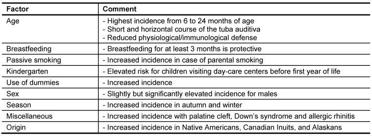
Anatomical, physiological, and immunological factors play a role, leading to the high incidence of AOM in childhood. The most important factor is dysfunction of the tuba auditiva (TA) [1]. Its course in infancy is short and horizontal and shows reduced rigidity. These characteristics forward ascending germs from the nasopharynx. Due to viral infections of the nasopharynx, its bacterial colonization and following ascend through the TA is promoted. Another important variable is the immune status, as the primary immune defense of the upper respiratory tract is dominated by Waldeyer’s tonsillar ring. Locally secreted antibodies represent the earliest defense against bacterial and viral adhesion and reduce local bacterial colonization [1]. While high concentrations of IgG2 can be measured in a newborn’s chord blood, the incidence of AOM from 6 to 11 months of age shows an opposing trend compared with the decrease of maternal antibodies. Besides these aspects, numerous studies have shown that there are predisposing risks for the development of AOM (Table 1 (Tab. 1); [1], [5], [8], [9], [10], [11], [12], [13]).
Table 1. Predisposing factors of acute otitis media [1, 5, 8–13].
2.1.4 Symptoms and diagnosis
AOM is characterized by acute symptoms of an ear infection. Older children report severe otalgia along with general symptoms of an infection, such as reduced general condition, fever, nausea, abdominal pain, absence of appetite, cough, and headache. Smaller children often touch their ears [14]. Yet, these symptoms are not specific for AOM [12] and therefore not reliable for diagnosis.
Otoscopic or microscopic visualization of the tympanic membrane (TM) is essential for diagnosis of AOM. Depending on the phase, the TM shows hyperemia or mild to severe bulging. In addition, there can be a spontaneous perforation with concomitant otorrhea [15]. Hyperemia alone is not sufficient for diagnosis due to its low positive predictive value (7%) [14] The newest guidelines of the American Academy of Pediatrics list the following criteria for the diagnosis of AOM [12], [16]:
Mild to severe bulging of the TM and confirmation of otorrhea that is not related to otitis externa
Mild bulging of the TM with otalgia or severe redness in the last 48 hours
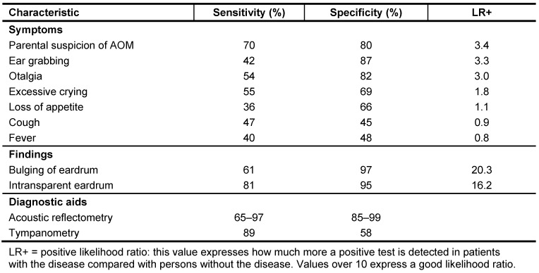
Furthermore, the American Academy of Pediatrics recommends the lack of middle ear effusion as a criterion for exclusion of AOM. The proper examination of babies and smaller children is often challenging, due to narrowed ear canals or non-removable cerumen. For these reasons, an appropriate assessment of the TM is not always possible [1], [12]. Also, distinguishing TM bulging from mild to severe remains unstandardized. According to the literature, additional diagnostic techniques are tympanometry and acoustic reflectometry [12]. An effusion in the tympanic cavity leads to a pathological pattern of reflected noise waves, which can be detected with the acoustic reflectometry. Both are tools for diagnosis of a middle ear effusion but are not sufficient for diagnosis of AOM. The relevance of clinical symptoms and diagnostic aids for AOM are summarized in Table 2 (Tab. 2).
Table 2. Relevance of clinical symptoms and diagnostic aids in diagnosis of acute otitis media (modified from [12]).
2.1.5 Microbiology
In most cases, a viral infection of the upper airways precedes AOM. The most common verified virus in middle ear effusions during AOM is Respiratory Syncytial Virus [1], [12], [14], [16]. Other important agents are influenza virus and parainfluenza virus, as well as adeno-, entero-, and rhinovirus [17]. Additional bacteria are verified in up to 90% of cases [18]. Here, the most important are Haemophilus influenzae, followed by Moraxella catarrhalis. Rather rare are Streptococcus pyogenes and Staphylococcus aureus (Table 3 (Tab. 3); [12], [14], [19], [20], [21], [22]). Some studies from the United States have shown that H. influenzae infections have risen since the introduction of Streptococcus pneumoniae vaccination [14], [19]. These data could not be confirmed in Germany.
Table 3. Spectrum of pathogens in children’s acute otitis media [12, 14, 19–22].
2.1.6 Conservative therapy
Goals in the management of AOM are controlling symptoms (especially otalgia), prophylaxis of relapse, and avoidance of otogenic complications. Otalgia and fever are the main symptoms of AOM that require appropriate therapy. Recommended therapies are paracetamol (rectal/oral; 60 mg/kg/day in 4–6 partial doses) or ibuprofen (oral, 20–30 mg/kg/day in 3–4 partial doses). Histamine antagonist could relieve nasal symptoms in case of concomitant rhinosinusitis but extend the duration of middle ear effusions [23]. There are no evidence-based data for the use of decongestant nasal spray or drops, although they could also relieve concomitant nasal symptoms [1], [24]. A systematic Cochrane meta-analysis from 2004 that included 2,695 patients did not show any benefit from decongestant nasal spray/drops regarding healing, complications, or prevention of surgical interventions [24].
Most children suffering from AOM show spontaneous remission after 10–14 days. For that reason, the indication of antibiotic therapy should be regarded critically [12]. The introduction of antibiotics was responsible for the reduction of AM in the past but could later not be verified for uncomplicated AOM. Thus, the incidence of AM is not different between a wait-and-see strategy (watchful waiting) and immediate antibiotic therapy in older children. In addition, several studies comparing different therapy schemes did not show significant differences in the incidence of AM [25], except in children younger than 2 years where wait and see increased the rate of AM [26]. According to the presented literature, a wait-and-see strategy seems to be maintainable in children older than 2 years suffering from AOM, but close clinical monitoring is essential. In case of no improvement, an antibiotic therapy should be initiated.
Since the 1980s, several studies have shown only a small difference between watchful waiting and immediate antibiotic therapy [27], [28], [29]. Randomized studies of the last 30 years have proved that the majority of children recover from AOM without antibiotic therapy [1]. A meta-analysis of randomized studies discovered that, in particular, children below 2 years with bilateral AOM and children with AOM and otorrhea benefit from antibiotic therapy. Watchful waiting should be conducted in all other children with a mild course of AOM [30]. A recent study from 2011 investigated the helpfulness of antibiotics (amoxicillin/clavulanic acid) versus placebo in children between the 6th and 23rd months of life. Here, the antibiotics group showed significant advantages regarding reduction of symptoms and rate of inflammatory signs on the TM [31].
In summary, current clinical studies show that immediate antibiotic therapy is not necessary in most children suffering from uncomplicated AOM. The disadvantages of antibiotic therapy and the little benefit versus clinical observation recommend an individual therapeutic strategy [4], [12], [19], [22], [31]. Here, age and severity of symptoms play a critical role. According to the available data, indications for immediate antibiotic therapy are [4], [7], [16], [31], [32]:
Age <6 months
Age <2 years with bilateral AOM, even with little otalgia and temperature <39.0°C
AOM with moderate to severe otalgia or temperature ≥39.0°C
Persistent purulent otorrhea
High-risk patients (intratemporal or intracranial complications, immunodeficiency, cleft lip and palate, Down’s syndrome, cochlea implant)
Clinical control in the first 3 days not reliably possible
The first-choice antibiotic is amoxicillin, 50–60 mg/kg/d in three single doses for 10 days [12], [33]. In children older than 6 years with mild to moderate AOM, therapy for 5–7 days is sufficient [16], [34]. The advantages of amoxicillin are good effectiveness, high safety slim microbiological spectrum, low rate of adverse effects, low cost, and acceptable taste. As water-soluble dry syrups increase oral bioavailability and reduce gastro-intestinal adverse effects, this pharmaceutical form is recommended [35]. In case of presumably participating β-lactamase-positive bacteria, amoxicillin and clavulanic acid should be applied [16], [36]. In the following patient populations, amoxicillin should be combined with clavulanic acid:
Children with concomitant conjunctivitis
Amoxicillin has been taken in the last 30 days
Children using amoxicillin for prophylaxis of recurrent urinary tract infections or recurrent AOMs
Alternatives in case of penicillin allergy are cephalosporins (cefuroxime, cefpodoxime) if there is no anaphylactic shock or urticaria in response to penicillin treatment in anamnesis [12]. Otherwise, erythromycin, clarithromycin, or azithromycin can be applied [16]. When therapy compliance is reduced or children suffer from strong vomiting, a parenteral single shot with ceftriaxone (50 mg/kg) could be another therapeutic strategy [12].
If there is no improvement of symptoms during the first 48–72 hours, the child should be examined again and the diagnosis should be re-evaluated. Clinically observed children should then be given antibiotic therapy as described above. Children that are already treated with antibiotic require a change of antibiotic. Parenteral application of ceftriaxone is recommended if there is resistance against amoxicillin and clavulanic acid or strong vomiting [12], [14], [16], [31]. Cochlear implant users in the first 2 months following implantation should be treated systemically with ceftriaxone. After that period, oral amoxicillin with or without clavulanic acid is sufficient [37].
2.1.7 Surgical therapy
In some countries, myringotomy is routine therapy for AOM. However, randomized clinical trials showed that there was no healing benefit for combined myringotomy and antibiotic therapy compared with antibiotic therapy alone [1]. Therefore, myringotomy should only be conducted in case of serious disease progression, failure of antibiotic treatment, or complications of AOM. The most important complication is AM (see 2.2) and its intracranial and intratemporal complications.
2.2 Acute mastoiditis
AM is defined as an acute inflammation of the mastoid with colliquation of the air-filled mastoidal bone [38]. This complication of AOM is rather rare today [39], but has been a life-threatening disease in the pre-antibiotic era, with high mortality and morbidity [40]. In those days, about 20% of AOM led to AM, with frequent intracranial complications [41], [42].
2.2.1 Epidemiology
Since the introduction of antibiotic therapy in the 1940s, the incidence of AM has been considerably reduced; yet, an increase has been detected over the last years [38], [40], [43], [44]. Today’s incidence is 1.2–6.1 per 100,000 inhabitants in developed countries [44], [45]. Serious progressions appear more frequently in young children [43], [46], [47]. The rising incidence is connected to restrained antibiotic therapy of AOM, inadequate dosing, choice of antibiotics, and increasing resistance of bacteria [38], [43], [48].
Another factor is the exposure of infants to day-care centers, which leads to an increased incidence of infections of the upper airways and AOM [49]. This issue is emphasized by Groth et al. [44], who investigated the age-dependent incidence of AM in Swedish children aged 0 to 16 years. The highest incidence of AM has been detected in the second year of life. This could be explained by the exposure of Swedish children to day-care centers after their first year of life [44]. Countries with different time points of starting child care in day-care centers show incidence peaks of AM at other time points of childhood [50], [51], [52].
The most frequent causative organism for AM is S. pneumoniae, followed by S. pyogenes and S. aureus [38], [44], [53]. With aging, the major pathogen shifts from S. pneumonia to S. aureus [44]. Despite being the second most frequent cause of AOM, H. influenzae is rare in AM of childhood [40].
2.2.2 Symptoms and diagnosis
The diagnosis of AM is concluded from anamnesis and clinical symptoms. Most AM follow a preliminary AOM [53]. Depending on severity and duration, the main symptoms are intense otalgia, pasty retroauricular swelling, and a protruding ear caused by subperiosteal abscess formation. Groth et al. [26] report that 70% of their group of 577 patients showed signs of AOM at the time of AM diagnosis. The most frequent retroauricular signs were redness or a protruding ear. Subperiosteal abscess formation was reported in 20% of all cases [26].
Laboratory values are dominated by increased C-reactive protein, leukocytosis, and increased blood sedimentation speed. The inflammation can spread to the soft tissues of the neck (Bezold mastoiditis) or to the fossa digastrica (Muret mastoiditis) [54].
The execution of additional radiological procedures should be decided, depending on the severity of AM. Especially in infants, diagnosis has to be determined by clinical examination and symptoms [38]. Here, additional radiological procedures are not obligatory. In suspicion of complications, the performance of CT or MRI is advisable, but ought to be decided on a case-by-case basis [55]. MRI is preferred regarding intracranial complications [56]. CT of the temporal bone is the method of choice if clinical symptoms are not specific, but the suspicion of occult AM cannot be rejected [38].
2.2.3 Conservative treatment
AM should be treated by systemic antibiotic therapy. The antibiotics of choice according to the guideline of the German ENT Society are aminopenicillin combined with β-lactamase inhibitor or 2nd or 3rd generation cephalosporin (cefuroxime, ceftriaxone). After receiving the intraoperative resistance, the antibiotic therapy has to be adjusted, if necessary. Concerning Pseudomonas aeruginosa, other alternatives are fosfomycin, piperacillin, or ciprofloxacin [33].
Despite generally accepted opinions, many studies showed that middle ear symptoms precede AM by 10–14 days [57]. Luntz et al. [57] reported that 32% of children had suffered from middle ear symptoms for shorter than 48 hours at the time of AM diagnosis. A masked or occult mastoiditis (normal TM findings combined with signs of mastoiditis) was reported in 3% of all cases. This rising subgroup deserves a watchful and extensive clinical examination in order not to be overlooked. In addition, Luntz et al. described that 54% had already taken antibiotics before the development of AM. Therefore, we can conclude that antibiotic therapy does not absolutely protect from AM and its complications [55], [57].
2.2.4 Surgical therapy
Immediate mastoidectomy is still the method of choice today to treat AM with subperiosteal abscess formation. Classical mastoidectomy technique with a retroauricular approach has been established worldwide over the last decades. Complete removal of swollen mucosa is not necessary, yet the opening of inflammatory foci and affected mastoidal cells is required. Adequate ventilation should be guaranteed by a small silicone catheter directed to the antrum [58], [59]. All surgically treated patients have to receive a paracentesis (PC) and, depending on middle ear findings, a tympanostomy tube in addition to mastoidectomy [53]. Inspection of the nasopharynx and possibly adenoidectomy ought to be performed routinely in this surgical intervention [38]. Some authors even perform a PC prior to mastoidectomy. Psarommatis et al. [55] reported that 70% of their patients could have been treated successfully by PC only.
For that reason, immediate mastoidectomy has been controversially discussed in publications of the last few years. Some authors suggest antibiotic therapy alone, PC alone, or combined with puncture of superiosteal abscess as methods of choice [40], [44], [55], [60]. However, most studies preferring conservative therapy rely on retrospectively analyzed data. Prospective clinical studies with randomized comparison of different therapeutic strategies are still missing. Current literature additionally shows that AM is still associated with extra- and intracranial complications, especially in infants. Therefore, mastoidectomy should be generously indicated in this subgroup. The low complication rate and the established technique of mastoidectomy justify its status as the therapy of choice [38], [48], [53].
2.2.5 Complications of acute mastoiditis and their treatment
Since the introduction of antibiotics and adequate therapy of AOM, complications of AM have been significantly reduced [44]. Huge studies have shown that intracranial complications have been reduced from 2.3% to 0.24% [61]. The total incidence of complications is described as between 7% and 35%, whereas subperiosteal abscess formation is listed as a complication in many studies [40], [44], [61], [62]. Table 4 (Tab. 4) shows incidence rates from the literature. In the following sections, complications and their therapy are described.
Table 4. Selected studies on acute mastoiditis and its complication in children (SPA: subperiosteal abscess; FP: facial palsy; SST: sinus sigmoideus thrombosis).
Labyrinthitis
Labyrinthitis is a rare complication of AM [54]. Sensorineural hearing loss, vertigo, and spontaneous nystagmus are pathbreaking for its diagnosis. Nevertheless, the diagnosis could be very challenging in childhood. Therapy depends on removing the inflammatory focus by mastoidectomy and PC.
Petrositis
Today, this complication is rare but could be part of Gradenigo’s syndrome (retrobulbar pain, abducens nerve palsy, and ipsilateral acute or chronic otitis media) [54], [63]. A combined therapy of mastoidectomy (including the opening of mastoid cells in the petrous apex) with high-dose intravenous (i.v.) antibiotics is sufficient [54].
Facial palsy
Facial palsy is also a rare complication of AM. In addition to antibiotics, a prompt surgical management consisting of mastoidectomy and PC is indicated. Further, decompression of the mastoid portion of the nerve and steroids are recommended [38]. In cases of facial palsy as a complication of AOM without secure signs of AM, a PC and ventilation tubes (VT) are advisable. If there is no improvement within 3 days, a mastoidectomy is indicated [64].
Sinu sigmoideus thrombosis
This complication could be asymptomatic or become clinical if a thrombotic obstruction of the internal jugular vein leads to an increased intracranial pressure. The diagnostic tool of choice is a MRI-angiography [62]. Therapeutically, the sinus is exposed from the sinus-dura angle to the mastoid tip during the mastoidectomy. In cases of sepsis or suspicion of thrombosis, the sinus is punctured. If there is sign of thrombosis, the sinus is opened and the thrombosis evacuated. Further, the sinus should be obliterated with muscle or Surgicel [2]. Surgical removal of the thrombus is nowadays controversial. Some authors recommend in these cases heparin [54], [65]. In cases of sepsis, a transcervical ligation of the internal jugular vein is recommended [54].
Intracranial complications
The following intracranial complications are described: epidural and subdural abscess, meningitis, and brain abscess. The diagnosis of an intracranial complication could be very challenging. The most common symptoms are fever, otalgia, cephalgia, and reduced general condition. An altered mental status in combination with an AM could also be a sign of intracranial complication [54], [61]. The diagnostic method of choice is CT or MRI. The two radiological techniques are regarded as equally effective [54], [56], [61]. The treatment of choice is mastoidectomy combined with antibiotics that penetrate the central nervous system (CNS), such as ceftriaxone. An epidural abscess can be drained during the mastoidectomy. The treatment of a brain abscess should be interdisciplinary, including neurosurgery [2].
2.3 Otitis media with effusion
2.3.1 Definition
OME is referred to as an accumulation of non-purulent fluid behind the intact eardrum. The eardrum itself usually shows no signs of inflammation. The fluid can be serous (thin) or mucoid (thick). If the fluid is very thick, it is also called a “glue-ear” [15], [66]. If the tympanic effusion persists for more than 3 months, it is called an OME.
2.3.2 Epidemiology
OME is the most common cause of hearing loss and the most common disease of the ear in childhood. Eighty percent of all children have had an episode of this disease by the age of 10 years, mostly by the age of 3 years [66]. The prevalence is about 20% at the age of 2 years. The prevalence of OME has two maxima, the first at the age of 2 years, the second at the age of 5 years [67]. The two maxima of prevalence coincide with the time of kindergarten and primary school. The prevalence decreases to 8% at the age of 8 years. There are significant seasonal differences in the level of prevalence; it can double during the winter period in the northern hemisphere [68]. More than half of these cases are preceded by an AOM. Children with a previous OME suffer from an AOM five times more often [66].
2.3.3 Pathogenesis and predisposing factors
Dysfunction of the TA plays a key role in the pathogenesis of OME [15]. Politzer postulated an “ex-vacuo” theory in 1867 [69]. It states that a chronic vacuum of the middle ear (as a consequence of a TA dysfunction) causes an effusion. Inflammatory processes of the TA mucosa lead to an edema, which causes contraction of the TA. Also, adenoids of the epipharynx can cause mechanical obstruction of the TA. All forms of obstruction can cause an inadequate equalization of pressure between the middle ear and the epipharynx. The consequence of a chronic vacuum of the middle ear favors the accumulation of effusion in the tympanic cavity [66].
Brieger postulated a second theory about the genesis of OME in 1914 [66]. It focused on an inflammatory etiology of the mucosa of the middle ear. This theory was confirmed by histomorphologic studies of Sadé [70]. He described inflammatory hypertrophy of the middle ear mucosa, combined with hyperplasia of secretory cells. Current studies reveal that an inflammatory process is not the primary cause of OME. It is rather a secondary reaction of the mucosa, caused by chronic obstruction of the tuba auditiva [15], [66], [71], [72].
Also, other etiologies and diseases are mentioned concerning the genesis of OME, including allergies, primary ciliary dyskinesia (PCD), chronic rhinosinusitis, cleft palate, and an immature immune system [66], [71], [73], [74]. Ninety percent of children with a cleft palate develop OME [74], [75]. The majority of patients with PCD also develop OME, which may last unusually long [73], [76].
Today, OME is considered to be a multifactorial disease, which clinically appears on different levels, caused by predisposing factors. Sequence of birth and gender are predisposing factors. Younger siblings as well as children visiting nursery school run a higher risk of an OME. It is interesting that smoking by the mother is a risk factor but smoking by the father is not [15], [66], [71].
The role of allergic disease in the pathogenesis of OME has been investigated intensively in the last years. There is a broad association from 5 to 80% in childhood [77]. It seems that inhalant allergy plays a bigger role than food allergy. An increase of serum IgE could not be proved so far [78]. An allergy-triggered cascade is supposed to cause obstruction of the TA, which again could lead to an OME.
The role of a bacterial infection of the middle ear in the pathogenesis of OME is currently unclear [79]. S. pneumoniae, H. influenzae, and M. catarrhalis are the most common bacteria in case of OME or AOM. These pathogens could only be found in half of the cases in most studies. However, with polymerase chain reaction (PCR), bacterial DNA could be found in more than 80% of cases [80]. This discrepancy is explained by bacterial colonization and biofilm, which are intended to stimulate an OME. Bacterial colonization and biofilm are favored as an etiological factor [79].
2.3.4 Symptoms and diagnosis
OME can remain asymptomatic for a long period. Some children complain about a pressure sensation in the ear [66]. The leading symptom is hearing loss on both sides. Younger children with an OME show reduced articulation and a delay of language development. Older children often show a deficit of attention in school [81].
The most important diagnostic method is microscopic examination of the eardrum. Usually, a retracted but intact eardrum with a shortened malleus handle can be seen. The effusion can gleam through clear or yellowish to bluish, depending on viscosity.
Tone audiometric measurement is usually possible with children around the age of 4 years. Depending on the level of liquid in the middle ear, a conductive hearing loss around 10–50 dB can be observed. The majority of children show a hearing loss about 20–30 dB. The reliability of tone audiometry is interpreted differently and studies on this topic are rare. The specificity is stated to be around 53–92%, and sensitivity around 52–88% [82], [83], [84]. The tympanometric measurement usually shows a flattening of the curve. In contrast to tone audiometry, there are a lot of studies showing the high reliability of tympanometry [85]. For the diagnosis of OME, sensitivity is stated to be around 93%, specificity around 70% [85], [86], [87].
2.3.5 Conservative therapy
There has been an intensive discussion on the treatment of OME over the last decades. Due to the high rate of spontaneous healing, the indication for conservative or surgical therapy should be weighed carefully. Many studies show that the majority of children with AOM develop a chronic tympanic effusion, but which can dissolve spontaneously [15]. The rate of spontaneous healing is stated to be 42% after 6 months and 33% after 12 months [72]. A newly diagnosed OME without any risk factors (craniofacial anomalies, cleft palate, PCD) should be treated conservatively for at least 3 months [15], [33], [66], [85].
In contrast to AOM, the benefit of antibiotic treatment of OME is disputed [88]. Some studies show that antibiotic treatment for 2–8 weeks could be useful [89], [90]. The benefit was often insignificant after the interruption of treatment within 2 weeks [91]. On the other hand, there are side effects, development of resistance, and high costs [88]. A recent meta-analysis examined 23 studies on the efficiency of antibiotic treatment, including 3,027 children [92]. It showed no benefit concerning hearing loss or reduction of the number of VTs.
A 2002 meta-analysis examined the efficiency of treatment of OME with oral or topic steroid in combination with or without antibiotics [93]. It showed a short-term benefit concerning the dissolving of a tympanic effusion. However, there was no long-term profit. Treatment with antibiotics in combination with or without steroids is recommended only in exceptional cases. These include children whose parents refuse surgical treatment. In these cases, an attempt with single antibiotic treatment for 10–14 days can be made. Long-term treatment with antibiotics in combination with or without steroids is not recommended [88].
Several studies did not show any effect of treatment with antihistamines or decongestants (also in combination); therefore, application for OME treatment is not recommended [88]. Further conservative options of therapy are the Valsalva maneuver and nasal inflation of a special balloon. These methods can be efficient if executed several times a day. Because of insufficient compliance, around 12% of children between 3–10 years of age are not able to use these methods [66].
2.3.6 Surgical therapy
General aspects
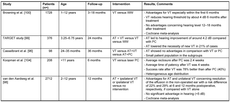
Surgical therapy is still one of the most discussed topics in the field of pediatric ENT medicine [94], [95], [96], [97], [98], [99]. In case of an OME persisting for over 3 months, the indication for a surgical therapy is given. The aims of this treatment are to improve hearing and to prevent a relapse. Surgical methods include PC, insertion of a VT, and AT and will be discussed later. These three methods have been used a lot over the last decades (alone or in combination), so there are a lot of data concerning the risks and benefits [85], [88], [95], [98], [99], [100], [101], [102], [103], [104]. Table 5 (Tab. 5) shows some important randomized clinical studies and meta-analyses on the efficiency of the mentioned methods of therapy.
Table 5. Current studies on effectiveness of surgical management for OME (AT: adenoidectomy; PC: paracentesis; VT: ventilation tube; WW: watchful waiting).
Insertion of ventilation tubes
The gold standard for surgical treatment of OME is the insertion of vent tubes in combination with an AT [15], [66]. A meta-analysis by Browning et al. [100] examined the efficiency of the insertion of VTs compared with conservative clinical observation. This meta-analysis underlines that the insertion of VTs within 6 months after surgical treatment has an advantage against clinical observation. The auditory threshold (6–9 months after treatment) was around 4.2 dB better in children with surgical treatment in contrast to conservative therapy. This difference disappeared after the period of 1 year [100]. There was no difference in language development after 6–9 months between surgical and conservative therapies.
The data of this meta-analysis and other studies were summarized and analyzed recently [105]. The insertion of VT reduced the duration of a tympanic effusion by 32% compared with clinical observation and by 42% compared with PC only. Further, the insertion of VT increased hearing by around 10 dB versus clinical observation or PC only 4–6 months after treatment.
Adenoidectomy
Presently, there is no consensus about performing an AT at the moment of VT insertion. The guidelines of the American Academy of Pediatrics (2004) do not recommend an AT in children with a first-time VT insertion because of an OME [88]. The reason for this decision was medicolegal rather than based on the small therapeutic benefit [66]. The current guidelines of the German ENT Society recommend a simultaneous PC or insertion of VT and AT. This combined treatment is superior to PC or VT only [106].
The randomized multi-center Trial of Alternative Regimens in Glue Ear Treatment (TARGET) examined the efficiency of AT [99]. This study included 376 children older than 3.5 years with an OME and a hearing loss over 20 dB from 11 hospitals. The study focused on the efficiency of combining AT and VT versus VT only or clinical observation. The patients were observed over 2 years, with five points of follow-up. The study showed an advantage of combining AT and VT. AT reduced the need for a second VT insertion by 21%. The major conclusion of this study is that combined treatment (AT and VT) has a long-term advantage by reducing the risk of a relapsing OME after VT extrusion [99].
A 2010 meta-analysis confirmed the benefit of an AT [98]. AT in combination with a single-sided VT showed an advantage concerning the dissolving of the tympanic effusion versus the un-operated ear at 6–12 months after diagnosis. Also, the combination of an AT and a double-sided VT showed advantages in contrast to VT only. For the following symptoms, an AT is indicated:
Nasal obstruction
Recurrent infections of the upper airways with chronic adenoiditis
Snoring
High-risk patients
The most important high-risk patients are children with a cleft palate, PCD, or Down’s syndrome. Currently, there are hardly any studies with high evidence level concerning this subpopulation [105]. The literature research by Campbell et al. [73] revealed that most of the studies on OME therapy for children with PCD had a retrospective design or were case studies (evidence level IV). The analysis by Campbell et al. showed that hearing loss persisted for the whole childhood and also that it showed a variation of severity and a bilateral appearance. The insertion of VTs can lead to a normal threshold of hearing in the critical phase of language development [73].
Children with Down’s syndrome are highly susceptible to OME and show specific problems like prolonged duration, earlier start, and a higher risk of complications [105]. Some cases of children with Down’s syndrome show a sensorineural hearing loss, which is difficult to diagnose. Therefore, an interdisciplinary consensus of therapy is essential [66], [105]. The current data show that a VT is effective concerning the reduction of conductive hearing loss, but can be difficult or impossible because of a narrow auditory canal. Further chronic otorrhea and higher rates of VT extrusion are observed. Based on these disadvantages, broad information for the parents and a hearing aid as an alternative need to be considered [85].
Complications
Numerous randomized studies and meta-analyses examined short- and long-term complications such as the side effects of VT [85], [99], [100], [101], [105], [107]. The TARGET study showed permanent eardrum perforation in less than 1% of cases [99]. Another meta-analysis showed that 16.6% of patients with long-lasting VTs and 2.2% of patients with short-term VTs suffered from eardrum perforation [85].
Chronic otorrhea is a further complication of VT insertion. The meta-analysis by Browning et al. [100] showed otorrhea in 2% of cases after 24-month follow-up. One problem in this study population is the heterogeneity of age and, as a consequence, poor comparability. The study by Rovers et al. [108], including only younger children, showed a higher incidence of otorrhea, in up to 49% of cases with VT. Patients with PCD showed otorrhea in 5% of cases [73]. A further complication of PR is tympanosclerosis. The TARGET study stated a frequency of 20% [99].
Conclusion
Under the following conditions, a combined surgical treatment consisting of AT and PR has an advantage concerning the hearing of children older than 3 years: OME without improvement within 3 months and double-sided hearing loss over 20 dB. In these cases, VT can effect an immediate increase of hearing. Improved sleep is a further advantage, if PR is combined with AT. There are no long-term advantages in terms of language development and quality of life. Further, there are no profound studies concerning children with unilateral OME. AT seems to reduce the incidence of OME for a short time compared with clinical observation only. The current data give no final statements concerning the long-term advantages of AT. The treatment of patients with Down’s syndrome or PCD is still problematic. There are currently no data comparing the different treatments in this subpopulation. An interdisciplinary strategy of therapy is essential in these cases.
2.4 Chronic suppurative otitis media
2.4.1 Definition
Chronic suppurative otitis media (CSOM) is defined as a chronic inflammation of the middle ear mucosa and submucosa. This chronic inflammation can also affect the eardrum and/or the auditory ossicles [109]. The World Health Organization (WHO) defined CSOM in 2004 as “a chronic inflammation of the middle ear and mastoid cavity, which presents with recurrent ear discharges or otorrhea through a tympanic perforation”. Based on the WHO definition, the CSOM has to last at least 2 weeks [110]. The 10th Conference on advances in research in otitis media set a duration of otorrhea of 2–6 weeks [111]. A tympanic perforation is considered to be chronic if its duration is longer than 3 months [112]. CSOM can involve classic tympanic perforation, eardrum retraction, or cholesteatoma. For a better overview, the last two diseases are discussed separately.
2.4.2 Epidemiology
The global incidence rate is estimated at 4.76 cases per 1,000 inhabitants, whereby 22.6% of cases concern U5 (6–7 months of age) children [7]. The highest incidence rate can be seen among children younger than 1 year (15.40 cases per 1,000 inhabitants); the lowest rate is among adults older than 65 years (2.51 cases per 1,000 inhabitants). The lowest incidence rates are in areas like the Andean (1.7 cases per 1,000 inhabitants), Asian-Pacific (3.02 cases per 1,000 inhabitants), and North America (3.06 cases per 1,000 inhabitants). The incident rate for West Europe is about 3.39 per 1,000 inhabitants. The highest incident rates are in Oceania (9.37 cases per 1,000 inhabitants) and Central and West Africa (7.56 and 7.22 cases per 1,000 inhabitants, respectively) [7].
Furthermore, it is known that specific ethnic groups show a higher prevalence of CSOM during childhood. These include Australian Aborigines, the Inuit, and Native American Indians. These subpopulations show CSOM with tympanic perforation and without the development of cholesteatoma, usually around the second year of life [113].
2.4.3 Pathogenesis
The pathogenesis of CSOM is multifactorial. Fliss et al. [114] determined the following risk factors concerning the genesis of CSOM:
Previous AOM or relapsing otitis media
CSOM in parental anamnesis
Families with a high number of siblings
Attendance in big nursery schools
Some of the mentioned risk factors also affect the pathogenesis of AOM. A connection between CSOM and allergies, relapsing infections of the upper airways, gender, breastfeeding, or secondhand smoke has not yet been found [115].
The tuba auditiva plays an important role in the development of OME. Infants and children are particularly susceptible to CSOM because of the horizontal direction of the TA [116]. Also, Down’s syndrome and craniofacial malformations are factors that predispose to CSOM [115], [117]. Ciliary dyskinesia of the middle ear mucosa leads to progression from AOM/OME to CSOM as well [118].
P. aeruginosa (18–67%), S. aureus (14–33%), and gram-negative bacteria (4–43%) are the most common pathogens of CSOM. H. influenzae is found in 1–11% of cases [115].
2.4.4 Symptoms and diagnosis
Otorrhea and hearing loss are the most important clinical symptoms of CSOM. Otorrhea occurs intermittently and shows an odorless mucous or serous consistence. Earache is not a typical symptom. If earache occurs, it is either a complication or a result of an otitis externa [2].
Microscopy of the ear is the gold standard of clinical examination and can provide information about the extent of the disease. The classic CSOM consists of a central eardrum perforation with an intact annulus fibrosus (Figure 1a (Fig. 1)). Sometimes, mucopurulent exudate can be found. Otorrhea often goes along with edema and inflammation of the middle ear mucosa. The malleus handle can be displaced towards the promontorium and can have contact with the middle ear mucosa. It is important to distinguish between CSOM with central eardrum perforation and cholesteatoma in childhood. Cholesteatoma is an indication for a surgical treatment, whereas classic CSOM can just be clinically observed, depending on its stage. The clinical microscopic examination can be complicated in younger children because of mucopurulent exudate or cerumen. If so, examination under general anesthesia should be considered [109].
Figure 1. Figure 1a: Central defect of the ear drum on a right ear in a 14-year-old child with tympanosclerosis at the anterior portion (*); b: Intraoperative removal of plaques from the eardrum; c: Reconstruction of the eardrum in underlay technique with cartilage-perichondrium-transplant.

Tone audiometry is part of the basic diagnostics of CSOM and is usually possible with children older than 4 years. Usually, conductive hearing loss under 20 dB is found, because there is no eroding of auditory ossicles and tympanic perforation is small at this age. The literature states a conductive hearing loss between 20–60 dB [119]. Only in a few cases does CSOM lead to sensorineural hearing loss. It is assumed that inflammatory mediators from the middle ear damage the hair cells of the inner ear [120]. Kaplan et al. [119] examined inner ear function in 127 ears from 87 children with CSOM. There was no significant difference compared with the control group.
2.4.5 Conservative therapy
A conservative treatment of CSOM is primarily intended to optimize the conditions for a tympanoplasty. This includes the reduction of pathogens and complete drying of the ear. Local treatment of CSOM with eardrops can reduce infections and chronic inflammation of the middle ear. In the developing world, antiseptic eardrops are preferably used because of their low costs [115]. The development of antibiotic eardrops with or without anti-inflammatory components has provided an effective method for local CSOM treatment. Today, the most common are fluoroquinolones (gyrase inhibitors). Among this group, ciprofloxacin is the gold standard. A Cochrane meta-analysis examined the effectiveness of local CSOM treatment with fluoroquinolones [121]. This analysis showed an advantage for local treatment with antibiotics versus treatment with antiseptic eardrops or cleaning only. There was no significant difference between fluoroquinolones and non-fluoroquinolone. Furthermore, the meta-analysis showed an advantage for the combination of local treatment with antibiotics and cleaning of the auditory canal. It is not at present possible to make a statement considering long-term positive consequences (avoiding complications, improvement of hearing) for treatment with ciprofloxacin and related antibiotics. Because of the heterogeneity of the study population, current data have to be treated with caution [121]. Routine oral antibiotic treatment of CSOM is not recommended. A Cochrane meta-analysis showed that local treatment with gyrase inhibitors had more influence on the reduction of otorrhea than systemic antibiotic therapy [121].
In summary, the following recommendations for CSOM treatment can be made based on the current study data:
A combination of antibiotic eardrops and regular cleaning of the auditory canal is recommended for local treatment of CSOM
Gyrase inhibitors are suitable for local treatment and have no ototoxic side effects
Routine oral antibiotic treatment of CSOM is not recommended
2.4.6 Surgical therapy
General aspects
Tympanoplasty, possibly combined with ossiculoplasty, is the treatment of choice for CSOM. The main therapeutic targets are:
Permanent closure of the eardrum
Healing of the chronic inflammation and thus drying of the ear
Improvement of hearing
Time of surgical intervention
The appropriate age for tympanoplasty varies in the literature, but represents an important parameter for the outcome of surgical intervention. A meta-analysis by Vrabec et al. [122] states that an increasing age from 6 to 13 years improves the chances of success of tympanoplasty. Previous AT, status of the contralateral ear, dimension of the perforation, and function of the TA do not influence the outcome of tympanoplasty. On the contrary, other studies showed an influence on the outcome of tympanoplasty [123]. Yung et al. [124] examined the outcome of tympanoplasty in 51 children, which were divided into two groups: 4–8 years and 9–13 years. Success was defined as the following criteria 1 year after surgical intervention: intact eardrum, no tympanic effusion, no retraction or otorrhea, and stable or improved hearing. There was no significant difference between younger children (success in 54.5% of cases) and older children (success in 68.8% of cases). From these data, the authors concluded that age did not affect the success of a surgical intervention. Other authors support an operation under the age of 5 years based on the following arguments [125]:
Reduction of medical consultation due to relapsing acute episodes
Decrease of quality of life due to hearing loss und reduced recreation
Higher incidence of severe secondary complications
Better protection of the inner ear by closure of the eardrum
Prevention of erosion of the auditory ossicles
In spite of these arguments, there is a general consensus among the experts that an age over 6 years is the ideal time for a tympanoplasty [109], [123], [125]. A general statement on the appropriate time of an operation is not possible. It is important to give an individual recommendation based on the experience of the surgeon and the current clinical status.
Surgical technique
The surgical approach of tympanoplasty in children depends on the education and experience of the surgeon. We prefer a retroauricular approach. It gives a very good overview, especially with an eardrum perforation in the front lower quadrant [126]. In every case, regardless of the approach, preparation of the perforation edges is necessary in order to remove squamous epithelium and scar tissue. Tympanosclerotic plaques (Figure 1b (Fig. 1)) that adjoin the perforation edges should be removed in order to guarantee better vascularization and a chance of healing of the transplant [127]. These are found especially in patients with long-lasting VT.
Removal of tympanosclerotic plaques can cause subtotal perforation of the eardrum. Different transplants can be used for the reconstruction of the eardrum in childhood. Recommended are temporalis fascia, perichondrium, and/or cartilage from the tragus or the cavum conchae [123], [128], [129], [130], [131]. We prefer the perichondrium alone for smaller and one perichondrium-cartilage-piece for bigger perforations in an underlay technique (Figure 1c (Fig. 1)) [132]. For subtotal eardrum perforations, the palisade technique by Heermann is recommended [133], [134]. To stabilize the transplant, in particular in the front quadrants, many techniques have been suggested [123], [132], [135]. After gaining experience with these methods, the Gelita sponge turned out to be the solution to this problem [132].
Results
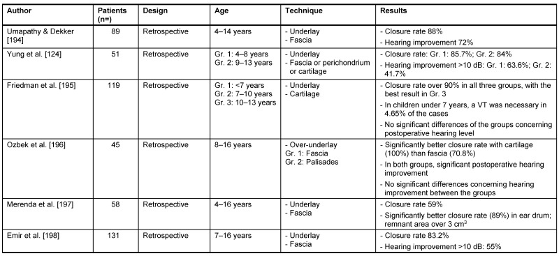
Studies on the success of tympanoplasty in children with CSOM with a high evidence level are rare. Due to the heterogeneity of study populations, a comparison between the related techniques is not possible. The available studies report success rates of tympanoplasty of around 35–95%. Factors like age, dimension and localization of the perforation, the surgical technique used, the transplant, function of the TA, and otorrhea play a role. Table 6 (Tab. 6) displays the results of some current studies.
Table 6. Selected studies concerning functional results after tympanoplasty in childhood (Gr.: Group; VT: Ventilation tube).
Conclusion
The success of tympanoplasty in children with CSOM depends on many different factors. Age seems to be the most important factor based on the current data. Generally, surgical intervention in children should not be executed before the age of 6 years. The decision to undergo an operation should be considered individually and after detailed informing of the parents.
2.5 Retraction pocket
2.5.1 Introduction
From a slight retraction of the TM in the direction towards the tympanon up to an extended cholesteatoma, all kinds of adhesive processes are possible, so that the spectrum of therapy can reach from clinical/conservative control up to extended middle ear surgery. In the following sections, some important aspects of clinical findings and treatment of these adhesive processes will be illustrated. In particular, it will be demonstrated whether a conservative therapy or a surgical therapy should be considered.
2.5.2 Classification and progress
Basically, an adhesive process or a retracted pouch is meant to be an invagination of the TM in the direction towards the middle ear (Figure 2 (Fig. 2)). In the literature, different classifications are recommended, which should be consulted for therapeutic decision-making. In this regard, the classifications orientate themselves, amongst others, along biological characteristics (self-cleaning-retained, aggressively growing), the place of origin (pars tensa-pars flaccida), its progress (age, existence of chronic otitis media), or along the performance during the Valsalva maneuver (fixated, non-fixated). A frequently applied classification was suggested by Tos and Poulsen [136] (Table 7 (Tab. 7)).
Figure 2. Advanced retraction pocket (Type III after Poulsen and Tos) of the right ear.
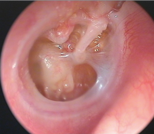
Table 7. Classification of epitympanic retraction pockets (after Tos and Poulsen [136]).
On the basis of the size of the retraction, the following general progress performances can be resumed in the literature: slight retractions (type I) are usually temporary appearances and often show a tendency to complete restitution; type II and type III show dynamic performance. Mostly, improvement occurs; ca. 15% show progress over a period of 3–5 years. Type IV retractions do not show a tendency for restitution and ca. 15% show perforation after 3–5 years [109].
2.5.3 Therapy
There are basically three courses of action in the treatment of TM retraction [137], [138]:
Continuous clinical observation (watchful waiting)
Minor procedures to influence the ventilation (Valsalva training, VT)
Surgery (tympanoplasty, atticoantrotomy, mastoidectomy)
In the literature, basic elements that can influence the strategy of treatment are mentioned (Table 8 (Tab. 8)). None of these elements on their own are a determining factor for surgical indication. Microscopic finding of the TM is an important criterion for decision-making. In case of non-observable retractions, the additional use of an endoscope is recommended [139]. Surgery should be performed if the base of the retraction is not visible with the microscope or detritus is found endoscopically. In comparison with adulthood, adhesive processes in childhood show a much higher proliferation tendency. In case of conductive hearing loss, surgical treatment is recommended.
Table 8. Selected important factors for surgical indication of retracted pockets (modified after [138]).
In case of beginning retractions, some authors recommend the insertion of VT [137]. Others think that VT can only bring improvement for a short time with regard to hearing and that they have no influence on the course of disease. In addition, VT can promote the development of atrophy and calcified platelets [109]. The final surgical therapy of adhesive processes includes definite excision or removal of the retraction, which is usually combined with an accidental perforation of the TM. Therefore, a tympanoplasty or reinforcement of the TM with an autogenetic transplant like fascia, perichondrium, or cartilage is required (see also 2.5). In particular, in case of extended adhesive processes (type III or IV), a tympanoplasty is recommended in the literature [137], [138], [140], [141].
2.6 Cholesteatoma in childhood
2.6.1 Classification and pathogenesis
A cholesteatoma is a form of chronic otitis media that also presents the appearance of squamous epithelium in parts of the middle ear. The term “cholesteatoma” was introduced in the year 1838 by Johannes Müller, a German physiologist [142]. Based on the pathogenesis and the symptoms, it is differentiated between the rare congenital cholesteatoma (CC) and the more common acquired cholesteatoma in childhood [109], [143].
Congenital cholesteatoma
Embryologically, a CC is a dermoid that is located medial of an intact TM. The CC is usually attached to the mucosa of the middle ear (anterior to the handle of the malleus) and has to match the following criteria according to Derlacki and Clemis [144]:
Whitish material medial of the intact TM
Regular pars tensa and flaccida
No case history regarding otorrhea, perforation, or ear surgery
After the modification of Levenson et al. [145], episodes of an AOM do not exclude a genuine cholesteatoma, because AOM appears endemically in the first 5 years of age. For the formation of the CC, the “epithelial rest theory” is actually favored. In human fetus, it could be demonstrated that epithelial fractions, which are usually resorbed, appear in middle ear cavities until the 33rd week of gestation. The theory indicates that genuine cholesteatoma develops due to the disposition and lack of resorption of these epithelial fractions [146].
Acquired cholesteatoma
The pathogenesis of acquired cholesteatoma has not been fully clarified yet. During the 20th century, four classical theories have been established [147]:
Immigration: growth of squamous epithelium of the auditory canal through a marginal perforation of the TM into the middle ear
Retraction: formation of an expansive retraction pouch due to a functional disturbance of the tube with loss of self-cleaning mechanism; development of tensa cholesteatoma
Proliferation: papillary growth of the basal cell layer in the area of the pars flaccida
Metaplasia: transformation of the mucous epithelium of the middle ear into squamous epithelium due to chronic inflammatory stimulus
For the formation of an attic cholesteatoma, nowadays a combination of the theories of retraction and proliferation is favored [147]. Besides these reasons, an acquired cholesteatoma can also be formed due to trauma (i.e., fracture of the petrous bone) or iatrogenically (VT insertion).
2.6.2 Epidemiology
The annual incidence of cholesteatoma in children is specified as 3 per 100,000 inhabitants. The prevalence in Europe is stated to be 0.1% [143]. However, there are considerable ethnic and regional differences. Among the Inuit, the disease is very rarely [148]. In children with palatine cleft, the risk of developing the disease is 20 times higher, so that the risk of the formation of cholesteatoma in these patients is 2.6% [149].
CC represents 4–28% of all cholesteatomas in children. A bilateral manifestation appears in 3% of all cases. Primary diagnosis of the CC is made at the age of 5 years (5.6 ± 2.8); that of acquired cholesteatoma is made at the age of 10 years (9.7 ± 3.3). With 72% of the cases the masculine gender is affected more often than the feminine gender [143].
2.6.3 Symptoms and diagnosis
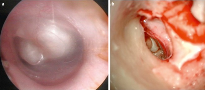
Small CCs are mainly asymptomatic and therefore appear mostly as an incidental finding. Microscopy of the ear shows a whitish globular growth behind an intact TM, which is generally located in the front upper quadrant, anterior to the handle of the malleus (Figure 3 (Fig. 3)). In the case of CC of a larger dimension, the disease can become noticeable by conductive hearing loss due to chain erosion or tympanic effusion (blockade of the tubes). A large number of CCs are diagnosed in the course of VT insertion. In these cases, placing of VT should be abandoned and prompt reconstructive surgery should be pursued. Nelson et al. [150] reported their experiences with CCs in 119 children with an average age of 5.6 years. Most frequently, the node of the cholesteatoma was located in the upper front quadrant. In 69% of the children, erosion of the incus occurred; in 57%, there was erosion of the stapes suprastructure. The average loss of hearing was 36.1 dB.
Figure 3. Figure 3a: Congenital cholesteatoma of the right ear in a 5-year-old child; b: Intraoperative situs showing a cholesteatom pearl anterior of the malleus.
In contrast, acquired cholesteatoma generally shows fetid otorrhea accompanied by conductive hearing loss. Microscopically, a marginal defect of the tympanum is found, usually in the pars flaccida with keratinoid scales. In the study of Iino et al. [151] in 123 children with cholesteatoma, an average conductive hearing loss of 34.7 dB was diagnosed.
Imaging
The significance of CT before surgery for cholesteatoma is discussed controversially in the literature [65]. For determination of the size and localization of the cholesteatoma, CT can be very helpful. Generally, cholesteatomas can be detected from a size of 3–5 mm. However differentiation from other soft tissue proliferations is very difficult [152]. Thus, differentiation between cholesteatoma and cholesterol granuloma in CT is not possible [148]. Radiation exposure of the eye lens, particularly in children, has to be considered. Further, general anesthesia or sedation might be necessary for an MRI. From our point of view, conventional X-ray diagnosis after Schüller is generally sufficient for preoperative assessment of the petrous bone. An indication for CT exists in following cases:
Revision surgery
Suspicion of fistula of the labyrinth
Preoperative paresis of the facial nerve
Suspicion of otogenic complications
2.6.4 Surgical therapy
General
Operative restoration is the definitive therapy for cholesteatoma. An immediate preoperative application of antibiotics (locally or systemically) can be reasonable in an acute superinfection. For successful surgical treatment, the following targets are defined:
Complete resection of the cholesteatoma
Prevention of relapse
Establishment and preservation of a physiological condition with a self-cleaning, dry ear
Preservation or improvement of hearing
Surgical techniques
In the course of the 20th century, two principal techniques for the resection of cholesteatomas were developed. They differ in removal or preservation of the posterior wall of the acoustic meatus. With the classic closed technique (“canal wall up”, CWU) the posterior wall of the external auditory canal (EAC) is preserved and the cholesteatoma is resected through the EAC and, if necessary, via the mastoid process. A great advantage of this technique is the preservation of the normal anatomy of the ECA, with a shorter healing process and prevention of durable secretion. The disadvantages are the restricted sight in the epitympanic cavity and the associated high recurrence rate. In contrast, the classic open technique offers an excellent exposure of the cholesteatoma, with a low recurrence rate after resection of the posterior canal wall (“canal wall down”, CWD). A substantial disadvantage is the establishment of an open mastoid cavity, which results in frequent lavation, with a reduced quality of life. Over the years, both techniques have been modified several times. One modification of the CWU technique is the reconstruction of the posterior canal wall, after it has been removed partly or completely, depending on the dimension of the cholesteatoma. This technique includes the technique of tracing (inside-out mastoidectomy) invented by Plester in which, through the adapted resection of the posterior canal wall, the bursa of the cholesteatoma is traced to its reverse side and resected under complete vision [153]. The posterior canal wall is subsequently reconstructed in the same session using bone, cartilage, or alloplastic materials [65], [153], [154], [155].
The techniques mentioned above have their eligibility in children as well as in adults. However, many auricular surgeons have abandoned the classic CWU technique due to the very high recurrence rate. However, according to Jahnke, a closed technique should be pursued if possible, particularly in children [156]. The CWD technique, with the establishment of an open mastoid cavity, requires continuous cleaning, which is tolerated little by children. Furthermore, assessment of the open mastoid cavity can be limited in the further course by the growth of bone in children [65]. Therefore, nowadays the technique of operative restoration should not be regarded dogmatically, but should be adapted individually to the pathology and patient. In children, a closed or a modified version of the closed technique should always be pursued. Independent of age, for us the above-mentioned technique of tracing for the restoration of cholesteatomas has proved to be valuable (Figure 4 (Fig. 4)).
Figure 4. Figure 4a: Aquired epitampanic cholesteatom on the left side in a 16-year-old child (C: Chorda tympani; +: Incus); b: Removal of cholesteatoma with the inside-out mastoidectomy technique through partial removal of the posterior ear canal; c: Condition after total removal with preservation of the ossicles.

CC is regarded as the classic indication for a CWU technique with a planned 2nd-look operation by many authors. As mentioned above, these cholesteatomas are generally located anterior to the handle of the malleus. Due to the lack of vision, this localization can make the resection difficult. In the last years, the use of microendoscopes, which allow an endoscopic assessment of hidden areas of the tympanic cavity, has proved its value [157].
For resection of CC, Nelson et al. [150] suggested a classification that described the dimension of the cholesteatoma and surgical strategy, depending on the type:
Type 1: cholesteatoma exclusively in the tympanic cavity without participation of the chain except the handle of the malleus; extended tympanotomy
Type 2: participation of the chain with extension to the posterior upper quadrant and in the attic cavity; extended tympanotomy and atticotomy with reconstruction of the chain if necessary
Type 3: extension into the mastoid process; tympanotomy and mastoidectomy
Potsic et al. [158] proposed a classification into four stages: in stage I, only one quadrant is affected; in stage II, multiple quadrants are affected; in stage III, the ossicles are eroded, hence surgical resection for reconstruction is necessary; in stage IV, every extension into the mastoid process is defined.
Functional results
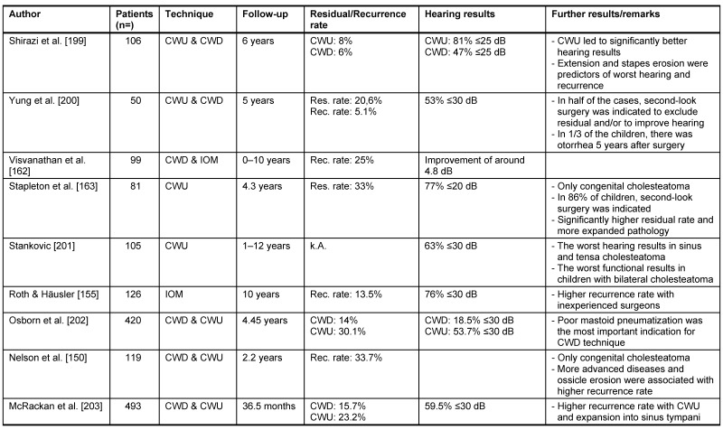
The most important functional results after cholesteatoma surgery are the recurrence rate and postoperative hearing. Due to different operation techniques, extension of cholesteatomas, follow-up times, and composition of patients in the studies, recurrence rates in cholesteatoma surgery in children in the literature vary between 5 and 71%. A cumulative composition of Dodson et al. [159] from studies of the years 1985–1995 showed an average recurrence rate of 42–44% for the CWU technique and 13–22 % for the CWD technique. On the other hand, Tos and Lau [160] could not find any significant differences between the two classical techniques regarding the recurrence rate, in study of over 700 patients. Factors such as experience of the surgeon, high facial nerve spur, condition of the auditory ossicles, and extension of the cholesteatoma are associated with high recurrence rates [161], [162].
In a recent study, Stapleton et al. [163] examined possible predictive factors for a residual/relapse and postoperative hearing in children with CC. Cholesteatomas with larger extensions, erosion of the ossicles, and those that required resection of the ossicles showed a higher rate of residuals/relapses. Furthermore, these factors were associated with significantly worse postoperative hearing. While recurrence rates vary greatly in the literature, information about postoperative hearing is more consistent. Thus, most studies report that in the majority of patients, acceptable hearing (<30 dB) could be achieved after surgery. In Table 9 (Tab. 9), the essential functional results from a selection of studies are outlined.
Table 9. Selected studies on functional results after cholesteatoma surgery in childhood (CWD: canal wall down; CWU: canal wall up; IOM: inside-out mastoidectomy).
Postoperative treatment
The postoperative treatment of cholesteatoma surgery includes, in particular, the control of clinical progress with frequent microscopic examinations of the ear and evaluation of whether a second operation (second-look) to exclude residuals/relapses is required. In many cases, a second operation in order to improve hearing is indicated. In particular, the indication for a second-look operation is questioned, as a result of current developments in imaging diagnostics, notably MRI [164], [165].
In recent years, due to a diffusion-based MRI technique, a new method for the detection of residual cholesteatomas could be introduced. The average sensitivity and specificity of this method is stated to be 81.6% and retrospectively 100% [166], [167]. The positive predictive value is 100%. With the refinement of this technique, the detection limit for residuals is 3–5 mm. The disadvantages of this method are a lack of experience with large groups of patients and deficient data records from prospective studies [168]. Furthermore, the length of the examination is a major problem, because particularly in infancy, motion artifacts can aggravate the interpretation of even an experienced radiologist [148], [169].
3 Bone anchored hearing aid BAHA®
The bone-anchored hearing aid (BAHA®; Company Cochlear, Australia) is one of the partly implantable hearing devices and includes three components: first, an external hearing aid composed of processor, microphone, battery, and electromagnetic transformer/converter; second, an implantable part consisting of an osseointegrated titanium screw; and third, the abutment that connects the hearing aid and screw. Commercial use of osseointegrated screws for sound transmission through the bone with the assistance of hearing aids was introduced in 1989 by Tiellström [170]. Since then, BAHA® is well-established as an alternative to other bone conduction hearing aids in childhood and in adulthood. Since several years ago, the model Ponto® from Oticon has also been available. The number of BAHA® users is estimated to be over 55,000 worldwide [171]. In the following sections, classical indication and functional results of the system will be presented.
3.1 Indications
Generally, BAHA® is indicated for pure middle ear hearing loss and/or mixed hearing loss if middle ear surgery has no chance of success and a conventional hearing aid is ineffective. At the beginning, mainly children with CSOM and therapy-resistant otitis externa were classical indications for BAHA®. In the meantime, there is an advanced range of indication in childhood. The current literature cites the following typical indications for BAHA® in childhood [172], [173]:
Congenital atresia of the auditory canal
Congenital microtia
Chronic otitis externa
Persistent chronic middle ear effusion and hearing aids do not help
Unilateral high-grade sensorineural hearing loss, to gain a pseudostereophonic hearing impression
Trauma/burn of the outer ear
Children with middle ear hearing loss/mixed hearing loss who do not tolerate a conventional hearing aid
Currently, three systems exist (BP 100, BP 110, and Cordelle II), the choice of which depends on auditory threshold in the bone conduction. The content of bone conduction for BP 100 are ≤45 dB , BP 110 ≤55 dB, and Cordelle II ≤65 dB. A bilateral supply is possible, if the average difference in bone conduction of both ears is >10 dB. In the USA, the BAHA® is licensed from the age of 5 years. In Europe, implantation generally takes places from a thickness of the cranium of 3 mm [173].
3.2 Surgical strategy
3.2.1 Techniques and results
The BAHA® is implanted at the bone of the mastoid in one or two sessions. It is typically situated 5 to 5.5 cm posterior and slightly superior to the acoustic meatus. In this position, the external component does not touch the auricle. The individual steps of the surgical procedure have already been extensively described elsewhere [174]. In the first session, the titanium screw is fixed ideally up to a minimum depth of 2.5 mm of the cortical bone. In children, the maximum depth of insertion is specified as 3 mm due to the thin cranium and the contour of the skull [175]. For the second surgical step, a waiting period of between 3 and 6 months is recommended, in order to achieve sufficient osseointegration. Furthermore, the risk of losing the implant due to trauma can be minimized [170], [172], [176]. In the second session, the titanium screw is exposed and the abutment is installed.
The risk of losing the implant, mainly by trauma, is estimated to be 5.8 to 15% in infancy [176]. This is typically in the early postoperative phase due to direct trauma, because osseointegration takes longer in children than in aduls. Possible irritations and subsequent development of infections and swelling of the skin in very young children are other problems.
The time for a BAHA® implant is being discussed controversially in the literature. Furthermore, there is no consensus on whether the operation should be carried out in one or two steps. Due to the reasons mentioned above, the age of 4 years for implantation is recommended by most of the authors [173]. Davis et al. [176] reported on their experiences with 40 children, who had an implant before or after their 5th year of life. The youngest age of implantation in their group of patients was 14 months. The decision for surgery in one or two steps was made during the operation, depending on the thickness of the cranium. In case of thickness of less than 2 mm, the operation was always in two steps, and with more than 4 mm, it was invariably in one step. At a thickness of between 3–4 mm, other factors (age, lag of growth, distance between place of residence and hospital) were considered for the decision. In the majority of the children (95%), implantation was in two steps. The interval to the second session was significantly longer (7.72 months) in children under 5 years than in children above 5 years (4.41 months). In both groups, the adoption of the BAHA® could be performed 6–8 weeks after the second operation. In summary, the study showed that successful implantation with a low rate of complications in children below 5 years is possible. However, here it should always be performed in two steps and the interval between the sessions should be extended compared with older children [176]. For MRI imaging (until 3 Tesla), children have to take out only the sound processor, since the implant and the abutment are not ferromagnetic [177], [178].
Actually, in older children one single procedure is favored. Saliba et al. [179] reported in a study in 26 children with an average age of 8.5 years implantation of BAHA® in one step. With regard to the rate of complications, there were no significant differences versus a comparison group in which implantation was carried out in two steps. The authors argued that the shorter overall duration of the operation and the passable rate of complications justified a single-step procedure in this age group.
With regard to quality of life after implantation of the BAHA®, de Wolf et al. [180] reported on 38 children, who were at least 4 years old at the time of implantation and who had worn the BAHA® for 1–4 years. Here, particularly in children with bilateral middle ear deafness, a benefit in emotional level and in learning appeared. The children especially underlined the benefit of the BAHA® in hearing situations with background noise. Children with loss of hearing on one side valued the beneficial effect of the BAHA® less [180]. In general, an acoustic threshold of 17.5 dB can be achieved in children with middle ear deafness and severe dysplasia [173]. Overall, major centers report success with up to 96% of implanted children. Long-term studies have shown that most of the children are using the system 5–10 years after the operation and are pleased with it [172].
3.2.2 Complications
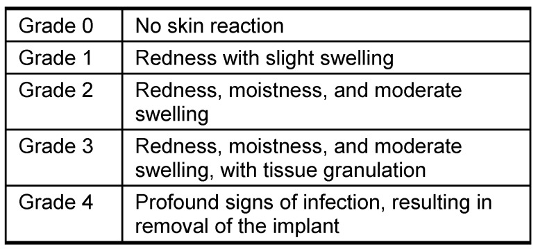
Compared with adults, more complications appeared after implantation of the BAHA® in children. The most important short-, middle-, and long-term complications were loosening of the screw and irritation of the skin [173]. Loss of the implant through a lack of osseointegration occurred most frequently in the first postoperative year [171]. In 2000, Holgers [181] presented a classification for inflammatory reactions of the skin after BAHA® implantation (Table 10 (Tab. 10)). After infantile implantation of the BAHA®, skin reactions appeared as the central issue. The main reasons were chronic grinding of the skin with the abutment and local infections of the skin in range of the implant [173]. Particularly due to deficient hygiene and cleaning by parents, this complication was detected very often in mentally retarded children. These aspects should be evaluated and considered before surgery [172]. In older children, a higher risk of formation of acne is described. Cutaneous hypertrophy can be opposed with a longer abutment (8.5 mm) [182].
Table 10. Holgers classification of skin reaction (around the abutment) after BAHA® implantation [181].
Altogether, the rate of dermal complications is stated to be between 2.4 and 44%. The rate of loss of the implant in children is 5.3 to 40% [171]. In the study of McDermott et al. [183], data of 182 children were analyzed retrospectively after implantation of the BAHA®. Here, loosening of the implant appeared in 14% of the patients. The highest rate of this complication (40%) was found in children below 5 years of age. In children between 5–10 years of age, the rate was 8%; in children above 10 years, the rate was 1%.
3.2.3 Further development
One enhancement of the actual BAHA® systems is the BAHA® BIA 300, which is coated with a rough layer of TiO2 (titanium dioxide). By the use of this material, the stability of the inserted screw is meant to be increased and the duration of complete osseointegration is meant to be accelerated. With a faster definite fixation of the system, the time until the first modulation of the hearing device can be reduced. D’Eredita et al. [184] reported on first clinical experiences with these systems in 12 patients. Three children with an average age of 7.7 years participated in the study. In the observation period of 12 months, barely any dermal complications appeared. Good stability of the implant could be achieved after 3 months. However, additional studies with a larger group of patients and longer follow-up times are necessary. In the newest development, it is possible to resign the thinning of the skin during the implantation.
The bone-anchored hearing system Bonebridge® (Fa. MED-EL, Innsbruck, Österreich) has been on the market for almost 1 year. So far it has not been approved for implantation in children. The receiving implant is placed completely under the skin, which could possibly prevent discomfort or complications due to penetrating parts. The first results in adults concerning quality of life and hearing improvement are very encouraging [185], and the system could also be applied in children in the future.
4 Vibrant Soundbridge® in childhood
The Vibrant Soundbridge® (VSB; MED-EL, Innsbruck, Austria) is an active partly implantable middle ear implant that was introduced clinically in 1996, and has become the most frequently inserted middle ear implant [186], [187], [188], [189]. The system consists of two components: the external component, which contains the audio processor, the energy source (battery), the microphone, the transmitter coil, and a magnet; and the inside component (vibrating ossicular prosthesis, VORP), containing the receiver, which is connected to an electromagnetic sound converter (floating mass transducer, FMT) via a cable. The external sound signals are absorbed by the microphone of the audio processor and transmitted as electric signals transcutaneously to the receiver. The absorbed signals are transmitted to the FMT and cause it to oscillate. This vibrating energy can be used for sound transmission in different ways (see below).
The implantation of the VSB in children was permitted in the EU and other countries, which received the CE label in June 2009 [188]. Therefore, the VSB is the only active middle ear implant that is also approved in children.
At the time of birth, the middle ear is fully developed. However, there is also a slight increase of the middle ear cavities up to the 5th year of age, which causes a minor lateral shift of the TM. Therefore, the use of active middle ear implants in childhood has been controversially discussed. Some argue that skeletal growth requires surgical readjustment of the implant. By single-point fixation of a VSB, the postnatal growth of cranium can be disregarded [190].
The recommendation to implant VSB was published in 2008 by an international conference [190]. With intent a certain minimum age for the date/time of implantation was not defined. Furthermore, middle ear anatomy should be analyzed concerning its dimension and the possibility of implantation, with the help of obligatory pre-surgical imaging (generally CT).
Sensorineural hearing loss is the conventional indication for VSB. In 2007, the indication in Europe was expanded to conductive and to mixed hearing loss. The most important indications for VSB in childhood are congenital external auditory canal atresia and some malformations of the ossicles, which are cause conductive hearing loss or mixed hearing loss. Here, the FMT can be connected to different locations of the ossicles [188] or to the round window [191]. For connection of the FMT, different solutions have been described:
Attachment of the FMT to the long process of the incus for pure sensorineural hearing loss
Connection via Bell- or Clip-Coupler to the stapes’ head
Connection to the oval window (Figure 5 (Fig. 5))
Connection to the round window
Figure 5. Figure 5a: Vibrant Soundbridge® Coupler for the ovale window; condition after creation of an open cavity left (yellow arrow: N. facialis; #: open cavity with intact horizontal semicircular canal); b: Attachement of the couplers with the Floating Mass Transducer (FMT); c: Placement of the FMT on the footplate.

Surgical access to the middle ear for the classical connection is carried out on the long process of the incus via a posterior tympanotomy. Intraoperative monitoring of the facial nerve is recommended by most auricular otosurgeons.
There are only few studies reporting on experiences with VSB in children. One of the most frequent indications in this age group is represented in children with congenital atresia of the acoustic meatus and microtia. Especially in patients with osseous atresia of the external auditory canal, the primary studies are very promising. Frenzel et al. [192] reported on their experiences in seven patients with a unilateral osseous atresia of the external auditory canal. The VSB implant was integrated in line with a total reconstruction of the auricle. Among them were three patients aged between 10 and 15 years. In two of these patients, the FMT was connected to the round window; in one patient, it was connected to the stapes. The average gain of hearing for the entire group was 45.5 dB. In free-field audiometry, patients postoperatively scored an average discrimination of speech of 99% on 65 dB with noise on the other side.
In a recently published study, Colletti et al. [193] reported on results after connection of the FMT to the round window in children (n=10; average age: 5.7 years) with congenital atresia of the external auditory canal. For optimization of the connection, the authors utilized an intraoperative electrocochleography and cartilage for stabilization of the FMT. At 1-year follow-up, the threshold of sound conduction caused by the VSB was 28 dB HL, which was significantly better than the preoperative hearing (84 dB HL). Furthermore, the study showed that migration of the FMT was unlikely in the long term. This complication was not observed in this study. The authors saw a contraindication for connection of the FMT in case of extremely overhanging facial nerve with an obstruction of the round window [193]. An MRI study is not recommended by the company in patients with a VSB.
As an active, partly implantable hearing device, the VSB has established itself very well in the rehabilitation of hearing in adults. Primary studies are available for diagnostic evaluation in children, which reported very good results. For a final conclusion about the effectiveness of VSB, more studies are necessary.
Abbreviations
AM – Acute mastoiditis
AOM – Acute otitis media
AT – Adenoidectomy
BAHA® – Bone anchored hearing aid
CC – Congenital cholesteatoma
CSOM – Chronic suppurative otitis media
CT – Computer tomography
CWD – Canal wall down
CWU – Canal wall up
EAC – External auditory canal
FP – Facial paralysis
MRI – Magnet resonance imaging
OME – Otitis media with effusion
PC – Paracentesis
PCD – Primary ciliary dyskinesia
SPA – Subperiosteal abscess
SST – Sigmoid sinus thrombosis
TA – Tuba auditiva
TARGET – Trial of Alternative Treatment in Glue Ear Treatment
VSB – Vibrant Soundbridge®
Notes
Competing interests
The authors declare that they have no competing interests.
Acknowledgement
We sincerely thank Dr. Carolina Morales-Minovi, Tobias Dombrowski, Dr. med. Jan Peter Thomas, and Dr. med. Stefan Volkenstein for assistance in literature research and for their contributions of discussions to the presentation.
References
- 1.Dhooge IJ. Acute Otitis Media in Children. In: Graham JM, Scadding GK, Bull PD, editors. Pediatric ENT. Heidelberg: Springer; 2007. pp. 399–420. [Google Scholar]
- 2.Strutz J, Mann W. Praxis der HNO-Heilkunde, Kopf- und Halschirurgie. Stuttgart: Thieme; 2009. [Google Scholar]
- 3.Koneczny N, Schmidt-Troschke S, Berger T, Isfort J, Floer B, Vollmar HC, Butzlaff M. Akute Otitis media (AOM) bei Kindern (ICD: H66.0): eine evidenzbasierte Leitlinie -- Neue Perspektiven für Kinderärzte in Deutschland. [Acute otitis media in children: an evidence-based practice guideline]. Klin Padiatr. 2004 Jul-Aug;216(4):215–224. doi: 10.1055/s-2004-44891. (Ger). Available from: http://dx.doi.org/10.1055/s-2004-44891. [DOI] [PubMed] [Google Scholar]
- 4.Wald ER. Acute otitis media and acute bacterial sinusitis. Clin Infect Dis. 2011 May;52 Suppl 4:S277–S283. doi: 10.1093/cid/cir042. Available from: http://dx.doi.org/10.1093/cid/cir042. [DOI] [PMC free article] [PubMed] [Google Scholar]
- 5.Teele DW, Klein JO, Rosner B. Epidemiology of otitis media during the first seven years of life in children in greater Boston: a prospective, cohort study. J Infect Dis. 1989 Jul;160(1):83–94. doi: 10.1093/infdis/160.1.83. Available from: http://dx.doi.org/10.1093/infdis/160.1.83. [DOI] [PubMed] [Google Scholar]
- 6.Vergison A, Dagan R, Arguedas A, Bonhoeffer J, Cohen R, Dhooge I, et al. Otitis media and its consequences: beyond the earache. Lancet Infect Dis. 2010;10:195–203. doi: 10.1016/S1473-3099(10)70012-8. Available from: http://dx.doi.org/10.1016/S1473-3099(10)70012-8. [DOI] [PubMed] [Google Scholar]
- 7.Monasta L, Ronfani L, Marchetti F, Montico M, Vecchi Brumatti L, Bavcar A, Grasso D, Barbiero C, Tamburlini G. Burden of disease caused by otitis media: systematic review and global estimates. PLoS ONE. 2012;7(4):e36226. doi: 10.1371/journal.pone.0036226. Available from: http://dx.doi.org/10.1371/journal.pone.0036226. [DOI] [PMC free article] [PubMed] [Google Scholar]
- 8.Daly KA, Brown JE, Lindgren BR, Meland MH, Le CT, Giebink GS. Epidemiology of otitis media onset by six months of age. Pediatrics. 1999 Jun;103(6 Pt 1):1158–1166. doi: 10.1542/peds.103.6.1158. Available from: http://dx.doi.org/10.1542/peds.103.6.1158. [DOI] [PubMed] [Google Scholar]
- 9.Heavner SB, Hardy SM, White DR, Prazma J, Pillsbury HC., 3rd Transient inflammation and dysfunction of the eustachian tube secondary to multiple exposures of simulated gastroesophageal refluxant. Ann Otol Rhinol Laryngol. 2001 Oct;110(10):928–934. doi: 10.1177/000348940111001007. [DOI] [PubMed] [Google Scholar]
- 10.Leach AJ, Morris PS. Perspectives on infective ear disease in indigenous Australian children. J Paediatr Child Health. 2001;37:529–530. doi: 10.1046/j.1440-1754.2001.00729.x. Available from: http://dx.doi.org/10.1046/j.1440-1754.2001.00729.x. [DOI] [PubMed] [Google Scholar]
- 11.Niemelä M, Pihakari O, Pokka T, Uhari M. Pacifier as a risk factor for acute otitis media: A randomized, controlled trial of parental counseling. Pediatrics. 2000 Sep;106(3):483–488. doi: 10.1542/peds.106.3.483. Available from: http://dx.doi.org/10.1542/peds.106.3.483. [DOI] [PubMed] [Google Scholar]
- 12.Ramakrishnan K, Sparks RA, Berryhill WE. Diagnosis and treatment of otitis media. Am Fam Physician. 2007 Dec;76(11):1650–1658. [PubMed] [Google Scholar]
- 13.Uhari M, Mäntysaari K, Niemelä M. A meta-analytic review of the risk factors for acute otitis media. Clin Infect Dis. 1996 Jun;22(6):1079–1083. doi: 10.1093/clinids/22.6.1079. Available from: http://dx.doi.org/10.1093/clinids/22.6.1079. [DOI] [PubMed] [Google Scholar]
- 14.Coker TR, Chan LS, Newberry SJ, Limbos MA, Suttorp MJ, Shekelle PG, Takata GS. Diagnosis, microbial epidemiology, and antibiotic treatment of acute otitis media in children: a systematic review. JAMA. 2010 Nov;304(19):2161–2169. doi: 10.1001/jama.2010.1651. Available from: http://dx.doi.org/10.1001/jama.2010.1651. [DOI] [PubMed] [Google Scholar]
- 15.Healy GB, Rosbe KW. Otitis Media and Middle Ear Effusions. In: Snow JB Jr, Ballenger JJ, editors. Ballenger's Otorhinolaryngology Head and Neck Surgery. Hamilton: BC Decker; 2003. pp. 249–260. [Google Scholar]
- 16.Lieberthal AS, Carroll AE, Chonmaitree T, Ganiats TG, Hoberman A, Jackson MA, Joffe MD, Miller DT, Rosenfeld RM, Sevilla XD, Schwartz RH, Thomas PA, Tunkel DE. The diagnosis and management of acute otitis media. Pediatrics. 2013 Mar;131(3):e964–e999. doi: 10.1542/peds.2012-3488. Available from: http://dx.doi.org/10.1542/peds.2012-3488. [DOI] [PubMed] [Google Scholar]
- 17.Heikkinen T, Thint M, Chonmaitree T. Prevalence of various respiratory viruses in the middle ear during acute otitis media. N Engl J Med. 1999 Jan;340(4):260–264. doi: 10.1056/NEJM199901283400402. Available from: http://dx.doi.org/10.1056/NEJM199901283400402. [DOI] [PubMed] [Google Scholar]
- 18.Turner D, Leibovitz E, Aran A, Piglansky L, Raiz S, Leiberman A, Dagan R. Acute otitis media in infants younger than two months of age: microbiology, clinical presentation and therapeutic approach. Pediatr Infect Dis J. 2002 Jul;21(7):669–674. doi: 10.1097/01.inf.0000021080.57835.8a. Available from: http://dx.doi.org/10.1097/01.inf.0000021080.57835.8a. [DOI] [PubMed] [Google Scholar]
- 19.Casey JR, Pichichero ME. Changes in frequency and pathogens causing acute otitis media in 1995-2003. Pediatr Infect Dis J. 2004 Sep;23(9):824–828. doi: 10.1097/01.inf.0000136871.51792.19. Available from: http://dx.doi.org/10.1097/01.inf.0000136871.51792.19. [DOI] [PubMed] [Google Scholar]
- 20.Arrieta A, Singh J. Management of recurrent and persistent acute otitis media: new options with familiar antibiotics. Pediatr Infect Dis J. 2004 Feb;23(2 Suppl):S115–S124. doi: 10.1097/01.inf.0000112525.88779.8b. Available from: http://dx.doi.org/10.1097/01.inf.0000112525.88779.8b. [DOI] [PubMed] [Google Scholar]
- 21.Hendley JO. Clinical practice. Otitis media. N Engl J Med. 2002 Oct;347(15):1169–1174. doi: 10.1056/NEJMcp010944. Available from: http://dx.doi.org/10.1056/NEJMcp010944. [DOI] [PubMed] [Google Scholar]
- 22.Rovers MM, Schilder AG, Zielhuis GA, Rosenfeld RM. Otitis media. Lancet. 2004 Feb 7;363(9407):465–473. doi: 10.1016/S0140-6736(04)15495-0. Available from: http://dx.doi.org/10.1016/S0140-6736(04)15495-0. [DOI] [PubMed] [Google Scholar]
- 23.McCormick DP, Saeed K, Uchida T, Baldwin CD, Deskin R, Lett-Brown MA, Heikkinen T, Chonmaitree T. Middle ear fluid histamine and leukotriene B4 in acute otitis media: effect of antihistamine or corticosteroid treatment. Int J Pediatr Otorhinolaryngol. 2003 Mar;67(3):221–230. doi: 10.1016/S0165-5876(02)00372-5. Available from: http://dx.doi.org/10.1016/S0165-5876(02)00372-5. [DOI] [PubMed] [Google Scholar]
- 24.Flynn CA, Griffin GH, Schultz JK. Decongestants and antihistamines for acute otitis media in children. Cochrane Database Syst Rev. 2004;(3):CD001727. doi: 10.1002/14651858.CD001727.pub2. Available from: http://dx.doi.org/10.1002/14651858.CD001727.pub2. [DOI] [PubMed] [Google Scholar]
- 25.Van Zuijlen DA, Schilder AG, Van Balen FA, Hoes AW. National differences in incidence of acute mastoiditis: relationship to prescribing patterns of antibiotics for acute otitis media? Pediatr Infect Dis J. 2001 Feb;20(2):140–144. doi: 10.1097/00006454-200102000-00004. Available from: http://dx.doi.org/10.1097/00006454-200102000-00004. [DOI] [PubMed] [Google Scholar]
- 26.Groth A, Enoksson F, Hermansson A, Hultcrantz M, Stalfors J, Stenfeldt K. Acute mastoiditis in children in Sweden 1993-2007--no increase after new guidelines. Int J Pediatr Otorhinolaryngol. 2011 Dec;75(12):1496–1501. doi: 10.1016/j.ijporl.2011.08.015. Available from: http://dx.doi.org/10.1016/j.ijporl.2011.08.015. [DOI] [PubMed] [Google Scholar]
- 27.Little P, Gould C, Williamson I, Moore M, Warner G, Dunleavey J. Pragmatic randomised controlled trial of two prescribing strategies for childhood acute otitis media. BMJ. 2001 Feb;322(7282):336–342. doi: 10.1136/bmj.322.7282.336. Available from: http://dx.doi.org/10.1136/bmj.322.7282.336. [DOI] [PMC free article] [PubMed] [Google Scholar]
- 28.Spiro DM, Tay KY, Arnold DH, Dziura JD, Baker MD, Shapiro ED. Wait-and-see prescription for the treatment of acute otitis media: a randomized controlled trial. JAMA. 2006 Sep;296(10):1235–1241. doi: 10.1001/jama.296.10.1235. Available from: http://dx.doi.org/10.1001/jama.296.10.1235. [DOI] [PubMed] [Google Scholar]
- 29.van Buchem FL, Peeters MF, van 't Hof MA. Acute otitis media: a new treatment strategy. Br Med J (Clin Res Ed) 1985 Apr 6;290(6474):1033–1037. doi: 10.1136/bmj.290.6474.1033. Available from: http://dx.doi.org/10.1136/bmj.290.6474.1033. [DOI] [PMC free article] [PubMed] [Google Scholar]
- 30.Rovers MM, Glasziou P, Appelman CL, Burke P, McCormick DP, Damoiseaux RA, Gaboury I, Little P, Hoes AW. Antibiotics for acute otitis media: a meta-analysis with individual patient data. Lancet. 2006 Oct 21;368(9545):1429–1435. doi: 10.1016/S0140-6736(06)69606-2. Available from: http://dx.doi.org/10.1016/S0140-6736(06)69606-2. [DOI] [PubMed] [Google Scholar]
- 31.Hoberman A, Paradise JL, Rockette HE, Shaikh N, Wald ER, Kearney DH, Colborn DK, Kurs-Lasky M, Bhatnagar S, Haralam MA, Zoffel LM, Jenkins C, Pope MA, Balentine TL, Barbadora KA. Treatment of acute otitis media in children under 2 years of age. N Engl J Med. 2011 Jan;364(2):105–115. doi: 10.1056/NEJMoa0912254. Available from: http://dx.doi.org/10.1056/NEJMoa0912254. [DOI] [PMC free article] [PubMed] [Google Scholar]
- 32.Amrhein P, Hospach A, Sittel C, Koitschev A. Akute Otitis media bei Kindern: Antibiose oder "watchful waiting". [Acute otitis media in children: antibiotic therapy or watchful waiting?]. HNO. 2013 May;61(5):374–379. doi: 10.1007/s00106-012-2486-1. (Ger). Available from: http://dx.doi.org/10.1007/s00106-012-2486-1. [DOI] [PubMed] [Google Scholar]
- 33.Deutsche Gesellschaft für Hals-Nasen-Ohren-Heilkunde, Kopf- und Hals-Chirurgie. Leitlinien der DGHNO. Antibiotikatherapie der Infektionen an Kopf und Hals. AWMF-Register Nr. 017/066. 2008. [Google Scholar]
- 34.Paradise JL. Short-course antimicrobial treatment for acute otitis media: not best for infants and young children. JAMA. 1997 Nov;278(20):1640–1642. doi: 10.1001/jama.1997.03550200016006. Available from: http://dx.doi.org/10.1001/jama.1997.03550200016006. [DOI] [PubMed] [Google Scholar]
- 35.Piglansky L, Leibovitz E, Raiz S, Greenberg D, Press J, Leiberman A, Dagan R. Bacteriologic and clinical efficacy of high dose amoxicillin for therapy of acute otitis media in children. Pediatr Infect Dis J. 2003 May;22(5):405–413. doi: 10.1097/01.inf.0000065688.21336.fa. Available from: http://dx.doi.org/10.1097/01.inf.0000065688.21336.fa. [DOI] [PubMed] [Google Scholar]
- 36.Dagan R, Hoberman A, Johnson C, Leibovitz EL, Arguedas A, Rose FV, Wynne BR, Jacobs MR. Bacteriologic and clinical efficacy of high dose amoxicillin/clavulanate in children with acute otitis media. Pediatr Infect Dis J. 2001 Sep;20(9):829–837. doi: 10.1097/00006454-200109000-00002. Available from: http://dx.doi.org/10.1097/00006454-200109000-00002. [DOI] [PubMed] [Google Scholar]
- 37.Rubin LG. Prevention and treatment of meningitis and acute otitis media in children with cochlear implants. Otol Neurotol. 2010 Oct;31(8):1331–1333. doi: 10.1097/MAO.0b013e3181f2f074. Available from: http://dx.doi.org/10.1097/MAO.0b013e3181f2f074. [DOI] [PubMed] [Google Scholar]
- 38.Fickweiler U, Müller H, Dietz A. Die akute Mastoiditis heute. [Acute mastoiditis today]. HNO. 2007 Jan;55(1):73–80. doi: 10.1007/s00106-006-1468-6. (Ger). Available from: http://dx.doi.org/10.1007/s00106-006-1468-6. [DOI] [PubMed] [Google Scholar]
- 39.Spratley J, Silveira H, Alvarez I, Pais-Clemente M. Acute mastoiditis in children: review of the current status. Int J Pediatr Otorhinolaryngol. 2000 Nov 30;56(1):33–40. doi: 10.1016/S0165-5876(00)00406-7. Available from: http://dx.doi.org/10.1016/S0165-5876(00)00406-7. [DOI] [PubMed] [Google Scholar]
- 40.Benito MB, Gorricho BP. Acute mastoiditis: increase in the incidence and complications. Int J Pediatr Otorhinolaryngol. 2007 Jul;71(7):1007–1011. doi: 10.1016/j.ijporl.2007.02.014. Available from: http://dx.doi.org/10.1016/j.ijporl.2007.02.014. [DOI] [PubMed] [Google Scholar]
- 41.Kangsanarak J, Navacharoen N, Fooanant S, Ruckphaopunt K. Intracranial complications of suppurative otitis media: 13 years' experience. Am J Otol. 1995 Jan;16(1):104–109. [PubMed] [Google Scholar]
- 42.Prellner K, Rydell R. Acute mastoiditis. Influence of antibiotic treatment on the bacterial spectrum. Acta Otolaryngol. 1986 Jul-Aug;102(1-2):52–56. doi: 10.3109/00016488609108646. Available from: http://dx.doi.org/10.3109/00016488609108646. [DOI] [PubMed] [Google Scholar]
- 43.Geva A, Oestreicher-Kedem Y, Fishman G, Landsberg R, DeRowe A. Conservative management of acute mastoiditis in children. Int J Pediatr Otorhinolaryngol. 2008 May;72(5):629–634. doi: 10.1016/j.ijporl.2008.01.013. Available from: http://dx.doi.org/10.1016/j.ijporl.2008.01.013. [DOI] [PubMed] [Google Scholar]
- 44.Groth A, Enoksson F, Hultcrantz M, Stalfors J, Stenfeldt K, Hermansson A. Acute mastoiditis in children aged 0-16 years--a national study of 678 cases in Sweden comparing different age groups. Int J Pediatr Otorhinolaryngol. 2012 Oct;76(10):1494–1500. doi: 10.1016/j.ijporl.2012.07.002. Available from: http://dx.doi.org/10.1016/j.ijporl.2012.07.002. [DOI] [PubMed] [Google Scholar]
- 45.Vassbotn FS, Klausen OG, Lind O, Moller P. Acute mastoiditis in a Norwegian population: a 20 year retrospective study. Int J Pediatr Otorhinolaryngol. 2002 Feb 25;62(3):237–242. doi: 10.1016/S0165-5876(01)00626-7. Available from: http://dx.doi.org/10.1016/S0165-5876(01)00626-7. [DOI] [PubMed] [Google Scholar]
- 46.Nussinovitch M, Yoeli R, Elishkevitz K, Varsano I. Acute mastoiditis in children: epidemiologic, clinical, microbiologic, and therapeutic aspects over past years. Clin Pediatr (Phila) 2004 Apr;43(3):261–267. doi: 10.1177/000992280404300307. Available from: http://dx.doi.org/10.1177/000992280404300307. [DOI] [PubMed] [Google Scholar]
- 47.Palma S, Fiumana E, Borgonzoni M, Bovo R, Rosignoli M, Martini A. Acute mastoiditis in children: the "Ferrara" experience. Int J Pediatr Otorhinolaryngol. 2007 Nov;71(11):1663–1669. doi: 10.1016/j.ijporl.2007.06.018. Available from: http://dx.doi.org/10.1016/j.ijporl.2007.06.018. [DOI] [PubMed] [Google Scholar]
- 48.Hoppe JE, Köster S, Bootz F, Niethammer D. Acute mastoiditis--relevant once again. Infection. 1994 May-Jun;22(3):178–182. doi: 10.1007/BF01716698. Available from: http://dx.doi.org/10.1007/BF01716698. [DOI] [PubMed] [Google Scholar]
- 49.Harley EH, Sdralis T, Berkowitz RG. Acute mastoiditis in children: a 12-year retrospective study. Otolaryngol Head Neck Surg. 1997 Jan;116(1):26–30. doi: 10.1016/S0194-5998(97)70347-4. Available from: http://dx.doi.org/10.1016/S0194-5998(97)70347-4. [DOI] [PubMed] [Google Scholar]
- 50.Bakhos D, Trijolet JP, Morinière S, Pondaven S, Al Zahrani M, Lescanne E. Conservative management of acute mastoiditis in children. Arch Otolaryngol Head Neck Surg. 2011 Apr;137(4):346–350. doi: 10.1001/archoto.2011.29. Available from: http://dx.doi.org/10.1001/archoto.2011.29. [DOI] [PubMed] [Google Scholar]
- 51.Oestreicher-Kedem Y, Raveh E, Kornreich L, Popovtzer A, Buller N, Nageris B. Complications of mastoiditis in children at the onset of a new millennium. Ann Otol Rhinol Laryngol. 2005 Feb;114(2):147–152. doi: 10.1177/000348940511400212. [DOI] [PubMed] [Google Scholar]
- 52.Pang LH, Barakate MS, Havas TE. Mastoiditis in a paediatric population: a review of 11 years experience in management. Int J Pediatr Otorhinolaryngol. 2009 Nov;73(11):1520–1524. doi: 10.1016/j.ijporl.2009.07.003. Available from: http://dx.doi.org/10.1016/j.ijporl.2009.07.003. [DOI] [PubMed] [Google Scholar]
- 53.Mross-Adam C, Klemm E. Die akute Mastoiditis im Kindesalter, eine Analyse des eigenen Krankengutes über 25 Jahre. [Acute mastoiditis in children, a retrospective analysis about a period of 25 years]. Laryngorhinootologie. 2005 Jul;84(7):497–502. doi: 10.1055/s-2005-861376. (Ger). Available from: http://dx.doi.org/10.1055/s-2005-861376. [DOI] [PubMed] [Google Scholar]
- 54.Arts HA, Neely JG. Intratemporal and intracranial complications of otitis media. In: Bailey BJ, editor. Head and neck surgery - otolaryngology. Philadelphia: Lippincott Williams & Wilkins; 2001. pp. 1759–1772. [Google Scholar]
- 55.Psarommatis IM, Voudouris C, Douros K, Giannakopoulos P, Bairamis T, Carabinos C. Algorithmic management of pediatric acute mastoiditis. Int J Pediatr Otorhinolaryngol. 2012 Jun;76(6):791–796. doi: 10.1016/j.ijporl.2012.02.042. Available from: http://dx.doi.org/10.1016/j.ijporl.2012.02.042. [DOI] [PubMed] [Google Scholar]
- 56.Vazquez E, Castellote A, Piqueras J, Mauleon S, Creixell S, Pumarola F, Figueras C, Carreño JC, Lucaya J. Imaging of complications of acute mastoiditis in children. Radiographics. 2003 Mar-Apr;23(2):359–372. doi: 10.1148/rg.232025076. Available from: http://dx.doi.org/10.1148/rg.232025076. [DOI] [PubMed] [Google Scholar]
- 57.Luntz M, Brodsky A, Nusem S, Kronenberg J, Keren G, Migirov L, Cohen D, Zohar S, Shapira A, Ophir D, Fishman G, Rosen G, Kisilevsky V, Magamse I, Zaaroura S, Joachims HZ, Goldenberg D. Acute mastoiditis--the antibiotic era: a multicenter study. Int J Pediatr Otorhinolaryngol. 2001 Jan;57(1):1–9. doi: 10.1016/S0165-5876(00)00425-0. Available from: http://dx.doi.org/10.1016/S0165-5876(00)00425-0. [DOI] [PubMed] [Google Scholar]
- 58.Helms J. Sanierende und rekonstruktive Operationen an Gehörgang, Mittelohr und Felsenbein. In: Helms J, Jahrsdoerfer RA, editors. Kopf- und Halschirurgie. Stuttgart: Thieme; 1997. pp. 67–130. [Google Scholar]
- 59.Tos M. Manual of Middle Ear Surgery. Volume 2: Mastoid Surgery and Recosntructive Proceduresery. Stuttgart: Thieme; 1995. [Google Scholar]
- 60.Quesnel S, Nguyen M, Pierrot S, Contencin P, Manach Y, Couloigner V. Acute mastoiditis in children: a retrospective study of 188 patients. Int J Pediatr Otorhinolaryngol. 2010 Dec;74(12):1388–1392. doi: 10.1016/j.ijporl.2010.09.013. Available from: http://dx.doi.org/10.1016/j.ijporl.2010.09.013. [DOI] [PubMed] [Google Scholar]
- 61.Go C, Bernstein JM, de Jong AL, Sulek M, Friedman EM. Intracranial complications of acute mastoiditis. Int J Pediatr Otorhinolaryngol. 2000 Apr 15;52(2):143–148. doi: 10.1016/S0165-5876(00)00283-4. Available from: http://dx.doi.org/10.1016/S0165-5876(00)00283-4. [DOI] [PubMed] [Google Scholar]
- 62.Zanetti D, Nassif N. Indications for surgery in acute mastoiditis and their complications in children. Int J Pediatr Otorhinolaryngol. 2006 Jul;70(7):1175–1182. doi: 10.1016/j.ijporl.2005.12.002. Available from: http://dx.doi.org/10.1016/j.ijporl.2005.12.002. [DOI] [PubMed] [Google Scholar]
- 63.Lutter SA, Kerschner JE, Chusid MJ. Gradenigo syndrome: a rare but serious complication of otitis media. Pediatr Emerg Care. 2005 Jun;21(6):384–386. doi: 10.1097/01.pec.0000166731.70847.d5. Available from: http://dx.doi.org/10.1097/01.pec.0000166731.70847.d5. [DOI] [PubMed] [Google Scholar]
- 64.Pellegrini S, Gonzalez Macchi ME, Sommerfleck PA, Bernáldez PC. Intratemporal complications from acute otitis media in children: 17 cases in two years. Acta Otorrinolaringol Esp. 2012 Jan-Feb;63(1):21–25. doi: 10.1016/j.otorri.2011.06.007. Available from: http://dx.doi.org/10.1016/j.otorri.2011.06.007. [DOI] [PubMed] [Google Scholar]
- 65.Hildmann H, Sudhoff H, Jahnke K. Principles of Individualized Approach to Cholesteatoma Surgery. In: Jahnke K, editor. Current Topics in Otolaryngology - Head and Neck Surgery Middle Ear Surgery Recent Advances and Future Directions. Stuttgart: Thieme; 2004. pp. 3–93. [Google Scholar]
- 66.Robb PJ. Otitis Media With Effusion. In: Graham JM, Scadding GK, Bull PD, editors. Pediatric ENT. Heidelberg: Springer; 2007. pp. 413–420. [Google Scholar]
- 67.Zielhuis GA, Rach GH, van den Broek P. Screening for otitis media with effusion in preschool children. Lancet. 1989 Feb 11;1(8633):311–314. doi: 10.1016/S0140-6736(89)91317-2. Available from: http://dx.doi.org/10.1016/S0140-6736(89)91317-2. [DOI] [PubMed] [Google Scholar]
- 68.Schilder AG, Zielhuis GA, Van Den Broek P. The otological profile of a cohort of Dutch 7.5-8-year-olds. Clin Otolaryngol Allied Sci. 1993 Feb;18(1):48–54. doi: 10.1111/j.1365-2273.1993.tb00809.x. Available from: http://dx.doi.org/10.1111/j.1365-2273.1993.tb00809.x. [DOI] [PubMed] [Google Scholar]
- 69.Politzer A. Diagnose und Theraphaie der AnsammIung seroeser Fluessigkeit in der Thrommelhoehle. Wien Med Wochenschr. 1867;17:224–247. [Google Scholar]
- 70.Sadé J, Eliezer N. Secretory otitis media and the nature of the mucociliaryy system. Acta Otolaryngol. 1970 Nov-Dec;70(5):351–357. doi: 10.3109/00016487009181896. Available from: http://dx.doi.org/10.3109/00016487009181896. [DOI] [PubMed] [Google Scholar]
- 71.Hoffman HJ, Daly KA, Bainbridge KE, Casselbrant ML, Homøe P, Kvestad E, Kvaerner KJ, Vernacchio L. Panel 1: Epidemiology, natural history, and risk factors. Otolaryngol Head Neck Surg. 2013 Apr;148(4 Suppl):E1–E25. doi: 10.1177/0194599812460984. Available from: http://dx.doi.org/10.1177/0194599812460984. [DOI] [PubMed] [Google Scholar]
- 72.Rosenfeld RM, Kay D. Natural history of untreated otitis media. Laryngoscope. 2003 Oct;113(10):1645–1657. doi: 10.1097/00005537-200310000-00004. Available from: http://dx.doi.org/10.1097/00005537-200310000-00004. [DOI] [PubMed] [Google Scholar]
- 73.Campbell RG, Birman CS, Morgan L. Management of otitis media with effusion in children with primary ciliary dyskinesia: a literature review. Int J Pediatr Otorhinolaryngol. 2009 Dec;73(12):1630–1638. doi: 10.1016/j.ijporl.2009.08.024. Available from: http://dx.doi.org/10.1016/j.ijporl.2009.08.024. [DOI] [PubMed] [Google Scholar]
- 74.Flynn T, Möller C, Jönsson R, Lohmander A. The high prevalence of otitis media with effusion in children with cleft lip and palate as compared to children without clefts. Int J Pediatr Otorhinolaryngol. 2009 Oct;73(10):1441–1446. doi: 10.1016/j.ijporl.2009.07.015. Available from: http://dx.doi.org/10.1016/j.ijporl.2009.07.015. [DOI] [PubMed] [Google Scholar]
- 75.Schönweiler R, Schönweiler B, Schmelzeisen R. Hörvermögen und Sprachleistungen bei 417 Kindern mit Spaltfehlbildungen. [Hearing capacity and speech production in 417 children with facial cleft abnormalities]. HNO. 1994 Nov;42(11):691–696. (Ger). [PubMed] [Google Scholar]
- 76.Prulière-Escabasse V, Coste A, Chauvin P, Fauroux B, Tamalet A, Garabedian EN, Escudier E, Roger G. Otologic features in children with primary ciliary dyskinesia. Arch Otolaryngol Head Neck Surg. 2010 Nov;136(11):1121–1126. doi: 10.1001/archoto.2010.183. Available from: http://dx.doi.org/10.1001/archoto.2010.183. [DOI] [PMC free article] [PubMed] [Google Scholar]
- 77.Bernstein JM. Immunologic reactivity in the middle ear in otitis media with effusion. Otolaryngol Clin North Am. 1991 Aug;24(4):845–858. [PubMed] [Google Scholar]
- 78.Bernstein JM, Doyle WJ. Role of IgE-mediated hypersensitivity in otitis media with effusion: pathophysiologic considerations. Ann Otol Rhinol Laryngol Suppl. 1994 May;163:15–19. doi: 10.1177/00034894941030s505. [DOI] [PubMed] [Google Scholar]
- 79.Daniel M, Imtiaz-Umer S, Fergie N, Birchall JP, Bayston R. Bacterial involvement in otitis media with effusion. Int J Pediatr Otorhinolaryngol. 2012 Oct;76(10):1416–1422. doi: 10.1016/j.ijporl.2012.06.013. Available from: http://dx.doi.org/10.1016/j.ijporl.2012.06.013. [DOI] [PubMed] [Google Scholar]
- 80.Post JC, Preston RA, Aul JJ, Larkins-Pettigrew M, Rydquist-White J, Anderson KW, Wadowsky RM, Reagan DR, Walker ES, Kingsley LA, Magit AE, Ehrlich GD. Molecular analysis of bacterial pathogens in otitis media with effusion. JAMA. 1995 May 24-31;273(20):1598–1604. doi: 10.1001/jama.1995.03520440052036. Available from: http://dx.doi.org/10.1001/jama.1995.03520440052036. [DOI] [PubMed] [Google Scholar]
- 81.Golz A, Angel-Yeger B, Parush S. Evaluation of balance disturbances in children with middle ear effusion. Int J Pediatr Otorhinolaryngol. 1998 Feb;43(1):21–26. doi: 10.1016/S0165-5876(97)00150-X. Available from: http://dx.doi.org/10.1016/S0165-5876(97)00150-X. [DOI] [PubMed] [Google Scholar]
- 82.Haapaniemi JJ. Pure-tone audiometric and impedance measurements in school-aged children in Finland. Eur Arch Otorhinolaryngol. 1997;254(6):269–273. doi: 10.1007/BF02905985. Available from: http://dx.doi.org/10.1007/BF02905985. [DOI] [PubMed] [Google Scholar]
- 83.Mitchell DB, Ford GR, Albert D, Waldron J. Acoustic reflectometry as an aid to the diagnosis of glue ear. Br J Clin Pract. 1990 Dec;44(12):557–559. [PubMed] [Google Scholar]
- 84.Vaughan-Jones R, Mills RP. The Welch Allyn Audioscope and Microtymp: their accuracy and that of pneumatic otoscopy, tympanometry and pure tone audiometry as predictors of otitis media with effusion. J Laryngol Otol. 1992 Jul;106(7):600–602. doi: 10.1017/S0022215100120298. Available from: http://dx.doi.org/10.1017/S0022215100120298. [DOI] [PubMed] [Google Scholar]
- 85.National Collaborating Centre for Women’s and Children’s Health (UK) Surgical Management of Otitis Media with Effusion in Children. London: RCOG Press; Feb, 2008. (NICE Clinical Guidelines; 60). [PubMed] [Google Scholar]
- 86.Sassen ML, van Aarem A, Grote JJ. Validity of tympanometry in the diagnosis of middle ear effusion. Clin Otolaryngol Allied Sci. 1994 Jun;19(3):185–189. doi: 10.1111/j.1365-2273.1994.tb01211.x. Available from: http://dx.doi.org/10.1111/j.1365-2273.1994.tb01211.x. [DOI] [PubMed] [Google Scholar]
- 87.Watters GW, Jones JE, Freeland AP. The predictive value of tympanometry in the diagnosis of middle ear effusion. Clin Otolaryngol Allied Sci. 1997 Aug;22(4):343–345. doi: 10.1046/j.1365-2273.1997.00023.x. Available from: http://dx.doi.org/10.1046/j.1365-2273.1997.00023.x. [DOI] [PubMed] [Google Scholar]
- 88.American Academy of Family Physicians; American Academy of Otolaryngology-Head and Neck Surgery; American Academy of Pediatrics Subcommittee on Otitis Media With Effusion. Otitis media with effusion. Pediatrics. 2004 May;113(5):1412–1429. doi: 10.1542/peds.113.5.1412. Available from: http://dx.doi.org/10.1542/peds.113.5.1412. [DOI] [PubMed] [Google Scholar]
- 89.Rosenfeld RM, Post JC. Meta-analysis of antibiotics for the treatment of otitis media with effusion. Otolaryngol Head Neck Surg. 1992 Apr;106(4):378–386. doi: 10.1177/019459989210600411. [DOI] [PubMed] [Google Scholar]
- 90.Williams RL, Chalmers TC, Stange KC, Chalmers FT, Bowlin SJ. Use of antibiotics in preventing recurrent acute otitis media and in treating otitis media with effusion. A meta-analytic attempt to resolve the brouhaha. JAMA. 1993 Sep;270(11):1344–1351. doi: 10.1001/jama.1993.03510110084037. Available from: http://dx.doi.org/10.1001/jama.1993.03510110084037. [DOI] [PubMed] [Google Scholar]
- 91.Mandel EM, Rockette HE, Bluestone CD, Paradise JL, Nozza RJ. Efficacy of amoxicillin with and without decongestant-antihistamine for otitis media with effusion in children. Results of a double-blind, randomized trial. N Engl J Med. 1987 Feb;316(8):432–437. doi: 10.1056/NEJM198702193160803. Available from: http://dx.doi.org/10.1056/NEJM198702193160803. [DOI] [PubMed] [Google Scholar]
- 92.van Zon A, van der Heijden GJ, van Dongen TM, Burton MJ, Schilder AG. Antibiotics for otitis media with effusion in children. Cochrane Database Syst Rev. 2012;9:CD009163. doi: 10.1002/14651858.CD009163.pub2. Available from: http://dx.doi.org/10.1002/14651858.CD009163.pub2. [DOI] [PubMed] [Google Scholar]
- 93.Butler CC, Van Der Voort JH. Oral or topical nasal steroids for hearing loss associated with otitis media with effusion in children. Cochrane Database Syst Rev. 2002;(4):CD001935. doi: 10.1002/14651858.CD001935. Available from: http://dx.doi.org/10.1002/14651858.CD001935. [DOI] [PubMed] [Google Scholar]
- 94.Hammarén-Malmi S, Saxen H, Tarkkanen J, Mattila PS. Adenoidectomy does not significantly reduce the incidence of otitis media in conjunction with the insertion of tympanostomy tubes in children who are younger than 4 years: a randomized trial. Pediatrics. 2005 Jul;116(1):185–189. doi: 10.1542/peds.2004-2253. Available from: http://dx.doi.org/10.1542/peds.2004-2253. [DOI] [PubMed] [Google Scholar]
- 95.Atkinson M. Surgical management of otitis media with effusion in children--NICE guideline: what paediatricians need to know. Arch Dis Child Educ Pract Ed. 2009 Aug;94(4):115–117. doi: 10.1136/adc.2009.164731. Available from: http://dx.doi.org/10.1136/adc.2009.164731. [DOI] [PubMed] [Google Scholar]
- 96.Casselbrant ML, Mandel EM, Rockette HE, Kurs-Lasky M, Fall PA, Bluestone CD. Adenoidectomy for otitis media with effusion in 2-3-year-old children. Int J Pediatr Otorhinolaryngol. 2009 Dec;73(12):1718–1724. doi: 10.1016/j.ijporl.2009.09.007. Available from: http://dx.doi.org/10.1016/j.ijporl.2009.09.007. [DOI] [PMC free article] [PubMed] [Google Scholar]
- 97.Popova D, Varbanova S, Popov TM. Comparison between myringotomy and tympanostomy tubes in combination with adenoidectomy in 3-7-year-old children with otitis media with effusion. Int J Pediatr Otorhinolaryngol. 2010 Jul;74(7):777–780. doi: 10.1016/j.ijporl.2010.03.054. Available from: http://dx.doi.org/10.1016/j.ijporl.2010.03.054. [DOI] [PubMed] [Google Scholar]
- 98.van den Aardweg MT, Schilder AG, Herkert E, Boonacker CW, Rovers MM. Adenoidectomy for otitis media in children. Cochrane Database Syst Rev. 2010;(1):CD007810. doi: 10.1002/14651858.CD007810.pub2. Available from: http://dx.doi.org/10.1002/14651858.CD007810.pub2. [DOI] [PubMed] [Google Scholar]
- 99.MRC Multicentre Otitis Media Study Group. Adjuvant adenoidectomy in persistent bilateral otitis media with effusion: hearing and revision surgery outcomes through 2 years in the TARGET randomised trial. Clin Otolaryngol. 2012 Apr;37(2):107–116. doi: 10.1111/j.1749-4486.2012.02469.x. Available from: http://dx.doi.org/10.1111/j.1749-4486.2012.02469.x. [DOI] [PubMed] [Google Scholar]
- 100.Browning GG, Rovers MM, Williamson I, Lous J, Burton MJ. Grommets (ventilation tubes) for hearing loss associated with otitis media with effusion in children. Cochrane Database Syst Rev. 2010;(10):CD001801. doi: 10.1002/14651858.CD001801.pub3. Available from: http://dx.doi.org/10.1002/14651858.CD001801.pub3. [DOI] [PubMed] [Google Scholar]
- 101.Diacova S, McDonald TJ. A comparison of outcomes following tympanostomy tube placement or conservative measures for management of otitis media with effusion. Ear Nose Throat J. 2007 Sep;86(9):552–554. [PubMed] [Google Scholar]
- 102.Dünne AA, Werner JA. Stand der kontroversen Diskussion um die Pathogenese und Behandlung des chronischen Paukenergusses im Kindesalter. [Status of the controversial discussion of the pathogenesis and treatment of chronic otitis media with effusion in childhood]. Laryngorhinootologie. 2001 Jan;80(1):1–10. doi: 10.1055/s-2001-11030. (Ger). Available from: http://dx.doi.org/10.1055/s-2001-11030. [DOI] [PubMed] [Google Scholar]
- 103.Vlastarakos PV, Nikolopoulos TP, Korres S, Tavoulari E, Tzagaroulakis A, Ferekidis E. Grommets in otitis media with effusion: the most frequent operation in children. But is it associated with significant complications? Eur J Pediatr. 2007 May;166(5):385–391. doi: 10.1007/s00431-006-0367-x. Available from: http://dx.doi.org/10.1007/s00431-006-0367-x. [DOI] [PubMed] [Google Scholar]
- 104.Koopman JP, Reuchlin AG, Kummer EE, Boumans LJ, Rijntjes E, Hoeve LJ, Mulder PG, Blom HM. Laser myringotomy versus ventilation tubes in children with otitis media with effusion: a randomized trial. Laryngoscope. 2004 May;114(5):844–849. doi: 10.1097/00005537-200405000-00010. Available from: http://dx.doi.org/10.1097/00005537-200405000-00010. [DOI] [PubMed] [Google Scholar]
- 105.Berkman ND, Wallace IF, Steiner MJ, Harrison M, Greenblatt AM, Lohr KN, Kimple A, Yuen A. Otitis Media With Effusion: Comparative Effectiveness of Treatments. Rockville, MD: Agency for Healthcare Research and Quality; 2013. (Comparative Effectiveness Review; 101). [PubMed] [Google Scholar]
- 106.Deutsche Gesellschaft für Hals-Nasen-Ohren-Heilkunde, Kopf- und Hals-Chirurgie. Leitlinien der DGHNO. Leitlinie Seromukotympanum. AWMF-Register Nr. 017/004. 2011. [Google Scholar]
- 107.Gunasekera H, Morris PS, McIntyre P, Craig JC. Management of children with otitis media: a summary of evidence from recent systematic reviews. J Paediatr Child Health. 2009 Oct;45(10):554–562. doi: 10.1111/j.1440-1754.2009.01564.x. Available from: http://dx.doi.org/10.1111/j.1440-1754.2009.01564.x. [DOI] [PubMed] [Google Scholar]
- 108.Ingels K, Rovers MM, van der Wilt GJ, Zielhuis GA. Ventilation tubes in infants increase the risk of otorrhoea and antibiotic usage. B-ENT. 2005;1(4):173–176. [PubMed] [Google Scholar]
- 109.Hamilton J. Chronic Otitis media. In: Graham JM, Scadding GK, Bull PD, editors. Pediatric ENT. Heidelberg: Springer; 2007. pp. 421–440. [Google Scholar]
- 110.World Health Organization. Chronic suppurative otitis media. Burden of Illness and Management Options. Genf: WHO; 2004. [Google Scholar]
- 111.Marchisio P, Chonmaitree T, Leibovitz E, Lieberthal A, Lous J, Mandel E, McCormick D, Morris P, Ruohola A. Panel 7: Treatment and comparative effectiveness research. Otolaryngol Head Neck Surg. 2013 Apr;148(4 Suppl):E102–E121. doi: 10.1177/0194599812465397. Available from: http://dx.doi.org/10.1177/0194599812465397. [DOI] [PubMed] [Google Scholar]
- 112.Gates GA, Klein JO, Lim DJ, Mogi G, Ogra PL, Pararella MM, Paradise JL, Tos M. Recent advances in otitis media. 1. Definitions, terminology, and classification of otitis media. Ann Otol Rhinol Laryngol Suppl. 2002 Mar;188:8–18. doi: 10.1177/00034894021110s304. [DOI] [PubMed] [Google Scholar]
- 113.Telian SA, Schmalbach CE. Chronic Otitis Media. In: Snow JB Jr, Ballenger JJ, editors. Ballenger's Otorhinolaryngology Head and Neck Surgery. Hamilton: BC Decker; 2003. pp. 261–293. [Google Scholar]
- 114.Fliss DM, Shoham I, Leiberman A, Dagan R. Chronic suppurative otitis media without cholesteatoma in children in southern Israel: incidence and risk factors. Pediatr Infect Dis J. 1991 Dec;10(12):895–899. doi: 10.1097/00006454-199112000-00003. Available from: http://dx.doi.org/10.1097/00006454-199112000-00003. [DOI] [PubMed] [Google Scholar]
- 115.Verhoeff M, van der Veen EL, Rovers MM, Sanders EA, Schilder AG. Chronic suppurative otitis media: a review. Int J Pediatr Otorhinolaryngol. 2006 Jan;70(1):1–12. doi: 10.1016/j.ijporl.2005.08.021. Available from: http://dx.doi.org/10.1016/j.ijporl.2005.08.021. [DOI] [PubMed] [Google Scholar]
- 116.Bluestone CD. Pathogenesis of otitis media: role of eustachian tube. Pediatr Infect Dis J. 1996 Apr;15(4):281–291. doi: 10.1097/00006454-199604000-00002. Available from: http://dx.doi.org/10.1097/00006454-199604000-00002. [DOI] [PubMed] [Google Scholar]
- 117.Nelson JD. Chronic suppurative otitis media. Pediatr Infect Dis J. 1988 Jun;7(6):446–448. doi: 10.1097/00006454-198806000-00033. Available from: http://dx.doi.org/10.1097/00006454-198806000-00033. [DOI] [PubMed] [Google Scholar]
- 118.Atef A, Ayad EE. Ciliary count in chronic suppurative otitis media: comparative quantitative study between mucosal and squamous types using scanning electron microscopy and image analysis. J Laryngol Otol. 2004 May;118(5):343–347. doi: 10.1258/002221504323086516. Available from: http://dx.doi.org/10.1258/002221504323086516. [DOI] [PubMed] [Google Scholar]
- 119.Kaplan DM, Fliss DM, Kraus M, Dagan R, Leiberman A. Audiometric findings in children with chronic suppurative otitis media without cholesteatoma. Int J Pediatr Otorhinolaryngol. 1996 Apr;35(2):89–96. doi: 10.1016/0165-5876(95)01283-4. Available from: http://dx.doi.org/10.1016/0165-5876(95)01283-4. [DOI] [PubMed] [Google Scholar]
- 120.Cureoglu S, Schachern PA, Paparella MM, Lindgren BR. Cochlear changes in chronic otitis media. Laryngoscope. 2004 Apr;114(4):622–626. doi: 10.1097/00005537-200404000-00006. Available from: http://dx.doi.org/10.1097/00005537-200404000-00006. [DOI] [PubMed] [Google Scholar]
- 121.Macfadyen CA, Acuin JM, Gamble C. Topical antibiotics without steroids for chronically discharging ears with underlying eardrum perforations. Cochrane Database Syst Rev. 2005;(4):CD004618. doi: 10.1002/14651858.CD004618.pub2. Available from: http://dx.doi.org/10.1002/14651858.CD004618.pub2. [DOI] [PMC free article] [PubMed] [Google Scholar]
- 122.Vrabec JT, Deskin RW, Grady JJ. Meta-analysis of pediatric tympanoplasty. Arch Otolaryngol Head Neck Surg. 1999 May;125(5):530–534. doi: 10.1001/archotol.125.5.530. Available from: http://dx.doi.org/10.1001/archotol.125.5.530. [DOI] [PubMed] [Google Scholar]
- 123.James AL, Papsin BC. Ten top considerations in pediatric tympanoplasty. Otolaryngol Head Neck Surg. 2012 Dec;147(6):992–998. doi: 10.1177/0194599812460497. Available from: http://dx.doi.org/10.1177/0194599812460497. [DOI] [PubMed] [Google Scholar]
- 124.Yung M, Neumann C, Vowler SL. A longitudinal study on pediatric myringoplasty. Otol Neurotol. 2007 Apr;28(3):353–355. doi: 10.1097/mao.0b013e318030d384. Available from: http://dx.doi.org/10.1097/mao.0b013e318030d384. [DOI] [PubMed] [Google Scholar]
- 125.Boronat-Echeverría NE, Reyes-García E, Sevilla-Delgado Y, Aguirre-Mariscal H, Mejía-Aranguré JM. Prognostic factors of successful tympanoplasty in pediatric patients: a cohort study. BMC Pediatr. 2012;12:67. doi: 10.1186/1471-2431-12-67. Available from: http://dx.doi.org/10.1186/1471-2431-12-67. [DOI] [PMC free article] [PubMed] [Google Scholar]
- 126.Gurr A, Sudhoff H, Hildmann H. Approaches to the Middle Ear. In: Hildmann H, Sudhoff H, editors. Middle Ear Surgery. Heidelberg: Springer; 2011. pp. 19–23. [Google Scholar]
- 127.Sanna M, Sunose H, Mancini F, Russo A, Taibah A. Middle Ear and Mastoid Microsurgery. Stuttgart: Thieme; 2003. [Google Scholar]
- 128.Sarkar S, Roychoudhury A, Roychaudhuri BK. Tympanoplasty in children. Eur Arch Otorhinolaryngol. 2009 May;266(5):627–633. doi: 10.1007/s00405-008-0908-1. Available from: http://dx.doi.org/10.1007/s00405-008-0908-1. [DOI] [PubMed] [Google Scholar]
- 129.Singh GB, Sidhu TS, Sharma A, Singh N. Tympanoplasty type I in children--an evaluative study. Int J Pediatr Otorhinolaryngol. 2005 Aug;69(8):1071–1076. doi: 10.1016/j.ijporl.2005.02.016. Available from: http://dx.doi.org/10.1016/j.ijporl.2005.02.016. [DOI] [PubMed] [Google Scholar]
- 130.Uyar Y, Keles B, Koç S, Oztürk K, Arbag H. Tympanoplasty in pediatric patients. Int J Pediatr Otorhinolaryngol. 2006 Oct;70(10):1805–1809. doi: 10.1016/j.ijporl.2006.06.007. Available from: http://dx.doi.org/10.1016/j.ijporl.2006.06.007. [DOI] [PubMed] [Google Scholar]
- 131.Zahnert T. Gestörtes Hören. Chirurgische Verfahren. [Hearing disorder. Surgical management]. Laryngorhinootologie. 2005 May;84 Suppl 1:S37–S50. doi: 10.1055/s-2005-861139. (Ger). Available from: http://dx.doi.org/10.1055/s-2005-861139. [DOI] [PubMed] [Google Scholar]
- 132.Dornhoffer JL. Cartilage tympanoplasty. Otolaryngol Clin North Am. 2006 Dec;39(6):1161–1176. doi: 10.1016/j.otc.2006.08.006. Available from: http://dx.doi.org/10.1016/j.otc.2006.08.006. [DOI] [PubMed] [Google Scholar]
- 133.Heermann J, Jr, Heermann H, Kopstein E. Fascia and cartilage palisade tympanoplasty. Nine years' experience. Arch Otolaryngol. 1970 Mar;91(3):228–241. doi: 10.1001/archotol.1970.00770040334004. Available from: http://dx.doi.org/10.1001/archotol.1970.00770040334004. [DOI] [PubMed] [Google Scholar]
- 134.Neumann A, Schultz-Coulon HJ, Jahnke K. Type III tympanoplasty applying the palisade cartilage technique: a study of 61 cases. Otol Neurotol. 2003 Jan;24(1):33–37. doi: 10.1097/00129492-200301000-00008. Available from: http://dx.doi.org/10.1097/00129492-200301000-00008. [DOI] [PubMed] [Google Scholar]
- 135.Carr MM, Poje CP, Nagy ML, Pizzuto MP, Brodsky LS. Success rates in paediatric tympanoplasty. J Otolaryngol. 2001 Aug;30(4):199–202. doi: 10.2310/7070.2001.19703. Available from: http://dx.doi.org/10.2310/7070.2001.19703. [DOI] [PubMed] [Google Scholar]
- 136.Tos M, Poulsen G. Attic retractions following secretory otitis. Acta Otolaryngol. 1980 May-Jun;89(5-6):479–486. doi: 10.3109/00016488009127165. Available from: http://dx.doi.org/10.3109/00016488009127165. [DOI] [PubMed] [Google Scholar]
- 137.Cassano M, Cassano P. Retraction pockets of pars tensa in pediatric patients: clinical evolution and treatment. Int J Pediatr Otorhinolaryngol. 2010 Feb;74(2):178–182. doi: 10.1016/j.ijporl.2009.11.004. Available from: http://dx.doi.org/10.1016/j.ijporl.2009.11.004. [DOI] [PubMed] [Google Scholar]
- 138.Neumann A, Dazert S. Empfehlungen zur Behandlung von Trommelfell-Retraktionstaschen. [Recommendations for treatment of retraction pockets]. Laryngorhinootologie. 2007 Feb;86(2):92–94. doi: 10.1055/s-2007-970221. (Ger). Available from: http://dx.doi.org/10.1055/s-2007-970221. [DOI] [PubMed] [Google Scholar]
- 139.Kakehata S, Hozawa K, Futai K, Shinkawa H. Evaluation of attic retraction pockets by microendoscopy. Otol Neurotol. 2005 Sep;26(5):834–837. doi: 10.1097/01.mao.0000185072.73446.09. Available from: http://dx.doi.org/10.1097/01.mao.0000185072.73446.09. [DOI] [PubMed] [Google Scholar]
- 140.Couloigner V, Molony N, Viala P, Contencin P, Narcy P, Van Den Abbeele T. Cartilage tympanoplasty for posterosuperior retraction pockets of the pars tensa in children. Otol Neurotol. 2003 Mar;24(2):264–269. doi: 10.1097/00129492-200303000-00022. Available from: http://dx.doi.org/10.1097/00129492-200303000-00022. [DOI] [PubMed] [Google Scholar]
- 141.Ramakrishnan Y, Kotecha A, Bowdler DA. A review of retraction pockets: past, present and future management. J Laryngol Otol. 2007 Jun;121(6):521–525. doi: 10.1017/S0022215106003550. Available from: http://dx.doi.org/10.1017/S0022215106003550. [DOI] [PubMed] [Google Scholar]
- 142.Müller J. Ueber den feineren Bau und die Formen der krankhaften Geschwulste. Berlin: G. Reimer; 1838. [Google Scholar]
- 143.Thomas JP, Volkenstein S, Minovi A, Dazert S. Aktuelle Aspekte des kindlichen Cholesteatoms. [Current aspects of paediatric cholesteatomas]. HNO. 2013 May;61(5):380–387. doi: 10.1007/s00106-012-2641-8. (Ger). Available from: http://dx.doi.org/10.1007/s00106-012-2641-8. [DOI] [PubMed] [Google Scholar]
- 144.Derlacki EL, Clemis JD. Congenital cholesteatoma of the middle ear and mastoid. Ann Otol Rhinol Laryngol. 1965 Sep;74(3):706–727. doi: 10.1177/000348946507400313. [DOI] [PubMed] [Google Scholar]
- 145.Levenson MJ, Parisier SC, Chute P, Wenig S, Juarbe C. A review of twenty congenital cholesteatomas of the middle ear in children. Otolaryngol Head Neck Surg. 1986 Jun;94(5):560–567. doi: 10.1177/019459988609400505. [DOI] [PubMed] [Google Scholar]
- 146.Sudhoff H, Liang J, Dazert S, Borkowski G, Michaels L. Zur Rolle der "Epidermoid Formations" in der Pathogenese des kongenitalen Cholesteatoms--eine aktuelle Ubersicht. [Epidermoid formation in the pathogenesis of congenital cholesteatoma--a current review]. Laryngorhinootologie. 1999 Feb;78(2):63–67. doi: 10.1055/s-2007-996832. (Ger). Available from: http://dx.doi.org/10.1055/s-2007-996832. [DOI] [PubMed] [Google Scholar]
- 147.Sudhoff H, Tos M. Pathogenesis of attic cholesteatoma: clinical and immunohistochemical support for combination of retraction theory and proliferation theory. Am J Otol. 2000 Nov;21(6):786–792. [PubMed] [Google Scholar]
- 148.Nevoux J, Lenoir M, Roger G, Denoyelle F, Ducou Le Pointe H, Garabédian EN. Childhood cholesteatoma. Eur Ann Otorhinolaryngol Head Neck Dis. 2010 Sep;127(4):143–150. doi: 10.1016/j.anorl.2010.07.001. Available from: http://dx.doi.org/10.1016/j.anorl.2010.07.001. [DOI] [PubMed] [Google Scholar]
- 149.Dominguez S, Harker LA. Incidence of cholesteatoma with cleft palate. Ann Otol Rhinol Laryngol. 1988 Nov-Dec;97(6 Pt 1):659–660. doi: 10.1177/000348948809700614. [DOI] [PubMed] [Google Scholar]
- 150.Nelson M, Roger G, Koltai PJ, Garabedian EN, Triglia JM, Roman S, Castellon RJ, Hammel JP. Congenital cholesteatoma: classification, management, and outcome. Arch Otolaryngol Head Neck Surg. 2002 Jul;128(7):810–814. doi: 10.1001/archotol.128.7.810. Available from: http://dx.doi.org/10.1001/archotol.128.7.810. [DOI] [PubMed] [Google Scholar]
- 151.Iino Y, Nagamine H, Sasaki Y, Kodera K. Hearing results of canal wall reconstruction tympanoplasty for middle ear cholesteatoma in children. Int J Pediatr Otorhinolaryngol. 2001 Jul 30;60(1):65–72. doi: 10.1016/S0165-5876(01)00514-6. Available from: http://dx.doi.org/10.1016/S0165-5876(01)00514-6. [DOI] [PubMed] [Google Scholar]
- 152.Lee DH, Kim CS, Park CW, Chung DY. Is preoperative computed tomographic density measurement of soft tissues helpful in the diagnosis of cholesteatoma? Ann Otol Rhinol Laryngol. 2012 Dec;121(12):792–797. doi: 10.1177/000348941212101204. [DOI] [PubMed] [Google Scholar]
- 153.Plester D, Hildmann H, Steinbach E. Atlas der Ohrchirurgie. Stuttgart: Kohlhammer; 1989. [Google Scholar]
- 154.Minovi A, Venjacob J, Volkenstein S, Dornhoffer J, Dazert S. Functional results after cholesteatoma surgery in an adult population using the retrograde mastoidectomy technique. Eur Arch Otorhinolaryngol. 2014 Mar;271(3):495–501. doi: 10.1007/s00405-013-2452-x. Available from: http://dx.doi.org/10.1007/s00405-013-2452-x. [DOI] [PubMed] [Google Scholar]
- 155.Roth TN, Haeusler R. Inside-out technique cholesteatoma surgery: a retrospective long-term analysis of 604 operated ears between 1992 and 2006. Otol Neurotol. 2009 Jan;30(1):59–63. doi: 10.1097/MAO.0b013e31818ee0a7. Available from: http://dx.doi.org/10.1097/MAO.0b013e31818ee0a7. [DOI] [PubMed] [Google Scholar]
- 156.Jahnke K, Khatib M, Rau U. Langzeitergebnisse nach Cholesteatomchirurgie. [Long-term Results after Cholesteatoma Surgery]. Laryngol Rhinol Otol (Stuttg) 1985;64(5):238–242. doi: 10.1055/s-2007-1008129. (Ger). Available from: http://dx.doi.org/10.1055/s-2007-1008129. [DOI] [PubMed] [Google Scholar]
- 157.Presutti L, Marchioni D, Mattioli F, Villari D, Alicandri-Ciufelli M. Endoscopic management of acquired cholesteatoma: our experience. J Otolaryngol Head Neck Surg. 2008 Aug;37(4):481–487. [PubMed] [Google Scholar]
- 158.Potsic WP, Samadi DS, Marsh RR, Wetmore RF. A staging system for congenital cholesteatoma. Arch Otolaryngol Head Neck Surg. 2002 Sep;128(9):1009–1012. doi: 10.1001/archotol.128.9.1009. Available from: http://dx.doi.org/10.1001/archotol.128.9.1009. [DOI] [PubMed] [Google Scholar]
- 159.Dodson EE, Hashisaki GT, Hobgood TC, Lambert PR. Intact canal wall mastoidectomy with tympanoplasty for cholesteatoma in children. Laryngoscope. 1998 Jul;108(7):977–983. doi: 10.1097/00005537-199807000-00005. Available from: http://dx.doi.org/10.1097/00005537-199807000-00005. [DOI] [PubMed] [Google Scholar]
- 160.Tos M, Lau T. Late results of surgery in different cholesteatoma types. ORL J Otorhinolaryngol Relat Spec. 1989;51(1):33–49. doi: 10.1159/000276030. Available from: http://dx.doi.org/10.1159/000276030. [DOI] [PubMed] [Google Scholar]
- 161.Stangerup SE, Drozdziewicz D, Tos M. Cholesteatoma in children, predictors and calculation of recurrence rates. Int J Pediatr Otorhinolaryngol. 1999 Oct 5;49 Suppl 1:S69–S73. doi: 10.1016/S0165-5876(99)00136-6. Available from: http://dx.doi.org/10.1016/S0165-5876(99)00136-6. [DOI] [PubMed] [Google Scholar]
- 162.Visvanathan V, Kubba H, Morrissey MS. Cholesteatoma surgery in children: 10-year retrospective review. J Laryngol Otol. 2012 May;126(5):450–453. doi: 10.1017/S0022215111003483. Available from: http://dx.doi.org/10.1017/S0022215111003483. [DOI] [PubMed] [Google Scholar]
- 163.Stapleton AL, Egloff AM, Yellon RF. Congenital cholesteatoma: predictors for residual disease and hearing outcomes. Arch Otolaryngol Head Neck Surg. 2012 Mar;138(3):280–285. doi: 10.1001/archoto.2011.1422. Available from: http://dx.doi.org/10.1001/archoto.2011.1422. [DOI] [PubMed] [Google Scholar]
- 164.Migirov L, Tal S, Eyal A, Kronenberg J. MRI, not CT, to rule out recurrent cholesteatoma and avoid unnecessary second-look mastoidectomy. Isr Med Assoc J. 2009 Mar;11(3):144–146. [PubMed] [Google Scholar]
- 165.Profant M, Sláviková K, Kabátová Z, Slezák P, Waczulíková I. Predictive validity of MRI in detecting and following cholesteatoma. Eur Arch Otorhinolaryngol. 2012 Mar;269(3):757–765. doi: 10.1007/s00405-011-1706-8. Available from: http://dx.doi.org/10.1007/s00405-011-1706-8. [DOI] [PubMed] [Google Scholar]
- 166.Dubrulle F, Souillard R, Chechin D, Vaneecloo FM, Desaulty A, Vincent C. Diffusion-weighted MR imaging sequence in the detection of postoperative recurrent cholesteatoma. Radiology. 2006 Feb;238(2):604–610. doi: 10.1148/radiol.2381041649. Available from: http://dx.doi.org/10.1148/radiol.2381041649. [DOI] [PubMed] [Google Scholar]
- 167.Vercruysse JP, De Foer B, Pouillon M, Somers T, Casselman J, Offeciers E. The value of diffusion-weighted MR imaging in the diagnosis of primary acquired and residual cholesteatoma: a surgical verified study of 100 patients. Eur Radiol. 2006 Jul;16(7):1461–1467. doi: 10.1007/s00330-006-0160-2. Available from: http://dx.doi.org/10.1007/s00330-006-0160-2. [DOI] [PubMed] [Google Scholar]
- 168.Koitschev A, Behringer P, Bögner D, Amrhein P, Winkler P, von Kalle T. Does diffusion-weighted MRI (DW-MRI) change treatment strategy in pediatric cholesteatoma? Acta Otolaryngol. 2013 May;133(5):443–448. doi: 10.3109/00016489.2012.743173. Available from: http://dx.doi.org/10.3109/00016489.2012.743173. [DOI] [PubMed] [Google Scholar]
- 169.Geoffray A, Guesmi M, Nebbia JF, Leloutre B, Bailleux S, Maschi C. MRI for the diagnosis of recurrent middle ear cholesteatoma in children--can we optimize the technique? Preliminary study. Pediatr Radiol. 2013 Apr;43(4):464–473. doi: 10.1007/s00247-012-2502-3. Available from: http://dx.doi.org/10.1007/s00247-012-2502-3. [DOI] [PubMed] [Google Scholar]
- 170.Tietze L, Papsin B. Utilization of bone-anchored hearing aids in children. Int J Pediatr Otorhinolaryngol. 2001 Apr 6;58(1):75–80. doi: 10.1016/S0165-5876(00)00472-9. Available from: http://dx.doi.org/10.1016/S0165-5876(00)00472-9. [DOI] [PubMed] [Google Scholar]
- 171.McDermott AL, Sheehan P. Bone anchored hearing aids in children. Curr Opin Otolaryngol Head Neck Surg. 2009 Dec;17(6):488–493. doi: 10.1097/MOO.0b013e32833237d7. Available from: http://dx.doi.org/10.1097/MOO.0b013e32833237d7. [DOI] [PubMed] [Google Scholar]
- 172.McDermott AL, Sheehan P. Paediatric Baha. Adv Otorhinolaryngol. 2011;71:56–62. doi: 10.1159/000323580. Available from: http://dx.doi.org/10.1159/000323580. [DOI] [PubMed] [Google Scholar]
- 173.Roman S, Nicollas R, Triglia JM. Practice guidelines for bone-anchored hearing aids in children. Eur Ann Otorhinolaryngol Head Neck Dis. 2011 Nov;128(5):253–258. doi: 10.1016/j.anorl.2011.04.005. Available from: http://dx.doi.org/10.1016/j.anorl.2011.04.005. [DOI] [PubMed] [Google Scholar]
- 174.Battista RA, Ho S. The Bone-Anchored Hearing Device. Oper Tech Otolaryngol Head Neck Surg. 2003;14(4):272–276. doi: 10.1053/S1043-1810(03)00095-2. Available from: http://dx.doi.org/10.1053/S1043-1810(03)00095-2. [DOI] [Google Scholar]
- 175.Priwin C, Stenfelt S, Granström G, Tjellström A, Håkansson B. Bilateral bone-anchored hearing aids (BAHAs): an audiometric evaluation. Laryngoscope. 2004 Jan;114(1):77–84. doi: 10.1097/00005537-200401000-00013. Available from: http://dx.doi.org/10.1097/00005537-200401000-00013. [DOI] [PubMed] [Google Scholar]
- 176.Davids T, Gordon KA, Clutton D, Papsin BC. Bone-anchored hearing aids in infants and children younger than 5 years. Arch Otolaryngol Head Neck Surg. 2007 Jan;133(1):51–55. doi: 10.1001/archotol.133.1.51. Available from: http://dx.doi.org/10.1001/archotol.133.1.51. [DOI] [PubMed] [Google Scholar]
- 177.Arndt S, Kromeier J, Berlis A, Maier W, Laszig R, Aschendorff A. Imaging procedures after bone-anchored hearing aid implantation. Laryngoscope. 2007 Oct;117(10):1815–1818. doi: 10.1097/MLG.0b013e3180f62b5e. Available from: http://dx.doi.org/10.1097/MLG.0b013e3180f62b5e. [DOI] [PubMed] [Google Scholar]
- 178.Fritsch MH, Naumann IC, Mosier KM. BAHA devices and magnetic resonance imaging scanners. Otol Neurotol. 2008 Dec;29(8):1095–1099. doi: 10.1097/MAO.0b013e31818201fd. Available from: http://dx.doi.org/10.1097/MAO.0b013e31818201fd. [DOI] [PubMed] [Google Scholar]
- 179.Saliba I, Froehlich P, Bouhabel S. One-stage vs. two-stage BAHA implantation in a pediatric population. Int J Pediatr Otorhinolaryngol. 2012 Dec;76(12):1814–1818. doi: 10.1016/j.ijporl.2012.09.007. Available from: http://dx.doi.org/10.1016/j.ijporl.2012.09.007. [DOI] [PubMed] [Google Scholar]
- 180.de Wolf MJ, Hol MK, Mylanus EA, Snik AF, Cremers CW. Benefit and quality of life after bone-anchored hearing aid fitting in children with unilateral or bilateral hearing impairment. Arch Otolaryngol Head Neck Surg. 2011 Feb;137(2):130–138. doi: 10.1001/archoto.2010.252. Available from: http://dx.doi.org/10.1001/archoto.2010.252. [DOI] [PubMed] [Google Scholar]
- 181.Holgers KM. Characteristics of the inflammatory process around skin-penetrating titanium implants for aural rehabilitation. Audiology. 2000 Sep-Oct;39(5):253–259. doi: 10.3109/00206090009073089. Available from: http://dx.doi.org/10.3109/00206090009073089. [DOI] [PubMed] [Google Scholar]
- 182.Monksfield P, Ho EC, Reid A, Proops D. Experience with the longer (8.5 mm) abutment for Bone-Anchored Hearing Aid. Otol Neurotol. 2009 Apr;30(3):274–276. doi: 10.1097/MAO.0b013e31819679ca. Available from: http://dx.doi.org/10.1097/MAO.0b013e31819679ca. [DOI] [PubMed] [Google Scholar]
- 183.McDermott AL, Williams J, Kuo M, Reid A, Proops D. The birmingham pediatric bone-anchored hearing aid program: a 15-year experience. Otol Neurotol. 2009 Feb;30(2):178–183. doi: 10.1097/MAO.0b013e31818b6271. Available from: http://dx.doi.org/10.1097/MAO.0b013e31818b6271. [DOI] [PubMed] [Google Scholar]
- 184.D'Eredità R, Caroncini M, Saetti R. The new Baha implant: a prospective osseointegration study. Otolaryngol Head Neck Surg. 2012 Jun;146(6):979–983. doi: 10.1177/0194599812438042. Available from: http://dx.doi.org/10.1177/0194599812438042. [DOI] [PubMed] [Google Scholar]
- 185.Mlynski R, Rak K, Harnisch W, et al. 84. Jahresversammlung der Deutschen Gesellschaft für Hals-Nasen-Ohren-Heilkunde, Kopf- und Hals-Chirurgie. Nürnberg, 08.-12.05.2013. Düsseldorf: German Medical Science GMS Publishing House; 2013. Funktionelle Ergebnisse bei der Versorgung mit dem aktiven Knochleitungsimplantat BoneBridge®. Available from: http://dx.doi.org/10.3205/13hnod470. [DOI] [Google Scholar]
- 186.Dazert S, Shehata-Dieler WE, Dieler R, Helms J. Das Mittelohrimplantat "Vibrant Soundbridge" zur Hörrehabilitation bei sensorischer Schwerhörigkeit. I. Klinik, Indikation und erste Ergebnisse. ["Vibrant Soundbridge" middle ear implant for auditory rehabilitation in sensory hearing loss. I. Clinical aspects, indications and initial results]. Laryngorhinootologie. 2000 Aug;79(8):459–464. doi: 10.1055/s-2000-5912. (Ger). Available from: http://dx.doi.org/10.1055/s-2000-5912. [DOI] [PubMed] [Google Scholar]
- 187.Lenarz T, Weber BP, Mack KF, Battmer RD, Gnadeberg D. Vibrant Soundbridge System: Ein neuartiges Hörimplantat für Innenohrschwerhörige. Teil 1: Funktionsweise und erste klinische Erfahrungen. [The Vibrant Soundbridge System: a new kind of hearing aid for sensorineural hearing loss. 1: Function and initial clinical experiences]. Laryngorhinootologie. 1998 May;77(5):247–255. doi: 10.1055/s-2007-996970. (Ger). Available from: http://dx.doi.org/10.1055/s-2007-996970. [DOI] [PubMed] [Google Scholar]
- 188.Sprinzl GM, Wolf-Magele A, Schnabl J, Koci V. Das aktive Mittelohrimplantat in der Rehabilitation von sensorineuralen, kombinierten und Schallleitungs-Hörstörungen. [The active middle ear implant for the rehabilitation of sensorineural, mixed and conductive hearing losses]. Laryngorhinootologie. 2011 Sep;90(9):560–572. doi: 10.1055/s-0031-1286321. (Ger). Available from: http://dx.doi.org/10.1055/s-0031-1286321. [DOI] [PubMed] [Google Scholar]
- 189.Zenner HP. Implantierbare Hörgeräte – der aktuelle Stand. Laryngorhinootologie. 2000;79(Suppl 2):S141–S161. [Google Scholar]
- 190.Cremers CW, O'Connor AF, Helms J, Roberson J, Clarós P, Frenzel H, Profant M, Schmerber S, Streitberger C, Baumgartner WD, Orfila D, Pringle M, Cenjor C, Giarbini N, Jiang D, Snik AF. International consensus on Vibrant Soundbridge® implantation in children and adolescents. Int J Pediatr Otorhinolaryngol. 2010 Nov;74(11):1267–1269. doi: 10.1016/j.ijporl.2010.07.028. Available from: http://dx.doi.org/10.1016/j.ijporl.2010.07.028. [DOI] [PubMed] [Google Scholar]
- 191.Colletti V, Soli SD, Carner M, Colletti L. Treatment of mixed hearing losses via implantation of a vibratory transducer on the round window. Int J Audiol. 2006 Oct;45(10):600–608. doi: 10.1080/14992020600840903. Available from: http://dx.doi.org/10.1080/14992020600840903. [DOI] [PubMed] [Google Scholar]
- 192.Frenzel H, Hanke F, Beltrame M, Steffen A, Schönweiler R, Wollenberg B. Application of the Vibrant Soundbridge to unilateral osseous atresia cases. Laryngoscope. 2009 Jan;119(1):67–74. doi: 10.1002/lary.20036. Available from: http://dx.doi.org/10.1002/lary.20036. [DOI] [PubMed] [Google Scholar]
- 193.Colletti L, Mandalà M, Colletti V. Long-term outcome of round window Vibrant SoundBridge implantation in extensive ossicular chain defects. Otolaryngol Head Neck Surg. 2013 Jul;149(1):134–141. doi: 10.1177/0194599813486255. Available from: http://dx.doi.org/10.1177/0194599813486255. [DOI] [PubMed] [Google Scholar]
- 194.Umapathy N, Dekker PJ. Myringoplasty: is it worth performing in children? Arch Otolaryngol Head Neck Surg. 2003 Oct;129(10):1053–1055. doi: 10.1001/archotol.129.10.1053. Available from: http://dx.doi.org/10.1001/archotol.129.10.1053. [DOI] [PubMed] [Google Scholar]
- 195.Friedman AB, Gluth MB, Moore PC, Dornhoffer JL. Outcomes of cartilage tympanoplasty in the pediatric population. Otolaryngol Head Neck Surg. 2013 Feb;148(2):297–301. doi: 10.1177/0194599812470434. Available from: http://dx.doi.org/10.1177/0194599812470434. [DOI] [PMC free article] [PubMed] [Google Scholar]
- 196.Ozbek C, Ciftçi O, Tuna EE, Yazkan O, Ozdem C. A comparison of cartilage palisades and fascia in type 1 tympanoplasty in children: anatomic and functional results. Otol Neurotol. 2008 Aug;29(5):679–683. doi: 10.1097/MAO.0b013e31817dad57. Available from: http://dx.doi.org/10.1097/MAO.0b013e31817dad57. [DOI] [PubMed] [Google Scholar]
- 197.Merenda D, Koike K, Shafiei M, Ramadan H. Tympanometric volume: a predictor of success of tympanoplasty in children. Otolaryngol Head Neck Surg. 2007 Feb;136(2):189–192. doi: 10.1016/j.otohns.2006.10.036. Available from: http://dx.doi.org/10.1016/j.otohns.2006.10.036. [DOI] [PubMed] [Google Scholar]
- 198.Emir H, Ceylan K, Kizilkaya Z, Gocmen H, Uzunkulaoglu H, Samim E. Success is a matter of experience: type 1 tympanoplasty : influencing factors on type 1 tympanoplasty. Eur Arch Otorhinolaryngol. 2007 Jun;264(6):595–599. doi: 10.1007/s00405-006-0240-6. Available from: http://dx.doi.org/10.1007/s00405-006-0240-6. [DOI] [PubMed] [Google Scholar]
- 199.Shirazi MA, Muzaffar K, Leonetti JP, Marzo S. Surgical treatment of pediatric cholesteatomas. Laryngoscope. 2006 Sep;116(9):1603–1607. doi: 10.1097/01.mlg.0000233248.03276.9b. Available from: http://dx.doi.org/10.1097/01.mlg.0000233248.03276.9b. [DOI] [PubMed] [Google Scholar]
- 200.Yung M, Jacobsen NL, Vowler SL. A 5-year observational study of the outcome in pediatric cholesteatoma surgery. Otol Neurotol. 2007 Dec;28(8):1038–1040. doi: 10.1097/mao.0b013e318159e799. Available from: http://dx.doi.org/10.1097/mao.0b013e318159e799. [DOI] [PubMed] [Google Scholar]
- 201.Stankovic MD. Audiologic results of surgery for cholesteatoma: short- and long-term follow-up of influential factors. Otol Neurotol. 2008 Oct;29(7):933–940. doi: 10.1097/MAO.0b013e31818201af. Available from: http://dx.doi.org/10.1097/MAO.0b013e31818201af. [DOI] [PubMed] [Google Scholar]
- 202.Osborn AJ, Papsin BC, James AL. Clinical indications for canal wall-down mastoidectomy in a pediatric population. Otolaryngol Head Neck Surg. 2012 Aug;147(2):316–322. doi: 10.1177/0194599812445539. Available from: http://dx.doi.org/10.1177/0194599812445539. [DOI] [PubMed] [Google Scholar]
- 203.McRackan TR, Abdellatif WM, Wanna GB, Rivas A, Gupta N, Dietrich MS, Haynes DS. Evaluation of second look procedures for pediatric cholesteatomas. Otolaryngol Head Neck Surg. 2011 Jul;145(1):154–160. doi: 10.1177/0194599810396624. Available from: http://dx.doi.org/10.1177/0194599810396624. [DOI] [PubMed] [Google Scholar]


