

Abstract
Objective
Information about maxillary arch and palatal dimensions in human populations is important for clinical orthodontics. This study was conducted to assess the determinants of maxillary arch dimensions in a sample of Yemeni individuals aged 18–25 years.
Materials and Methods
The study sample comprised 214/765 adults (101 women, 113 men) who underwent clinical examination and fulfilled the study criteria. Study models were constructed and evaluated to measure maxillary arch and palatal dimensions.
Results
The majority of mean maxillary arch dimensions were significantly greater in men than in women, with inter–second molar distance showing the greatest difference and palatal depth showing the least difference.
Conclusion
Measurements of palatal depth and relationships of the canines to one another and to other teeth thus had the widest ranges, implying that these dimensions are the strongest determinants of maxillary arch size.
Keywords: Maxillary arch dimensions, Arch length, Arch width, Yemeni norms
1. Introduction
Dental arch size and form vary among individuals according to tooth size and position, craniofacial growth pattern, and several genetic and environmental factors (Ferrario et al., 1994; Harris and Smith, 1982).
A survey of dental arch size and form could aid clinicians’ selection of stock trays, artificial tooth sizes, and artificial dental arches used as wax mock-ups and modified by dental surgeons and orthodontists (Knott, 1961; Mack, 1981).
Given its morphology and position, the palate is a key anatomical structure determining skeletal patterns. The palate can be affected by orthodontic treatment (Harris and Smith, 1982).
As orthodontics has advanced as a specialty, increasing numbers of adults seek orthodontic care. Thus, an understanding of the changes that normally occur in adult craniofacial structures is critical (Bishara et al., 1989).
Orthodontic practice and education remain relatively new in Yemen. A systematic and well-organized dental care program for any target population requires basic information, such as the prevalence of dental conditions. In the more developed parts of the world, where orthodontics has been established, adequate baseline information is available (Barrow and White, 1952; Bishara et al., 1997, 1998; Buschang et al., 1994; Lavelle et al., 1971; Mills, 1964; Raberin et al., 1993; Warren and Bishara, 2001).
Despite efforts in recent decades to make health systems more equitable in the Arab world (Al-Khateeb and Abu Alhaija, 2006; Diwan and Elahi, 1990; Eid et al., 1987; Ismail et al., 1996; Younes, 1984), access to dental health care remains far from adequate, especially in poor communities.
No previous study has examined maxillary arch or palate dimensions in the Yemeni population. Thus, this study was conducted to provide baseline data on these dimensions in Yemeni adults aged 18–25 years.
2. Materials and methods
The Ethics Committee of the Faculty of Dentistry, University of Sana’a, Yemen, approved this study. The study design and purpose were explained to all potential participants, who provided consent prior to participation.
The study sample comprised 214 adults (113 men, 101 women) aged 18–25 years selected from a population of 765 Yemeni adults (387 men, 378 women) who had undergone clinical examination. Eligible subjects met the following criteria:
-
1.
complete permanent dentition (excluding third molars)
-
2.
class I molar and canine occlusion (Angle, 1889; Houston et al., 1996)
-
3.
class I skeletal relationship, determined visually using the two-finger technique (Mills, 1987)
-
4.
absence of local factors that compromised dental arch integrity (e.g., congenitally absent teeth, deciduous tooth retention, supernumerary teeth)
-
5.
normally shaped teeth
-
6.
normal vertical and horizontal dental relationships (no overjet or overbite)
-
7.
absence of large fillings that may affect dental arch size and form
-
8.
no previous orthodontic, orthopedic, or facial surgical treatment
-
9.
well-aligned arches with < 3 mm space and no crowding (Staley et al., 1985)
-
10.
no history of bad oral habits, such as thumb sucking or mouth breathing.
All individuals were examined under natural light with interchangeable plane mouth mirrors. During examinations, each individual was seated on an ordinary chair with the head positioned so that the Frankfort horizontal plane was parallel to the floor.
Selected individuals underwent thorough clinical examination to ensure fulfillment of the inclusion criteria.
Certain tooth-related points visible in occlusal view were marked bilaterally with a sharp pencil on maxillary study casts to facilitate the identification of landmarks used to measure dental arch dimensions. Great care was taken to ensure that the landmarks were located accurately on the study casts.
Measurements were recorded on 214 maxillary casts made of dental stone, with bases made of plaster of Paris. The bases were trimmed as in orthodontics and numbered to correspond to study subjects. Dental arch dimensions and palatal length and width were measured using a modified sliding caliper gauge (accurate to 0.02 mm). Palatal depth was measured using a palatometer.
2.1. Landmarks
The following landmarks were used:
-
1.
incisal point: the point midway between the incisal edges of the two central incisors (Younes, 1984)
-
2.
canine cusp tips: the cusp tips of the right and left permanent canines (Staley et al., 1985)
-
3.
premolar cusp tips: the buccal cusp tips of the right and left second premolars (Bishara et al., 1989)
-
4.
mesiobuccal first molar cusp tips: the mesiobuccal cusp tips of the right and left permanent first molars (Kuntz, 1993)
-
5.
mesiolingual first molar cusp tips: the mesiolingual cusp tips of the right and left permanent first molars (Ghafari et al., 1994)
-
6.
distobuccal second molar cusp tips: the distobuccal cusp tips of the right and left permanent second molars (Raberin et al., 1993).
2.2. Maxillary arch width(Fig. 1)
Figure 1.
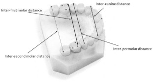
Maxillary arch width.
-
1.
intercanine distance: the linear distance between canine cusp tips
-
2.
interpremolar distance: the linear distance between the buccal cusp tips of the second premolars
-
3.
inter–first molar distance: the distance between the mesiobuccal cusp tips of the first molars
-
4.
inter–second molar distance: the distance between the distobuccal cusp tips of the second molars.
2.3. Maxillary arch length (Fig. 2)
Figure 2.
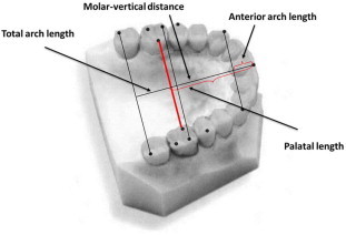
Maxillary arch and palatal length.
-
1.
anterior arch length: the vertical distance from the incisal point to the intercanine distance line
-
2.
molar-vertical distance: the vertical distance from the incisal point perpendicular to a line between the mesiolingual cusp tips of the first molars
-
3.
total arch length: the vertical distance from the incisal point to the midpoint of a line between the distobuccal cusp tips of the second molars.
2.4. Palatal width, length, and depth (Fig. 3)
Figure 3.
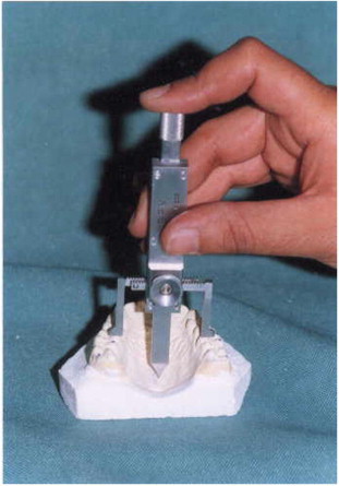
Measurement of palatal width and depth by Palatometer.
-
1.
palatal width: the linear distance between the mesiolingual cusp tips of the right and left first molars
-
2.
palatal length: equivalent to the molar-vertical distance
-
3.
palatal depth: the vertical distance from a point on the palatal width line to the palatal vault in the midline.
2.5. Statistical analysis
All statistical analyses were performed using SPSS software (version 13.0; SPSS Inc., Chicago, IL, USA). Descriptive statistics were obtained by calculating mean, minimum, and maximum values; standard deviations; ranges; and coefficients of variation (CVs). Differences in palatal and maxillary arch dimensions between men and women were examined using t-tests. P values < 0.05 were considered to be significant.
3. Results
Maxillary and palatal dimensions are shown in Table 1. The inter–second molar distance had the widest range.
Table 1.
Maxillary arch widths and lengths for the total sample.
| Meana | S.D. | Min. | Max. | Range | |
|---|---|---|---|---|---|
| Inter-canine distance | 34.2 | 2.04 | 30.0 | 39.0 | 9.0 |
| Interpremolar distance | 46.4 | 2.6 | 40.0 | 52.0 | 12.0 |
| Inter-first molar distance | 51.2 | 2.7 | 44.0 | 57.3 | 13.3 |
| Inter-second molar distance | 56.9 | 3.3 | 49.4 | 64.7 | 15.3 |
| Anterior arch length | 8.7 | 1.2 | 5.0 | 11.4 | 6.4 |
| Molar-vertical distance | 30.0 | 2.1 | 24.9 | 34.9 | 10.0 |
| Total arch length | 42.6 | 2.4 | 38.4 | 47.6 | 9.2 |
Measurements are in mm.
Dimensions are presented according to sex in Table 2. The majority of mean maxillary arch values were significantly larger in men than in women, with the inter–second molar distance showing the greatest difference and palatal depth showing the least difference. Mean molar-vertical distance and palatal length were significantly greater among women than among men.
Table 2.
Maxillary arch and Palatal dimensions according to gender.
| Females n = 101 | Males n = 113 | T-value | |||||
|---|---|---|---|---|---|---|---|
| Mean a | S.D. | C.V | Mean | S.D. | C.V | ||
| Inter-canine distance | 33.27 | 1.78 | 5.86 | 35.06 | 1.89 | 5.40 | 6.29⁎⁎ |
| Inter-premolar distance | 45.41 | 2.32 | 5.10 | 47.29 | 2.45 | 5.19 | 4.97⁎⁎ |
| Inter-first molar distance | 49.94 | 2.19 | 4.39 | 52.53 | 2.62 | 4.99 | 6.93⁎⁎ |
| Inter-second molar distance | 55.27 | 2.82 | 5.10 | 58.51 | 3.03 | 5.18 | 7.15⁎⁎ |
| Anterior arch length | 8.54 | 1.35 | 15.76 | 8.88 | 1.08 | 12.19 | 1.81 |
| Molar-vertical distance | 30.39 | 2.12 | 6.69 | 29.71 | 2.02 | 6.79 | 2.13⁎⁎ |
| Total arch length | 42.30 | 2.43 | 5.74 | 42.62 | 2.32 | 5.43 | 0.86 |
| Palatal width | 39.06 | 2.32 | 5.93 | 41.66 | 2.82 | 6.77 | 6.49⁎⁎ |
| Palatal length | 30.42 | 2.04 | 6.70 | 29.73 | 2.01 | 6.75 | 2.21⁎⁎ |
| Palatal depth | 20.71 | 1.39 | 6.73 | 21.17 | 1.51 | 7.12 | 2.04⁎⁎ |
Measurements are in mm.
Statistically significant (p < 0.05).
Correlation coefficients between arch widths and lengths were calculated. Some measurements of arch width and length were significantly, positively, and directly correlated, whereas others showed moderate, weak, and/or negative correlation. Maxillary arch dimensions were correlated more strongly in women than in men (Table 3).
Table 3.
Correlation Coefficient between the Maxillary widths and lengths.
| Anterior arch length | Molar-vertical distance | Total arch length | ||||
|---|---|---|---|---|---|---|
| F | M | F | M | F | M | |
| Inter-canine distance | 0.47 | 0.13 | 0.35 | -0.11 | 0.30 | -0.07 |
| Inter-premolar distance | 0.62 | 0.15 | 0.38 | 0.09 | 0.30 | 0.11 |
| Inter-first molar distance | 0.56 | 0.20 | 0.32 | 0.01 | 0.24 | 0.10 |
| Inter-second molar distance | 0.18 | -0.12 | 0.24 | 0.21 | 0.10 | 0.05 |
Values more than 0.21 were significant at p < 0.05.
4. Discussion
4.1. Sample selection
Oral health, particularly the treatment of malocclusion, is not currently a high priority in Yemen. However, for future planning purposes, valid and reliable information about norms (normal skeletodental relationships) is needed. Such information enables the establishment of useful guidelines for orthodontic diagnosis and treatment planning.
Dental arch integrity can be disturbed by many local factors affecting dental arch size and form, such as heavy fillings, spacing or crowding, abnormally shaped teeth, and bad oral habits (e.g., thumb sucking or mouth breathing). Individuals affected by any of these factors were excluded from participation in the present study.
Although precise skeletal relationships can be determined using lateral cephalometric radiographs, the ability to assess these relationships clinically is important due to the unavailability of cephalometric equipment in many practices. The two-finger technique of skeletal pattern assessment has been validated (Mills, 1987; Singh, 2007; Cobourne and DiBiase, 2010).
4.2. Sex differences
As confirmed in many previous studies, maxillary arch widths were greater in men than in women in the present study. Clinicians have speculated that women have smaller bony ridges and alveolar processes and the average weakness of musculature males, which have important effects on measurements of facial breadth and dental arch height and width, and the later growth period in males than females (Younes, 1984).
The observation of significant sex differences only in transverse dimensions is in agreement with many previous reports (Cohen, 1940; Moorrees and Reed, 1965;; Alvaran, 2009) and contrasts with reports with nearly similar maxillary widths in men and women (Knutz 1993). Ismail et al. (1996) reported a greater width in females than in males, but this difference was not statistically significant. In the present study, the inter–second molar distance showed the largest difference between sexes, which may be attributed to the difference in arch form.
In contrast with the findings of Raberin et al. (1993) and Borgan (2001), no sex difference in anterior or total maxillary arch length was observed in this study.
Mean molar-vertical distance and palatal length were greater in women than in men, contradicting the accepted view that maxillary arch dimensions are larger in men. Cohen (1940) reported similar results. This difference may be attributable to differences in ethnicity, sample size, tooth size (women have larger teeth), and/or environmental factors.
The observed greater palatal width and depth in men than in women is in agreement with the findings of Borgan (2001) and contrasts with the absence of a sex difference in these dimensions reported by Al-Mulla et al. (1997).
4.3. Determinants of Yemeni maxillary arch dimensions
Correlations between all maxillary dental arch widths and lengths were weak in men, but most correlations (except that between inter–second molar distance and total arch length) were strong in women.
CV values for all measurements of maxillary arch width and length were nearly close to each other, with intercanine distance and anterior arch length, respectively, producing the largest CVs among these values. These results are to be expected, as these two dimensions contribute to differences in arch form. These findings are in agreement with those of Andria and Carlos (1978).
CV values for all palatal measurements were also nearly close to each other, with palatal depth showing the largest CV.
Measurements of palatal depth and relationships of the canines to one another and to other teeth thus had the widest ranges, implying that these dimensions are the strongest determinants of maxillary arch size.
5. Conclusion
-
•
Maxillary arch width was greater in Yemeni men than in women, whereas molar-vertical distance and palatal length were greater in Yemeni women than in men.
-
•
Measurements related to the canines and palatal depth had the widest ranges implying that these dimensions are the strongest determinants of maxillary arch size.
Conflict of interest
The author declare no conflict of interest.
Footnotes
Peer review under responsibility of King Saud University.
References
- Al-Khateeb S., Abu Alhaija E. Tooth Size Discrepancies and Arch Parameters among Different Malocclusions in a Jordanian Sample. Angle Orthod. 2006;76:459–465. doi: 10.1043/0003-3219(2006)076[0459:TSDAAP]2.0.CO;2. [DOI] [PubMed] [Google Scholar]
- Al-Mulla A., Al-Bashir A. A new method for determination of highest point of the palate and its correlation. Iraqi D J. 1997;19:71–82. [Google Scholar]
- Alvaran N., Roldan S., Buschang P. Maxillary and mandibular arch widths of Colombians. Am J Orthod Dentofacial Orthop. 2009;135:649–656. doi: 10.1016/j.ajodo.2007.05.023. [DOI] [PubMed] [Google Scholar]
- Andria L.M., Carlos J. Relation of maxillary and mandibular intercuspid width to bizygomatic and bigonial breadth. Angle Orthod. 1978;48:154–162. doi: 10.1043/0003-3219(1978)048<0154:ROMAMI>2.0.CO;2. [DOI] [PubMed] [Google Scholar]
- Angle G.M. Classification of Malocclusion. Dental Cosmos. 1889;41:248–464. [Google Scholar]
- Barrow G.V., White J.R. Developmental changes of the maxillary and mandibular dental arches. Angle Orthod. 1952;22(1):41–46. [Google Scholar]
- Bishara S.E. Arch width changes from 6 weeks to 45 years of age. Am J Orthod Dentofacial Orthop. 1997;111:401–409. doi: 10.1016/s0889-5406(97)80022-4. [DOI] [PubMed] [Google Scholar]
- Bishara S.E. Changes in the maxillary and mandibular tooth size-arch length relationship from early adolescence to early adulthood “A longitudinal study”. Am J Orthod Dentofacial Orthop. 1989;92(1):46–59. doi: 10.1016/0889-5406(89)90135-2. [DOI] [PubMed] [Google Scholar]
- Bishara S.E., Jakonsen J.R., Nowak A. Arch length changes from 6 weeks to 45 years. Angle Orthod. 1998;68:69–74. doi: 10.1043/0003-3219(1998)068<0069:ALCFWT>2.3.CO;2. [DOI] [PubMed] [Google Scholar]
- Borgan, B.E., 2001. Dental arch dimensions analysis among Jordanian school children, Master Thesis, Cairo University-Egypt.
- Buschang P.H., Stroun B., Alexander R.G. Difference in dental arch morphology among adult females with untreated class I and II malocclusion. E J Orthod. 1994;16:47–52. doi: 10.1093/ejo/16.1.47. [DOI] [PubMed] [Google Scholar]
- Cobourne M.T., DiBiase A.T. first ed. Mosby; 2010. Handbook of orthodontics; p. 133. [Google Scholar]
- Cohen J. Growth and development of dental arches in children. JADA. 1940;27:1250–1260. [Google Scholar]
- Diwan R., Elahi J.M. A comparative study between three ethnic groups to derive some standards for maxillary arch dimensions. J Oral Rehab. 1990;17:43–48. doi: 10.1111/j.1365-2842.1990.tb01392.x. [DOI] [PubMed] [Google Scholar]
- Eid A., El-Namrawy M., Kadry W. The relationship between the width, depth and circumference of the dental arch for a group of Egyptian school children. Egyptian Orthod J. 1987;1:113–136. [Google Scholar]
- Ferrario V. Mathematical definition of the shape of dental arches in human permanent healthy dentitions. E J Orthod. 1994;16:287–294. doi: 10.1093/ejo/16.4.287. [DOI] [PubMed] [Google Scholar]
- Ghafari J. Changes of arch width in the early treatment of class II, division 1 malocclusion. Am J Orthod. Dentofac Orthop. 1994;106(5):496–501. doi: 10.1016/S0889-5406(94)70072-9. [DOI] [PubMed] [Google Scholar]
- Harris E.F., Smith R.J. Occlusion and arch size in families. Angle Orthod. 1982;52(2):135–142. doi: 10.1043/0003-3219(1982)052<0135:OAASIF>2.0.CO;2. [DOI] [PubMed] [Google Scholar]
- Houston, W.J., Stephens, C.D., Tulley, W.J., 1996. A textbook of Orthodontics. Wright PSG, P. 119.
- Ismail A.M., Hissain N., Hatem S. Maxillary arch dimensions in Iraqi population sample. Iraqi D J. 1996;8:111–120. [Google Scholar]
- Knott V.B. Size and form of the dental arches. Am J phys Anthropo. 1961;19:263–278. doi: 10.1002/ajpa.1330190308. [DOI] [PubMed] [Google Scholar]
- Knutz, T.R., 1993. An anthropometric comparison of cephalometric and dental arch measurements in classes I normal, class I crowded and class III individuals, Master Thesis, University of Iowa.
- Lavelle C.L., Foster T.D., Flinn R.M. Dental arches in various ethnic groups. Angle Orthod. 1971;41:293–299. doi: 10.1043/0003-3219(1971)041<0293:DAIVEG>2.0.CO;2. [DOI] [PubMed] [Google Scholar]
- Mack P.J. Maxillary arch and central incisor dimensions in a Nigerian and British population sample. J Dent Res. 1981;9(1):67–70. doi: 10.1016/0300-5712(81)90037-3. [DOI] [PubMed] [Google Scholar]
- Mills J.R. second ed. Churchil Livingstone; 1987. Principles and practice of orthodontics. Pp. 16-18, 23. [Google Scholar]
- Mills L.F. Arch width, arch length, and tooth size in young adult males. Angle Orthod. 1964;34:124–129. [Google Scholar]
- Moorrees C.F.A., Reed R.B. Change in the dental arch dimensions expressed on the basis of tooth eruption as a measure of biologic age. J D Res. 1965;44(1):129–140. doi: 10.1177/00220345650440010601. [DOI] [PubMed] [Google Scholar]
- Raberin M. Dimensions and form of dental arches in subjects with normal occlusion. Am J Orthod. Dentofacial Orthop. 1993;103:67–72. doi: 10.1016/0889-5406(93)70029-N. [DOI] [PubMed] [Google Scholar]
- Singh G. second ed. Jaypee Brothers Medical Publishers; 2007. Textbook of orthodontics; pp. 69–70. [Google Scholar]
- Staley R.N., Stuntz W.R., Peterson L. A comparison of arch widths in adults with normal occlusion and adults with class II division 1 occlusion. Am J Orthod. 1985;88:163–169. doi: 10.1016/0002-9416(85)90241-6. [DOI] [PubMed] [Google Scholar]
- Warren J.J., Bishara S.E. Comparison of dental arch measurement in the primary dentition between contemporary and historic samples. Am J Orthod Dentofacial Orthop. 2001;119:211–215. doi: 10.1067/mod.2001.112260. [DOI] [PubMed] [Google Scholar]
- Younes S.A. Maxillary arch dimensions in Saudi and Egyptian population sample. Am J Orthod Dentofacial Orthop. 1984;85:83–87. doi: 10.1016/0002-9416(84)90126-x. [DOI] [PubMed] [Google Scholar]