Abstract
Identifying inter-area communication in terms of the hierarchical organization of functional brain areas is of considerable interest in human neuroimaging. Previous studies have suggested that the direction of magneto- and electroencephalography (MEG, EEG) source currents depends on the layer-specific input patterns into a cortical area. We examined the direction in MEG source currents in a visual object recognition experiment in which there were specific expectations of activation in the fusiform region being driven by either feedforward or feedback inputs. The source for the early non-specific visual evoked response, presumably corresponding to feedforward driven activity, pointed outward, i.e., away from the white matter. In contrast, the source for the later, object-recognition related signals, expected to be driven by feedback inputs, pointed inward, toward the white matter. Associating specific features of the MEG/EEG source waveforms to feedforward and feedback inputs could provide unique information about the activation patterns within hierarchically organized cortical areas.
Keywords: magnetoencephalography, cerebral cortex, current dipole, top-down, bottom-up
Introduction
Non-invasive methods such as magneto- and electroencephalography (MEG, EEG) and functional magnetic resonance imaging (fMRI) provide means to examine neural activity in various ways, for example, by determining locations, sequences, and connectivity patterns among regions in the human brain [39]. In addition to these measures, identifying inter-area communication in terms of the hierarchical organization of functional brain areas would be highly relevant in the quest for understanding the operation of the brain. Characteristic anatomical laminar distributions of input and output connections between cortical areas have been described as being of feedforward, feedback, or lateral type, thereby defining a hierarchical organization among the areas [15, 36]. However, this type of information is not readily available in human imaging data. The spatial resolution of fMRI is approaching the level at which laminar distributions of cortical activity can be detected [35]. The direction of the MEG and EEG source currents is another piece of information that may help to characterize layer-specific input patterns into a cortical area, thereby providing cues about the flow and the function of the detected neural activity in terms of feedforward (bottom-up) and feedback (top-down) of inputs [18, 23].
MEG and EEG signals originate mainly from post-synaptic currents in cortical pyramidal cells [33], and the direction of the source current depends on the type and the dendritic location of the synaptic input [5, 27]. In event-related response waveforms, an initial deflection can often be associated with feedforward input, followed by broader feedback-related activity [3]. In somatosensory, auditory, and visual evoked MEG data, early biphasic or triphasic responses, presumably driven by feedforward inputs, and later uniphasic feedback driven responses have been observed [18-20]. Given the different laminar distributions of feedforward and feedback type inputs, it is conceivable that the direction of the initial phase of a feedforward driven response is opposite to that of a feedback driven response. Biophysically realistic computational neural modeling incorporating detailed physiology of the laminar structure in cortical circuits has been successfully applied to interpret the directionality of neural current sources underlying MEG signals during a somatosensory detection task [22, 23, 41]. In the present study, we examined the direction in MEG source currents in the fusiform region in a visual object detection experiment in which there were specific expectations of activation being driven by either feedforward or feedback inputs [9].
Methods
The present results were derived from new analyses applied to previously published MEG data from a visual object recognition experiment [9]. In this experiment, the experimental evidence supported specific theoretical predictions regarding latency- and condition-dependent feedforward and feedback inputs to the inferior occipitotemporal (fusiform) region. According to the model of Bar [7, 8] (Fig. 1A), low spatial frequency information about the visual object is quickly passed to the orbitofrontal cortex. Previously, activity associated with successful recognition of objects was found to occur earlier in the orbitofrontal cortex than in the fusiform region [9]. This is consistent with the orbitofrontal cortex enabling top-down facilitation of object recognition by sending predictions about the object identity to the fusiform cortex. Therefore, feedback-type input into the fusiform region is expected for those trials in which the subject recognized the object. The fusiform region is also expected to receive feedforward-type input through a bottom-up route along the ventral visual pathway. Thus, the fusiform region is expected to receive both top-down feedback-type input as well as bottom-up feedforward-type input. Here, we determined the direction of the MEG source current in these cases to evaluate whether the source direction is dependent on the input type.
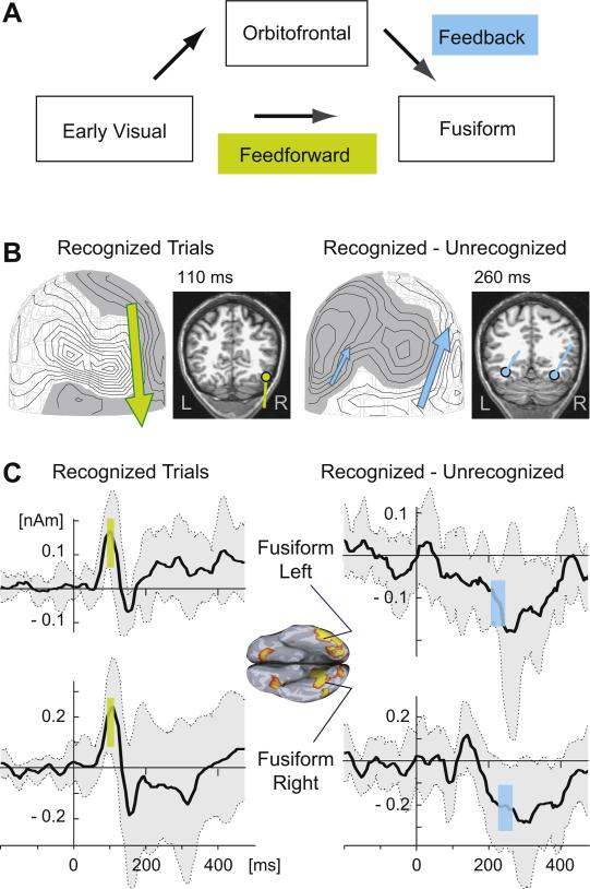
Figure 1. Direction of MEG source currents in a visual object recognition experiment.
A. Sequence of cortical activation during visual object recognition, according to Bar et al. [9]. The fusiform area is expected to receive inputs from both higher and lower areas in the hierarchical organization: feedback-type input from the orbitofrontal cortex representing top-down facilitation and feedforward-type input from the early visual areas.
B. MEG data from a single subject, depicting visual evoked response at the latency of 110 ms, in data averaged over all trials in which the subject recognized the object (left), and differential object recognition specific response at 260 ms in subtraction data (recognized vs. unrecognized object trials; right). The isocontour maps (40 fT between lines) depict the normal component of the magnetic field over the back of the head. The arrows represent equivalent current dipoles fitted to the MEG. The dots indicate the dipole locations in the inferior occipitotemporal area on magnetic resonance images. Note that the direction of the dipoles reversed between these two cases in which feedforward and feedback inputs are expected: dipoles pointing outward inward are shown in green and blue, respectively.
C. Estimated MEG source waveforms for inferior occipitotemporal (fusiform) regions-of-interest in the left (top) and right (bottom) hemispheres, obtained using a distributed source model. The shading indicates one standard deviation above and below the mean value across 9 subjects. For the early visual evoked activity (left), positive amplitude, corresponding to outward polarity, was observed in both hemispheres at 100-120 ms (green bars). For the difference data, object recognition specific response (right) the estimated source had negative, inward direction at 210-250 ms (blue). These data are consistent with the source currents being of opposite direction feedforward and feedback inputs.
Nine healthy volunteers (6 females, age range 22-30 years) performed a visual object recognition task during the MEG recordings. The protocols were approved by the Internal Review Board at Massachusetts General Hospital; written informed consent was obtained from all subjects. Line drawings of familiar objects were presented on a computer screen for 63 ms, preceded and followed by random-dot mask patterns for 27 ms and 108 ms, respectively. Subjects were instructed to recognize each of the objects and to indicate their level of knowledge about the identity of the object by pressing one of four response buttons. MEG signals were obtained using a 306 channel Vectorview system (Elekta Neuromag, Finland), comprising of 204 planar gradiometers and 102 magnetometers. The sampling frequency was 600 Hz with a 0.1-200 Hz band-pass filter. Responses were low-pass filtered off-line at 20 Hz. Epochs were baseline corrected by subtracting the mean of the 500 ms pre-stimulus interval in each sensor. For details of the experimental setup, see [9].
The direction of the source currents were examined using a distributed source model, the minimum-norm estimate (MNE) [17]. The MNE-based estimates of the time course of the source currents in the left and right hemisphere fusiform gyrus regions-of-interest (ROIs) were obtained [9]. The MNE was computed by assuming that all sources were located on the cortical surface extracted from anatomical MR images; a loose orientation constraint and depth-weighting were applied [25]. To determine the direction of the source currents, the source components normal to the cortical surface was extracted. The MNEs were constructed for each individual subject; the waveforms were computed as the mean value of the amplitude of the discretized source elements within the ROIs. In addition to the MNE analysis, the location and direction of the fusiform sources in individual subjects were illustrated with equivalent current dipoles.
For the practical estimation of the MEG and EEG source direction, it is helpful to make a distinction between the physiological direction and the physical orientation of the source current. MEG and EEG are highly sensitive to the physical orientation of the source [1], which usually can be reliably determined [29]. However, identifying the physiological direction of the source (i.e., outward vs. inward with respect to the white matter), accurate localization of the source with respect to the cortical anatomy is essential: if the source is mis-localized to the opposite bank of a sulcus, an erroneously reversed direction will be inferred. Here the tangentially oriented fusiform source currents were mainly on gyral parts of the inferior surface of the occipitotemporal region [9]; thus, they were well suited for reliable determination of the physiological source direction using MEG.
Two specific cases of fusiform activation were examined. The first was non-specific early evoked activity, obtained from all recognized trials in the latency window 100-120 ms after the appearance of the first visual masking stimulus. This early activity is assumed to results from feedforward input to the fusiform region, presumably form the occipital visual cortices. The second case was the later, recognition-related activity, obtained from the difference between conditions (recognized minus unrecognized trials, 210-250 ms). This recognition-related would be consistent with resulting from feedback-type top-down facilitatory inputs from the orbitofrontal cortex, which showed activation around 130 ms in the previous study [9]. For statistical analysis, a t-test was performed for the MNE-amplitude of the left and right hemisphere fusiform ROIs for the two cases against the null hypothesis of the mean amplitude across the subjects being zero.
Results
Measured MEG field maps and the corresponding equivalent current dipoles for one subject are shown in Fig. 1B. The early visual evoked response at 110 ms suggested a source in the right inferior occipitotemporal cortex, pointing outward, i.e., away from the white matter. In contrast, the differential signals for recognized and not recognized trials at 260 ms suggested later bilateral inferior occipitotemporal sources pointing inward, toward the white matter.
Results from a distributed source analysis of the MEG data confirmed the reversal of the source direction between the two conditions. Source waveforms for the left and right fusiform ROIs, averaged over 9 subjects, are shown in Fig. 1C. A t-test indicated significant positive (outward direction) source amplitude for the 100-120 ms latency window of the initial visual response (right hemisphere: tdf=9 = 4.01, p = 0.003, uncorrected; left: tdf=9 = 5.26, p = 0.0005), and negative (inward) amplitude for the 210-250 ms window of the recognition-specific subtraction data (right: tdf=9 = 2.58, p = 0.03; left: tdf=9 = 4.21, p = 0.002).
Discussion
The opposite directions of the estimated MEG source currents for the fusiform gyrus in the two experimental conditions are consistent with a dependence between the source direction and the type of information flow within a hierarchically organized network of cortical areas. Feedforward inputs connect to the middle parts (granular layer 4), whereas feedback type inputs connect mainly superficially (supragranular layers 1-3) and also to some extent to the deep layers (Fig. 2A) [10, 15, 36]. Biophysical computational modeling studies have suggested that excitatory input to granular layers (feedforward type input) subsequently propagating to supraand infragranular layers can create outward directing current source, whereas input to supragranular layers (feedback) creates a source in the opposite, inward direction [22, 23]. The present results are in accordance with this interpretation, with feedforward and feedback inputs creating outward and inward currents, respectively. Thus, the direction of MEG and EEG source currents may contain information about whether the observed activity is due to inputs from a cortical area lower or higher in the hierarchical organization.
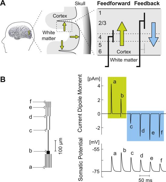
Figure 2. Dependence of the current dipole direction on the spatial location of synaptic input.
A. Schematic illustration of the laminar distribution of feedforward and feedback synaptic input (thick lines) into a cortical area (adapted from [37]). The hypothesized direction of the resulting macroscopic current dipole is outward, i.e., away from the white matter, for feedforward input (green arrow) and inward for feedback input (blue arrow). The cross-section of a region of the cortex on the left illustrates three dipoles (small green arrows) with different physical orientations: the two dipoles depicted in the sulcal walls are tangential with respect to the skull, the middle one in the gyrus is radial; however, the physiological direction for all of these is outwards. Note that in Fig. 1B the dipoles and the cortical lamina are “upside down” compared with this laminar diagram, because those sources are located in the inferior surface of the occipitotemporal cortex.
B. A simple computational model of a pyramidal neuron to demonstrate how the direction of the current dipole can depend on the location of synaptic input. The compartmental model, built using the NEURON software (www.neuron.yale.edu/neuron/), consisted of cylindrical segments representing the soma, five basal dendrites, and an apical dendrite with a trunk and three levels of bifurcations (left). All segments were assumed to be oriented perpendicular to the cortical surface. Passive membrane properties were assumed [38]. Synaptic inputs were modeled as changes in the transmembrane conductivity in the form of the alpha function with time constant τ = 0.7 ms. The synapses were placed at the end points of the dendritic segments (labeled “a” to “f”) and activated one at a time. The net current dipole was calculated by summing the dipole moment, obtained by multiplying the intracellular axial current by the length of the segment, for each segment of the pyramidal cell model [30]. When excitatory input was located at a basal dendrite or the soma, the corresponding current dipole (top) was in the upwards direction, i.e., positive values corresponding to outward currents (“a” and “b”, green), whereas for distal apical inputs the direction was reversed (“c”, “d”, “e”, and “f”, blue). The change in the trans-membrane potential in the soma (bottom) was in the same direction in each case.
A simple model to demonstrate how the direction of the current dipole can depend on the pattern of synaptic inputs is shown in Fig. 2B. Excitatory synapses in the distal parts of the branches representing the apical dendrite of a pyramidal cell resulted in a current dipole pointing towards the white matter (negative signals in Fig. 2B; labels c to f), whereas input close to the soma resulted in the opposite dipole direction (positive signals; labels a and b). The direction of the change in the somatic potential was the same in each case, i.e., depolarization. Similar reversal of source direction as a function of the dendritic input location has been shown previously using models with unbranched [6, 23] as well as realistically-shaped [2, 26] dendritc trees.
Using a detailed network model of the primary somatosensory cortex that contained many cell types in multiple layers, Jones et al. have shown that excitatory drive to different layers could create current flow in different directions [21-23, 41]. The model could reproduce a sequence of outward-inward-outward directed currents (M25, M35, M50) in the MEG data with a single thalamic feedforward event that predominantly drove supragranular layer (layer 2/3) pyramidal neurons. This sequence started with excitatory current up the apical dendrites due in part to back propagation of action potentials (i.e., outward current, M25), followed by local network recruited inhibition near the somas creating inward current (M35), followed by recurrent excitation that created outward current (M50). For the later part of the somatosensory evoked response, a large early inward peak occurred at ~70ms (M70); in the computational model this arose from a feedback type input to the supragranular layers that resulted in current flow down the supra- and infragranular pyramidal neuron dendrites [22, 23]. This peak was dominated by the activity in the larger layer 5 pyramidal neurons due to their dendritic lengths. This feedback drive also created a subsequent outward peak at ~100 ms (M100) from recurrent excitation in the network and back propagating action potentials and a later outward peak (M135) was recreated with a second thalamic feedforward drive, as might arise from a thalamocortical loop of input. During a tactile detection task the magnitude and timing of the feedback induced peaks, M70 and M100, depended on the percept of the subject, and could be modulated with the feedback drive in the model. This result is consistent with the idea of inward currents reflecting higher order feedback inputs [11].
Inward source currents associated with top-down influence have also been reported in scalp EEG studies of object perception using fragmented line drawings or illusory contours, showing surface negative responses in ventral visual areas [13, 31]. In a recent study of visual shape discrimination, late inward source currents related to target detection were found in area V4 that were consistent with feedback input from the lateral occipital region [4]. These surface negative responses were suggested to be related to the selection negativity event-related response component, which in primate recordings have been associated with attentional modulation of activity in area V4 mediated by inputs to feedback recipient layers [28]. Indeed, surface negative responses are commonly associated with top-down attentional modulatory inputs [40]. For example, although the N1 component of the auditory evoked response is considered to be “exogenous”, reflecting bottom-up influences in the cognitive sense, feedback to auditory cortex during dichotic listening tasks is observed as enhanced “negative difference/processing negativity”, reflecting enhanced inward currents, in N1 responses to attended vs. ignored responses [32].
To predict the source current direction on the basis of anatomical connections, it is essential to identify the location of the synaptic inputs with respect to the dendritic morphology of the receiving cell. For example, excitatory inputs to basal dendrites are expected to result in currents opposite to those resulting for excitatory inputs to distal parts of the apical dendrite (cf. Fig. 2B). Relating the laminar input patterns to the source current direction is challenging because only limited information about both the laminar pattern of synaptic connections and the laminar location of the soma and dendrites of the receiving neurons is currently simultaneously available [34].
The source direction also depends on whether the synaptic inputs are excitatory or inhibitory; the same spatial distribution of excitatory and inhibitory inputs would result in source currents of opposite directions [5]. The vast majority of inputs from other cortical areas are excitatory, but the ensuing activity within the local cortical circuitry is likely to involve activation of inhibitory interneurons. However, the spatial distribution and receptor properties of inhibitory synapses, in particular those of GABAA type, give reasons to expect that they contribute less than excitatory ones to the source currents. GABAA receptors are mostly at or near the soma [14], where the ensuing post-synaptic axial currents flow in opposite directions in the basal and apical dendrites, and the net source current largely cancels out [2, 26]. Furthermore, the reversal potential of GABAA receptors is typically close to the membrane resting potentials (shunting inhibition), and therefore, there will be little synaptic currents without simultaneous excitatory inputs. However, some contributions to the source currents can be expected from hyperpolarizing inhibitory GABAB type synapses, which are also more distally located [14].
A prior computational modeling study investigated the impact of somatic targeting GABAA synaptic inhibition on source currents in the context of gamma rhythms [24]. That study found that strong somatic GABAA inhibition could induce inward-directed dipole currents. In the model, these inward currents required strong spiking and synchrony in the inhibitory neuron population, which induced fast inward current flow in the apical dendrites across the pyramidal neuron population. Similarly, strong synchronous inhibition distally in the apical dendrites induced fast outward current flow. A rapid deflection in the dipole current waveform was suggested as a means to distinguish GABA mediated influences from more diffuse excitatory synaptic influences. Under this framework, the slower deflections in Fig. 1C are more consistent with diffuse excitatory synaptic inputs. In general, multi-feature dynamic patterns of activation could potentially be used to better resolve different types of inputs, including also, for example, lateral connections between areas at the same level of hierarchy [12]. For this, intracranial recordings with multi-contact electrodes provide invaluable data for relating laminar input patterns and the MEG and EEG source waveforms [16, 37].
The dependence of source direction on the type of input could provide a means for obtaining non-invasive information about the activation patterns among hierarchically organized cortical areas. Interpretation of the activation patterns in terms of hierarchical organization of the functional neuroimaging data is generally based on a priori assumptions about the hierarchical levels [12]. Relating features of the MEG/EEG source waveforms to feedforward and feedback inputs could provide unique information about the activation patterns within hierarchically organized cortical areas in the human brain.
Highlights.
Fusiform sources in a visual object recognition MEG experiment were examined
Directions of source currents were opposite for expected feedforward and feedback inputs
MEG and EEG source direction may depend on hierarchical organization
Acknowledgements
We thank Avniel Ghuman and Karim Kassam for help with the MEG data analysis, Christopher Wreh II for assistance with the computational modeling, and Maria Mody for useful discussions.
Financial Disclosure
This work was supported by NIH grants NS57500 (SPA), NS37462 (JWB/SPA), EY019477 (MB). This work was supported in part by The National Center for Research Resources (P41EB015896). The funders had no role in study design, data collection and analysis, decision to publish, or preparation of the manuscript.
Footnotes
Publisher's Disclaimer: This is a PDF file of an unedited manuscript that has been accepted for publication. As a service to our customers we are providing this early version of the manuscript. The manuscript will undergo copyediting, typesetting, and review of the resulting proof before it is published in its final citable form. Please note that during the production process errors may be discovered which could affect the content, and all legal disclaimers that apply to the journal pertain.
Author Contributions:
Conceived and developed the conceptual framework: SA SRJ JA JWB MB. Analyzed the data: SPA MB. Contributed analysis tools: MSH. Wrote the manuscript: SPA SRJ JA MB.
References
- 1.Ahlfors SP, Han J, Belliveau JW, Hamalainen MS. Sensitivity of MEG and EEG to source orientation. Brain Topogr. 2010;23:227–232. doi: 10.1007/s10548-010-0154-x. [DOI] [PMC free article] [PubMed] [Google Scholar]
- 2.Ahlfors SP, Wreh CI. Modeling the Effect of Dendritic Input Location on MEG and EEG Source Dipoles. doi: 10.1007/s11517-015-1296-5. (Submitted) [DOI] [PMC free article] [PubMed] [Google Scholar]
- 3.Aine CJ, Stephen JM, Christner R, Hudson D, Best E. Task relevance enhances early transient and late slow-wave activity of distributed cortical sources. J Comput Neurosci. 2003;15:203–221. doi: 10.1023/a:1025864825200. [DOI] [PubMed] [Google Scholar]
- 4.Ales JM, Appelbaum LG, Cottereau BR, Norcia AM. The time course of shape discrimination in the human brain. Neuroimage. 2013;67:77–88. doi: 10.1016/j.neuroimage.2012.10.044. [DOI] [PubMed] [Google Scholar]
- 5.Allison T, Puce A, McCarthy G. Category-sensitive excitatory and inhibitory processes in human extrastriate cortex. J Neurophysiol. 2002;88:2864–2868. doi: 10.1152/jn.00202.2002. [DOI] [PubMed] [Google Scholar]
- 6.Avitan L, Teicher M, Abeles M. EEG generator--a model of potentials in a volume conductor. J Neurophysiol. 2009;102:3046–3059. doi: 10.1152/jn.91143.2008. [DOI] [PubMed] [Google Scholar]
- 7.Bar M. A cortical mechanism for triggering top-down facilitation in visual object recognition. J Cogn Neurosci. 2003;15:600–609. doi: 10.1162/089892903321662976. [DOI] [PubMed] [Google Scholar]
- 8.Bar M. Visual objects in context. Nat Rev Neurosci. 2004;5:617–629. doi: 10.1038/nrn1476. [DOI] [PubMed] [Google Scholar]
- 9.Bar M, Kassam KS, Ghuman AS, Boshyan J, Schmidt AM, Dale AM, Hamalainen MS, Marinkovic K, Schacter DL, Rosen BR, Halgren E. Top-down facilitation of visual recognition. Proc Natl Acad Sci U S A. 2006;103:449–454. doi: 10.1073/pnas.0507062103. [DOI] [PMC free article] [PubMed] [Google Scholar]
- 10.Barbas H, Zikopoulos B. The prefrontal cortex and flexible behavior. Neuroscientist. 2007;13:532–545. doi: 10.1177/1073858407301369. [DOI] [PMC free article] [PubMed] [Google Scholar]
- 11.Cauller LJ, Kulics AT. The neural basis of the behaviorally relevant N1 component of the somatosensory-evoked potential in SI cortex of awake monkeys: evidence that backward cortical projections signal conscious touch sensation. Exp Brain Res. 1991;84:607–619. doi: 10.1007/BF00230973. [DOI] [PubMed] [Google Scholar]
- 12.David O, Harrison L, Friston KJ. Modelling event-related responses in the brain. Neuroimage. 2005;25:756–770. doi: 10.1016/j.neuroimage.2004.12.030. [DOI] [PubMed] [Google Scholar]
- 13.Doniger GM, Foxe JJ, Murray MM, Higgins BA, Snodgrass JG, Schroeder CE, Javitt DC. Activation timecourse of ventral visual stream object-recognition areas: high density electrical mapping of perceptual closure processes. J Cogn Neurosci. 2000;12:615–621. doi: 10.1162/089892900562372. [DOI] [PubMed] [Google Scholar]
- 14.Douglas R, Markram H, Martin K. Neocortex. In: Shepherd GM, editor. The Synaptic Organization of the Brain. Oxford University Press; New York: 2004. pp. 499–558. [Google Scholar]
- 15.Felleman DJ, Van Essen DC. Distributed hierarchical processing in the primate cerebral cortex. Cereb Cortex. 1991;1:1–47. doi: 10.1093/cercor/1.1.1-a. [DOI] [PubMed] [Google Scholar]
- 16.Halgren E, Wang C, Schomer DL, Knake S, Marinkovic K, Wu J, Ulbert I. Processing stages underlying word recognition in the anteroventral temporal lobe. Neuroimage. 2006;30:1401–1413. doi: 10.1016/j.neuroimage.2005.10.053. [DOI] [PMC free article] [PubMed] [Google Scholar]
- 17.Hamalainen MS, Ilmoniemi RJ. Interpreting magnetic fields of the brain: minimum norm estimates. Med Biol Eng Comput. 1994;32:35–42. doi: 10.1007/BF02512476. [DOI] [PubMed] [Google Scholar]
- 18.Inui K, Kakigi R. Temporal analysis of the flow from V1 to the extrastriate cortex in humans. J Neurophysiol. 2006;96:775–784. doi: 10.1152/jn.00103.2006. [DOI] [PubMed] [Google Scholar]
- 19.Inui K, Okamoto H, Miki K, Gunji A, Kakigi R. Serial and parallel processing in the human auditory cortex: a magnetoencephalographic study. Cereb Cortex. 2006;16:18–30. doi: 10.1093/cercor/bhi080. [DOI] [PubMed] [Google Scholar]
- 20.Inui K, Wang X, Tamura Y, Kaneoke Y, Kakigi R. Serial processing in the human somatosensory system. Cereb Cortex. 2004;14:851–857. doi: 10.1093/cercor/bhh043. [DOI] [PubMed] [Google Scholar]
- 21.Jones SR, Kerr CE, Wan Q, Pritchett DL, Hamalainen M, Moore CI. Cued spatial attention drives functionally relevant modulation of the mu rhythm in primary somatosensory cortex. J Neurosci. 2010;30:13760–13765. doi: 10.1523/JNEUROSCI.2969-10.2010. [DOI] [PMC free article] [PubMed] [Google Scholar]
- 22.Jones SR, Pritchett DL, Sikora MA, Stufflebeam SM, Hamalainen M, Moore CI. Quantitative analysis and biophysically realistic neural modeling of the MEG mu rhythm: rhythmogenesis and modulation of sensory-evoked responses. J Neurophysiol. 2009;102:3554–3572. doi: 10.1152/jn.00535.2009. [DOI] [PMC free article] [PubMed] [Google Scholar]
- 23.Jones SR, Pritchett DL, Stufflebeam SM, Hamalainen M, Moore CI. Neural correlates of tactile detection: a combined magnetoencephalography and biophysically based computational modeling study. J Neurosci. 2007;27:10751–10764. doi: 10.1523/JNEUROSCI.0482-07.2007. [DOI] [PMC free article] [PubMed] [Google Scholar]
- 24.Lee S, Jones SR. Distinguishing mechanisms of gamma frequency oscillations in human current source signals using a computational model of a laminar neocortical network. Front Hum Neurosci. 2013;7:869. doi: 10.3389/fnhum.2013.00869. [DOI] [PMC free article] [PubMed] [Google Scholar]
- 25.Lin FH, Witzel T, Ahlfors SP, Stufflebeam SM, Belliveau JW, Hamalainen MS. Assessing and improving the spatial accuracy in MEG source localization by depth-weighted minimum-norm estimates. Neuroimage. 2006;31:160–171. doi: 10.1016/j.neuroimage.2005.11.054. [DOI] [PubMed] [Google Scholar]
- 26.Linden H, Pettersen KH, Einevoll GT. Intrinsic dendritic filtering gives low-pass power spectra of local field potentials. J Comput Neurosci. 2010;29:423–444. doi: 10.1007/s10827-010-0245-4. [DOI] [PubMed] [Google Scholar]
- 27.Lopes da Silva FH. Electrophysiological Basis of MEG Signals. In: Hansen P, Kringelbach M, Salmelin R, editors. MEG: An Introduction to Methods. Oxford University Press; New York: 2010. pp. 1–23. [Google Scholar]
- 28.Mehta AD, Ulbert I, Schroeder CE. Intermodal selective attention in monkeys. II: physiological mechanisms of modulation. Cereb Cortex. 2000;10:359–370. doi: 10.1093/cercor/10.4.359. [DOI] [PubMed] [Google Scholar]
- 29.Mosher JC, Spencer ME, Leahy RM, Lewis PS. Error bounds for EEG and MEG dipole source localization. Electroencephalogr Clin Neurophysiol. 1993;86:303–321. doi: 10.1016/0013-4694(93)90043-u. [DOI] [PubMed] [Google Scholar]
- 30.Murakami S, Okada Y. Contributions of principal neocortical neurons to magnetoencephalography and electroencephalography signals. J Physiol. 2006;575:925–936. doi: 10.1113/jphysiol.2006.105379. [DOI] [PMC free article] [PubMed] [Google Scholar]
- 31.Murray MM, Wylie GR, Higgins BA, Javitt DC, Schroeder CE, Foxe JJ. The spatiotemporal dynamics of illusory contour processing: combined high-density electrical mapping, source analysis, and functional magnetic resonance imaging. J. Neurosci. 2002;22:5055–5073. doi: 10.1523/JNEUROSCI.22-12-05055.2002. [DOI] [PMC free article] [PubMed] [Google Scholar]
- 32.Naatanen R, Teder W. Attention effects on the auditory event-related potential. Acta Otolaryngol Suppl. 1991;491:161–166. doi: 10.3109/00016489109136794. discussion 167. [DOI] [PubMed] [Google Scholar]
- 33.Okada YC, Wu J, Kyuhou S. Genesis of MEG signals in a mammalian CNS structure. Electroencephalogr Clin Neurophysiol. 1997;103:474–485. doi: 10.1016/s0013-4694(97)00043-6. [DOI] [PubMed] [Google Scholar]
- 34.Petreanu L, Mao T, Sternson SM, Svoboda K. The subcellular organization of neocortical excitatory connections. Nature. 2009;457:1142–1145. doi: 10.1038/nature07709. [DOI] [PMC free article] [PubMed] [Google Scholar]
- 35.Polimeni JR, Fischl B, Greve DN, Wald LL. Laminar analysis of 7T BOLD using an imposed spatial activation pattern in human V1. Neuroimage. 2010;52:1334–1346. doi: 10.1016/j.neuroimage.2010.05.005. [DOI] [PMC free article] [PubMed] [Google Scholar]
- 36.Rockland KS, Pandya DN. Laminar origins and terminations of cortical connections of the occipital lobe in the rhesus monkey. Brain Res. 1979;179:3–20. doi: 10.1016/0006-8993(79)90485-2. [DOI] [PubMed] [Google Scholar]
- 37.Schroeder CE, Foxe JJ. The timing and laminar profile of converging inputs to multisensory areas of the macaque neocortex. Brain Res Cogn Brain Res. 2002;14:187–198. doi: 10.1016/s0926-6410(02)00073-3. [DOI] [PubMed] [Google Scholar]
- 38.Segev I, Burke RE. Compartmental Models of Complex Neurons. In: Koch C, Segev I, editors. Methods in Neuronal Modeling. MIT Press; Cambridge, MA: 1998. pp. 93–136. [Google Scholar]
- 39.Supek S, Aine CJ. Magnetoencephalography. From signals to dynamic cortical networks. Springer; Berlin: 2014. [Google Scholar]
- 40.Vaughan HG., Jr. The neural origins of human event-related potentials. Ann. N. Y. Acad. Sci. 1982;388:125–138. doi: 10.1111/j.1749-6632.1982.tb50788.x. [DOI] [PubMed] [Google Scholar]
- 41.Ziegler DA, Pritchett DL, Hosseini-Varnamkhasti P, Corkin S, Hamalainen M, Moore CI, Jones SR. Transformations in oscillatory activity and evoked responses in primary somatosensory cortex in middle age: a combined computational neural modeling and MEG study. Neuroimage. 2010;52:897–912. doi: 10.1016/j.neuroimage.2010.02.004. [DOI] [PMC free article] [PubMed] [Google Scholar]


