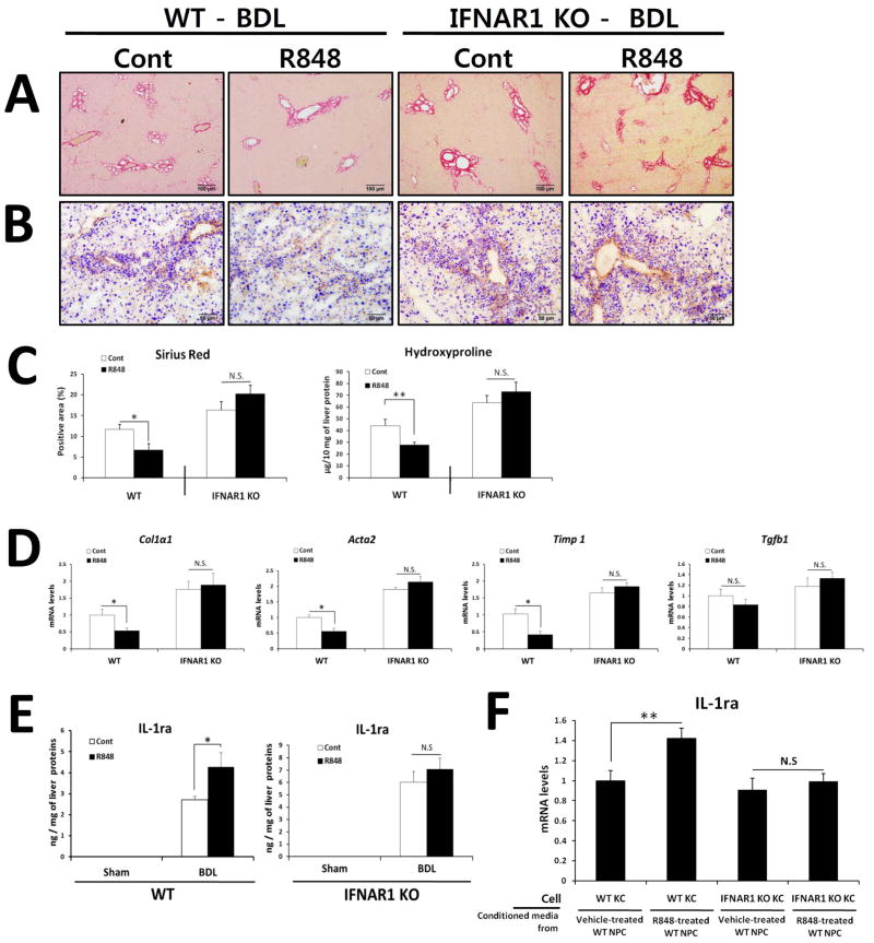
Fig. 7. R848-induced protection of liver fibrosis is dependent on type I IFN signaling.
(A–E) R848 or Saline-injected WT and IFNAR1-deficient mice underwent BDL (n = 12–14 per group). After 21 days, mice were evaluated as follows: (A) Fibrillar collagen deposition was determined by Sirius red staining and (C) quantification of the Sirius red–positive area. (B) The expression of α-SMA was determined by immunohistochemistry. (C) Hydroxyproline levels were determined. (D) Expression of pro-fibrogenic markers was determined by qRT-PCR and shown as fold change compared with saline-treated mice. (E) Hepatic IL-1ra was quantified by ELISA and expressed as ng of IL-1ra protein per mg of liver protein. (F) Gene expression was determined by qRT-PCR analysis of KCs isolated from WT or IFNAR1 KO mice and stimulated for 6 h by conditioned media from vehicle- or R848-treated WT NPCs. Values are presented from three independent experiments and each sample was assayed in duplicate. Data are presented as means ± SEM per group. Two-tailed Student’s t-test, *P < 0.05, **P < 0.01. Original magnification, ×100 (Sirius red), ×200 (α-SMA).

