Sir,
Giant cell arteritis (GCA) or temporal arteritis can cause profound and irreversible visual loss through anterior ischemic optic neuropathy (AION), posterior ischemic optic neuropathy, central retinal artery occlusion (CRAO), branch retinal artery occlusion (BRAO), choroidal infarction, and central nervous system stroke.1 We present a case where permanent vision loss was prevented by prompt recognition of the condition seen on fluorescein angiography (FA). To our knowledge, this is the first report of fluorescein angiographic evidence of reversible retinal circulatory abnormalities associated with GCA.
Case report
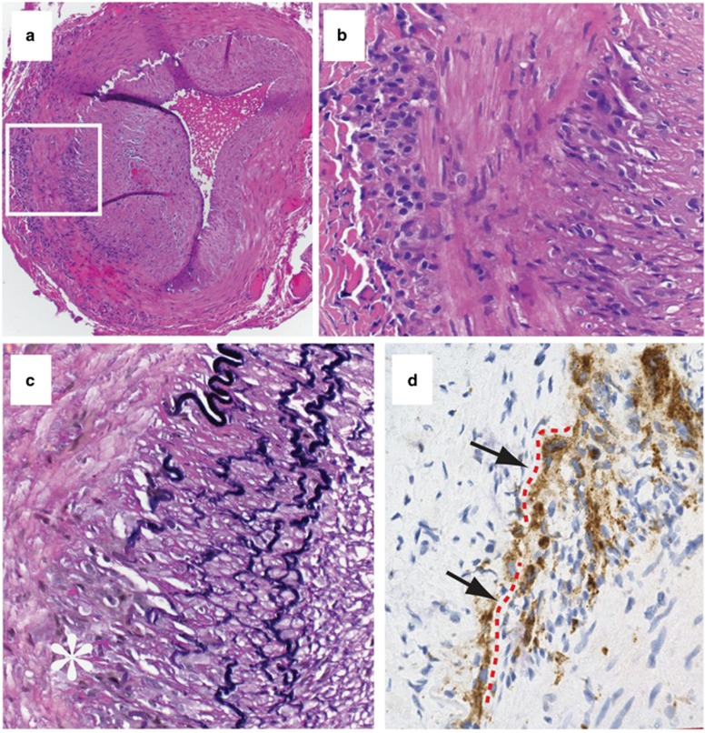
A 62-year-old woman presented with complaints of complete loss of vision in the right eye on waking in the morning, followed by gradual improvement over few hours 3 days before presentation. She continued to have transient episodes of blurry and distorted vision associated with a dull ache in the right eye for the next 3 days. She denied any systemic symptoms often seen in GCA including scalp tenderness, headache, jaw claudication, proximal muscle weakness, myalgia, weight loss, or fatigue. She had hypercholesterolemia, but no history of diabetes or hypertension. BCVA was 20/25 in the right eye (OD) and 20/20 in the left eye (OS). The anterior segment and funduscopy were normal in both eyes. FA of the OD revealed normal choroidal circulation, but delayed and sluggish filling of retinal arterioles (Figures 1a–c). Choroidal and retinal perfusion was normal in the fellow eye. Westergren erythrocyte sedimentation rate (ESR) and C-reactive protein (CRP) levels were elevated to 86 and 2.5 (normal<0.8 mg/dl), respectively. Ultrasound carotid duplex revealed 60–79% stenosis of the internal carotid arteries (ICA) bilaterally, with a mild heterogenous plaque in the right ICA and a moderate plaque in the left ICA. The patient was immediately treated with intravenous methylprednisone 1 g daily for 3 days, followed by a tapering course of oral prednisone. A biopsy of the right superficial temporal artery confirmed the diagnosis of temporal arteritis (Figure 2). Two weeks following treatment, the BCVA OD was 20/20. ESR and CRP returned to normal levels of 6 and 0.1 mg/dl respectively. FA repeated 2 weeks following treatment revealed normalization of retinal arterial circulation (Figure 3). There was no recurrence of the disease at 6 months following treatment.
Figure 1.
Fluorescein angiogram of the right eye at initial presentation. (a) Arrow points to the initial appearance of dye in the retinal arteries at 24 s. (b) Slow filling of retinal arteries at 37 s. Arrows point to the leading end of the fluorescein column in the retinal arteries around the macula. (c) Arrows point to the irregular filling and sludging of dye in the retinal arteries. Note the normal choroidal and papillary perfusion.
Figure 2.
Temporal artery histopathology. (a) Complete cross-section showing intimal hyperplasia, luminal narrowing, and inflammation of the external and internal elastic lamina regions (hematoxylin and eosin stain, × 10 magnification). (b) Magnified photomicrograph of the boxed in region shows histiocytes admixed with lymphocytes along the elastic lamina (hematoxylin and eosin stain, × 40 magnification). (c) Disruption of the elastic lamina at asterisk (Verhoeff–Van Gieson elastin stain, × 40 magnification). (d) CD68+ macrophages are abundant in the inflammatory infiltrate. Portions of the elastic lamina visible under differential interference contrast (not illustrated) are marked with red dashed lines (arrows) (avidin–biotin complex peroxidase immunohistochemistry using diaminobenzidine as the chromagen responsible for the brown color, × 40 magnification).
Figure 3.
Fluorescein angiogram of the right eye 2 weeks following treatment. (a) Appearance of dye in the retinal arteries 15 s after injection (note the dye in arteries near the disc shown by the arrow. (b) Complete filling of the retinal arteries at 24 s. (c) Complete filling of retinal veins at 34 s.
Comment
The incidence of occult GCA, ie, patients having GCA and visual loss but no systemic symptoms, is 21.2%.2 Of those, 94.4% present with AION and CRAO.2 Typically, findings on FA include absent or markedly delayed filling of the choroidal, papillary, and peripapillary circulation.3, 4, 5, 6 In contrast, the retinal circulation generally remains normal except in arterial occlusion.3 In a prospective study by Hayreh et al,7 FA of all seven patients with CRAO and GCA revealed occlusion or poor filling of the retinal circulation along with the posterior ciliary artery circulation abnormalities. These circulatory changes were attributed to thrombotic occlusion of either the posterior ciliary arteries, or the common trunk from the ophthalmic artery that gives off the central retinal artery and the posterior ciliary arteries. However, in our patient, presence of sluggish and stagnant retinal circulation, and normal choroidal perfusion was most likely secondary to partial and reversible thrombotic occlusion or inflammatory vasospasm of the central retinal artery.
Doppler studies in GCA patients have shown significant reduction in the mean flow velocity in the central retinal artery and short posterior ciliary arteries, along with changes in the ophthalmic artery flow.8 A trend toward normalization of blood-flow velocities is seen following treatment with systemic steroid in patients without clinical progression of the disease.
In our patient, FA was performed due to high suspicion of GCA despite lack of obvious fundus findings. It revealed retinal perfusion abnormalities before permanent visual loss. It also demonstrated normalization of retinal circulation with systemic steroid treatment. This case highlights the possibility that GCA may have an occult presentation, with disturbances in retinal artery filling being the sole demonstrable abnormality. It also emphasizes the value of fluorescein angiographic imaging in evaluating a patient with transient visual loss and a normal funduscopic appearance.
Acknowledgments
This work was supported in part by NIH R01 EY09412; Merit Review Award I01BX007080 from the Biomedical Laboratory Research & Development Service of the Veterans Affairs Office of Research and Development; Unrestricted Grant from Research to Prevent Blindness to the Department of Ophthalmology at SUNY Buffalo. The opinions expressed herein do not necessarily reflect those of the Veterans Administration or the United States Government.
The authors declare no conflict of interest.
References
- Hayreh SS, Zimmerman B, Kardon RH. Visual improvement with corticosteroid therapy in giant cell arteritis. Report of a large study and review of literature. Acta Ophthalmol Scand. 2002;80:355–367. doi: 10.1034/j.1600-0420.2002.800403.x. [DOI] [PubMed] [Google Scholar]
- Hayreh SS, Podhajsky PA, Zimmerman B. Occult giant cell arteritis:ocular manifestations. Am J Ophthalmol. 1998;125:521–526. doi: 10.1016/s0002-9394(99)80193-7. [DOI] [PubMed] [Google Scholar]
- Hayreh SS. Anterior ischemic optic neuropathy. Differentiation of arteritic from non arteritic type and its management. Eye. 1990;4:25–41. doi: 10.1038/eye.1990.4. [DOI] [PubMed] [Google Scholar]
- Hayreh SS. Anterior ischaemic optic neuropathy. II. Fundus on ophthalmoscopy and fluorescein angiography. Br J Ophthalmol. 1974;58:964–980. doi: 10.1136/bjo.58.12.964. [DOI] [PMC free article] [PubMed] [Google Scholar]
- Mack HG, O'Day J. Delayed choroidal perfusion in giant cell arteritis Currie. JNJ Clin Neuroophthalmol. 1991;11:221–227. [PubMed] [Google Scholar]
- Siatkowski RM, Gass JD, Glaser JS, Smith JL, Schatz NJ, Schiffman J. Fluorescein angiography in the diagnosis of giant cell arteritis. Am J Ophthalmol. 1993;115:57–63. doi: 10.1016/s0002-9394(14)73525-1. [DOI] [PubMed] [Google Scholar]
- Hayreh SS, Podhajsky PA, Zimmerman B. Ocular manifestations of giant cell arteritis. Am J Ophthalmol. 1998;125:509–520. doi: 10.1016/s0002-9394(99)80192-5. [DOI] [PubMed] [Google Scholar]
- Ho AC, Sergott RC, Regillo CD, Savino PJ, Lieb WE, Flaharty PM, et al. Color Doppler hemodynamics of giant cell arteritis. Arch Ophthalmol. 1994;112:938–945. doi: 10.1001/archopht.1994.01090190086026. [DOI] [PubMed] [Google Scholar]


