

Abstract
Glomus tumor is a rare, benign neoplasm rising from the glomus apparatus of the skin. It occurs most frequently on fingers and toes and accounts for 1.6% of all soft tissue tumors. Clinical diagnosis may prove difficult if the tumor occurs on an extra digital location. We report a case of a vascular-type glomus tumor (glomangioma) found in an atypical location, namely the lateral aspect of the knee joint.
Key words: glomangioma, glomus tumour, knee, extradigital
Case Report
A 51-year-old male was initially treated in our emergency department on April 2013 as a result of a bicycle fall and subsequent hip trauma. He presented a left subcapital fracture (Garden Stage III) and was treated with open reduction and internal fixation (Figure 1). At that time, he also complained of an extremely tender area on the lateral aspect of the ipsilateral knee that existed before the accident. The area had been under observation for the past 8 years. Even light contact from clothing exacerbated the knee pain, and it was hypersensitive to cold. The symptoms were not reduced with conservative treatments provided by previous physicians (non-steroidal anti-inflammatory drugs, corticoid injections or physical therapies). On physical examination, a small, faint reddish macule on the lower lateral portion of the left knee was noted. No inflammatory joint signs were observed, and the patient was very reluctant to allow knee inspection. Standard radiographs were normal, and MRI confirmed a solid, well defined, round mass that is adjacent to the lateral collateral ligament. The lesion was hypointense on T1-weighted sequences and hyperintense on T2-weighted images (Figure 2). An ultrasound study demonstrated a hypervascularised solid nodule. The lesion was excised en bloc from the subcutaneous tissue (Figure 3), with complete remission of the symptoms. Histopathological examination confirmed the initial suspicion of an extra-digital glomus tumor (round glomus cells with lightly stained cytoplasm and uniform, centrally located oval nuclei) with a prominent vascular component (glomangioma) and positive immunostaining for smooth muscle actin (Figure 4).
Figure 1.
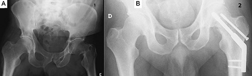
Pre- and postoperative antero-posterior bilateral hip view radiographs.
Figure 2.
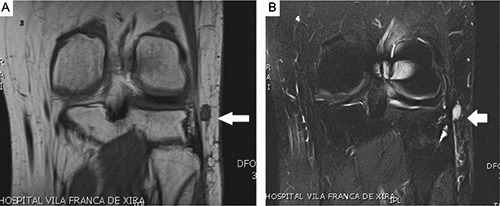
Coronal magnetic resonance images of the left knee presenting a hypointense mass (white arrow) on T1-weighted sequences and a hyperintense mass on T2-weighted
Figure 3.
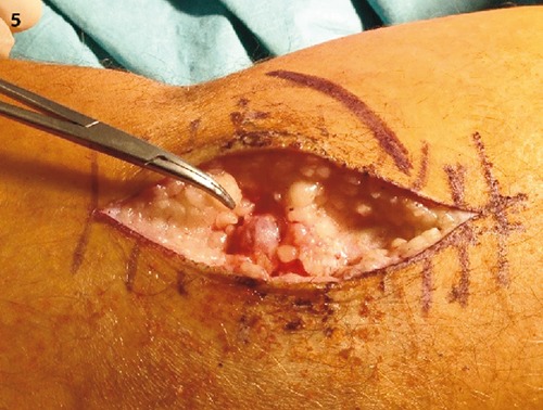
Intra-operative picture showing a well-defined, round mass that is superficial to the lateral collateral ligament.
Figure 4.
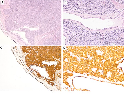
Glomus cell aggregates surrounding small vessels. Hematoxylin and Eosin stain; magnification ×50 (A) and ×200 (B). The tumor cells were immunoreactive for smooth muscle actin (SMA); magnification ×50 (C) and ×200 (D)
Discussion
Glomus tumor is a rare, benign neoplasm rising from the glomus apparatus in the skin. It occurs most frequently on fingers and toes and accounts for 1.6% of all soft tissue tumours.1 Clinical diagnosis may prove difficult if the tumor occurs on an extra digital location.2
They were first clinically described by Wood in 1812,3 and later explained in detail by Masson;4 they are rare benign tumors of the Sucquet-Hoyer canals within the glomus body (a neuromyoarterial canal system that is responsible for blood flow to the skin and thermoregulation). Local hyperplasia or overgrowth of this neuroangiomatous tissue may result in tumors that produce paroxysmal pain induced either by a mechanical or thermal stimulus. These tumors occur most frequently in distal extremities, particularly in the subungual area;5 however, atypical extradigital sites, including the head and neck, lungs, mediastinum, stomach, mesentery, bowels, bone and venous system, may be observed.6 There are reports of multiple possible locations around the knee: subcutaneous, sub-synovial, in the patellar ligament, within the fat pad, in the popliteal area or at the fibular head.7-9 Reviews of glomus tumors in the hand have indicated a 2:1 female predominance,10 but extradigital glomus tumors appear to be more common in males.11 These tumors are typically solitary and accompanied by the classic triad of pain, cold sensitivity and point tenderness.2,8,10,12 Symptoms are typically disproportional to the size of the tumor, and the pain mechanism still requires further elucidation.13 Studies suggest that the contraction of the myofilaments of the glomus cells due to cold temperatures may result in an increased intracapsular pressure that can lead to the perception of pain via transmission through unmyelinated nerve fibres.14 Extradigital tumours are frequently more difficult to diagnose due to the absence of classic symptoms (only 9% to 20% are initially correctly diagnosed).2,15
Relevant differences are noted in glomus tumors in children compared with adults. In children, tumors are frequently multifocal, infiltrative, and less commonly observed in subungual locations; superficial soft tissues of the extremities are often involved. In addition, an autosomal dominant inheritance pattern may be observed, and some tumors in children may be associated with neurofibromatosis type 1.16 According to the World Health Organization, 3 distinctive types of glomus tumor patterns are recognized: solid type, with scarce vasculature and a minimal muscle component (the most common variant); an angiomatous type called glomangioma comprised of a prominent vascular component; and a solid type with myomatous spindle cell differentiation referred to as glomangiomyoma (with prominent vascular and smooth muscle components).17 Glomangiomas represent approximately 20% of all glomus tumors and 1-5% of all soft tissue tumors of the hand.6,17 Preoperative imaging studies with color Doppler ultrasonography and magnetic resonance may provide information on tumor size, shape and precise anatomic location.18,19 The differential diagnoses (both clinical and histological) may include neuromas, hemangiopericytomas, angioleiomyomas, hemangiomas and other hamartomas of the cutaneous adnexa.11 Immunohistochemistry can be extremely helpful, especially in tumors with an atypical histological appearance.12 Treatment consists of complete surgical excision.
Recurrence rates for solitary tumors range from 12% to 33%.2,15 Malignancy is extremely rare and based mainly in histologic features (atypical mitotic figures, moderate to high nuclear grade and ≥5 mitotic figures per 50 high-power field), rather than clinical findings (deep location and size >2 cm), emphasising the importance of surgical marginal resection.20
Conclusions
Patients with glomus tumors may present to a variety of health care professionals, and an accurate and prompt diagnosis is fundamental to avoid treatment delays, chronic pain syndromes or psychiatric misdiagnosis. The occurrence of glomus tumors in extradigital locations provides an even greater challenge. In the present case, the knee pain was reminiscent of many common musculoskeletal conditions, such as meniscus tears, synovitis or ligamentous strains and sprains, thereby misleading clinicians and delaying the correct diagnosis. This illustrates the importance of maintaining a high index of suspicion for the diagnosis of an extradigital glomus tumor. It also reiterates the fact that the unusual distribution of clinical signs and symptoms provides the most important clue for the precis e diagnosis.
References
- 1.Soule EH, Ghormley RK, Bulbulian AH. Primary tumors of the soft tissues of the extremities exclusive of epithelial tumors: an analysis of five hundred consecutive cases. AMA ARCH Surg 1955;70:462-74. [DOI] [PubMed] [Google Scholar]
- 2.Schiefer TK, Parker WL, Anakwenze OA, et al. Extradigital glomus tumors: a 20-year experience. Mayo Clin Proc 2006; 81:1337-44. [DOI] [PubMed] [Google Scholar]
- 3.Wood W. On painful subcutaneous tubercle. Edin Med J 1812;8:283-91. [PMC free article] [PubMed] [Google Scholar]
- 4.Masson P. Le glomos neuromyoarterial des regions tactile et les tumeurs. Lyon Chir 1924;21:257-80. [Google Scholar]
- 5.Kale SS, Rao VK, Bentz ML. Glomus tumor of the index finger. J Craniofac Surg 2006;17:801-4. [DOI] [PubMed] [Google Scholar]
- 6.Tuncali D, Yilmaz AC, Terzioglu A, et al. Multiple occurrences of diferente histologic types of the glomus tumor. J Hand Surg 2004;1:161. [DOI] [PubMed] [Google Scholar]
- 7.Kato S, Fujii H, Yoshida A, Hinoki S. Glomus tumor beneath the plica synovialis in the knee: a case report. Knee 2007;14:164-6. [DOI] [PubMed] [Google Scholar]
- 8.Waseem M, Jari S, Paton RW. Glomus tumor, a rare cause of knee pain: a case report. Knee 2002;9:161-3. [DOI] [PubMed] [Google Scholar]
- 9.Okahashi K, Sugimoto K, Iwai M, et al. Glomus tumor of the lateral aspect of the knee joint. Arch Orthop Trauma Surg 2004;124:636-8. [DOI] [PubMed] [Google Scholar]
- 10.Maxwell GP, Curtis RM, Wilgis EF. Multiple digital glomus tumors. J Hand Surg (Am) 1979;4:363-7. [DOI] [PubMed] [Google Scholar]
- 11.Tsuneyoshi M, Enjoji M. Glomus tumor. A clinicopathologic and electron microscopicb study. Cancer 1982;50:1601-7. [DOI] [PubMed] [Google Scholar]
- 12.Van Geertruyden J, Lorea P, Goldschmidt D, et al. Glomus tumor of the hand: a retrospective study of 51 cases. J Hand Surg (Br) 199621:257-60. [DOI] [PubMed] [Google Scholar]
- 13.Weiss SW, Goldblum JR, Perivascular tumors. Enzinger and Weiss’s Soft Tissue tumors. 5thMaryland Heights: Mosby; 2007. 751-756. [Google Scholar]
- 14.Rohrich RJ, Hochstein LM, Millwee RH. Subungual glomus tumors: an algorithmic approach. Ann Plast Surg 1994;33:300-4. [DOI] [PubMed] [Google Scholar]
- 15.Lee DW, Yang JH, Chang S, et al. Clinical and pathological characteristics of extradigital and digital tumours: a retrospective comparative study. J Eur Acad Dermatol Venereol 2011;25:1392-7. [DOI] [PubMed] [Google Scholar]
- 16.Harrison B, Moore AM, Calfee R, et al. The association between glomus tumors and neurofibromatosis. JHS 2013;38:1571-4. [DOI] [PubMed] [Google Scholar]
- 17.Folpe AL. Glomus tumor. Barnes L, Everson JW, Reichart P, et al., World health organization classification of tumours: pathology and genetics of head and neck tumours. Lyon: IARC Publications; 2005. 136-137. [Google Scholar]
- 18.Glazebrook Kn, Laundre BJ, Schiefer TK, et al. Imaging features of glomus tumors. Skeletal Radiol 2011;40:855-62. [DOI] [PubMed] [Google Scholar]
- 19.Park HJ, Jeon YH, Kim SS, et al. Gray-scale and color doppler sonographic appearances of nonsubungual soft-tissue glomus tumors. J Clin Ultrasound 2011;39:305-9. [DOI] [PubMed] [Google Scholar]
- 20.Folpe AL, Fanburg-Smith JC, Miettinen M, et al. Atypical and malignant glomus tumors: analysis of 52 cases, with proposal for the reclassification of glomus tumors. Am J Surg Pathol 2011;25:1-12. [DOI] [PubMed] [Google Scholar]