Fig. 4.
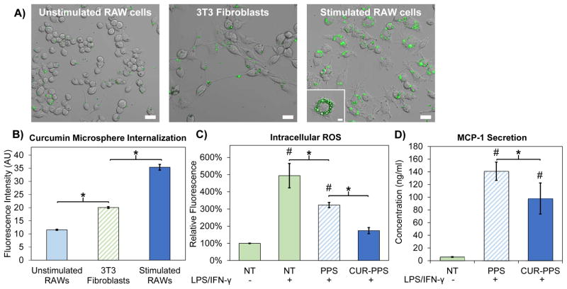
Curcumin-PPS microspheres are preferentially internalized by activated macrophages and exert functional effects on ROS generation and MCP-1 secretion. A) Confocal microscopy revealed that curcumin-PPS microspheres were internalized to a greater degree by LPS/IFN-γ stimulated RAW cells relative to non-activated macrophages and fibroblasts, suggesting size dependent targeting of the microparticles to pro-inflammatory macrophages. Scale bar is 20 μm. Inset in stimulated RAW macrophages is the center slice of a z-stack, confirming cell internalization of microspheres. Inset scale bar is 5 μm. B) Quantitative analysis of microsphere uptake was performed using flow cytometry to measure intracellular curcumin fluorescence, and significant differences were observed between all groups (p<0.05). C) Intracellular ROS levels are reduced in LPS/IFN-γ-stimulated RAW macrophages by treatment with PPS microspheres and Cur-PPS microspheres (p<0.05). Intracellular ROS levels in activated macrophages treated with CUR-PPS microspheres were statistically equivalent to the non-activated RAW cells. D) Secretion of MCP-1 is reduced in LPS/IFN-γ-stimulated RAW macrophages by treatment with Cur-PPS microspheres relative to blank PPS microspheres (p<0.05). Microsphere doses contain 3.4 μM curcumin or the equivalent polymer dose. *p<0.05 for differences between indicated groups. #p<0.05 relative to unstimulated macrophages (LPS/IFN-γ(−)/NT group).

