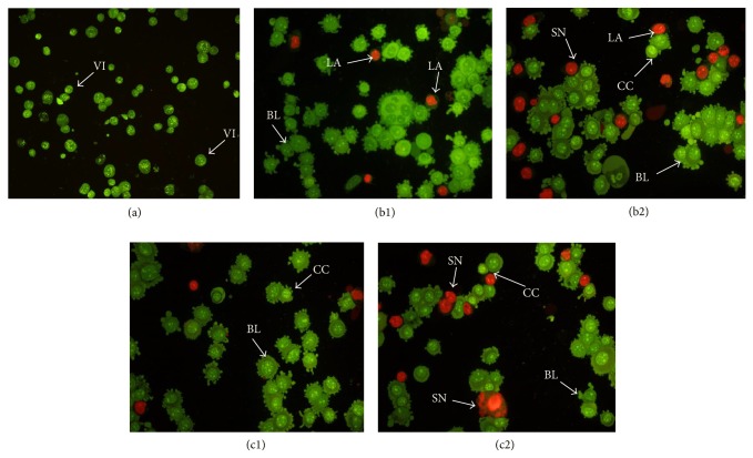
Figure 11.
Fluorescent micrographs of AO/PI-double-stained MCF-7 cells. (a) Untreated MCF-7 cells exhibit normal structures. (b1) and (c1) Early apoptosis features, namely, blebbing and chromatin condensation as well as late apoptotic cells, were detected after 24 h of treatment with (1) and (2). (b2) and (c2) Late apoptosis and secondary necrosis were obsereved after 48 h of treatment with (1) and (2), respectively (magnification: 200x). VI: viable cells; CC: chromatin condensation; BL: blebbing of the cell membrane; LA: late apoptosis; SN: secondary necrosis.

