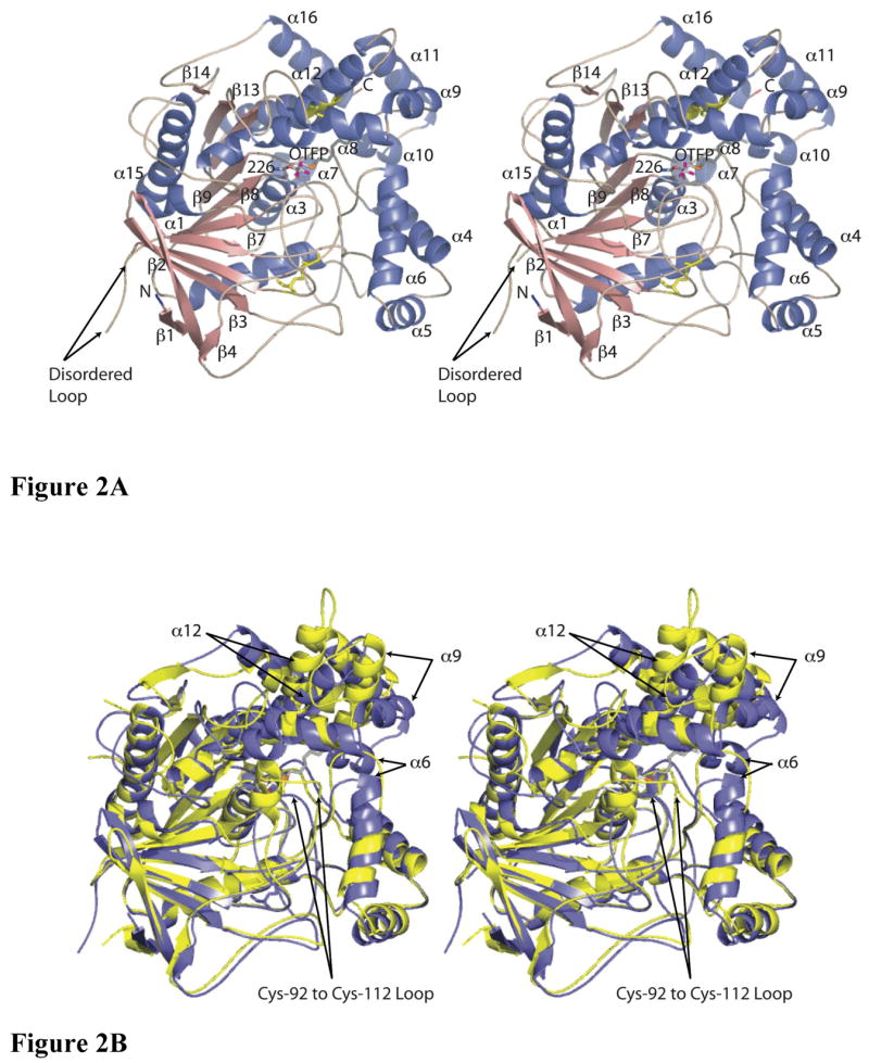
Figure 2.

Crystal structures of MsJHE and AChE. A) Stereo view of a cartoon representation of the secondary structure of JHE with OTFP covalently bound. Helices are colored blue, sheets are salmon, and loops and turns are tan. The N-terminus is labeled and is colored blue. The C-terminus is labeled and is colored red. The disulfide bonds are in yellow and are shown with the atoms as stick figures. OTFP and Ser226 (to which OTFP is covalently attached) are shown as a stick representation, with carbons colored grey, oxygen red, nitrogen blue, sulfur orange, and fluorine magenta. Helices and strands that are referred to in the text are labeled. B) Overlay of MsJHE and AChE from Torpedo californica. MsJHE is shown in blue and AChE is shown in yellow. Secondary structural elements that contribute to the binding pocket of MsJHE and which differ significantly from the corresponding elements in AChE are indicated with arrows. The labeling of the arrows indicates the secondary structural elements as found in MsJHE. This figure and all other figures of JHE structure were produced using the program PyMol ((DeLano, W.L., The PyMOL Molecular Graphics System (2002) DeLano Scientific, San Carlos, CA, USA.).
