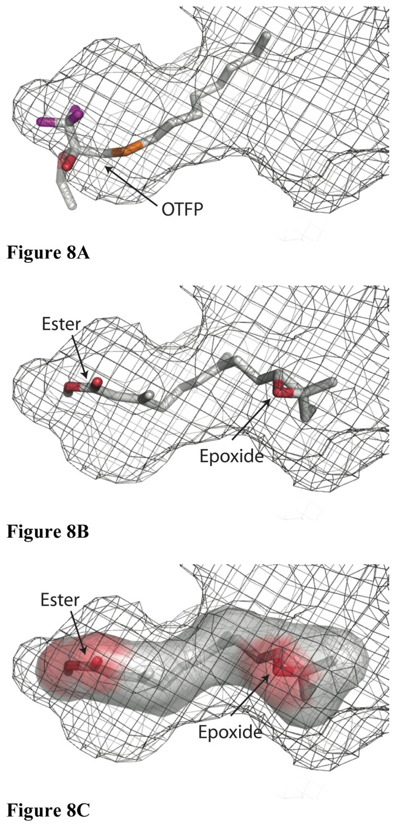
Figure 8.

A model of JH binding to substrate. Orientation is identical to the top panel in Figure 4, and the surface is again shown as a mesh representation, though it is not color coded. A) OTFP is shown as a stick representation using the same color scheme as in Figure 2. B) JH II is shown as a stick representation using the same color scheme as in Figure 2 and with the same orientation as in Fig. 8A. C) A surface representation of JH II was produced in the absence of JHE and then shown inside the binding pocket.
