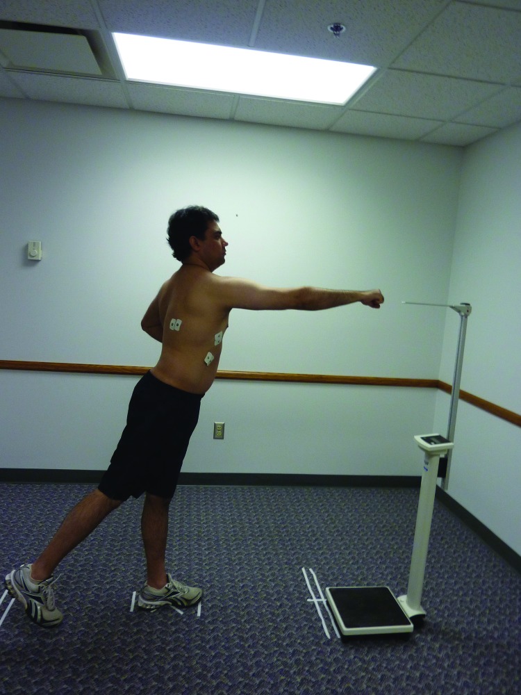
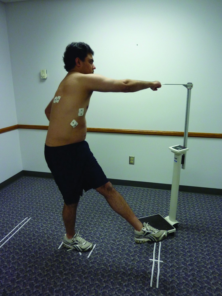
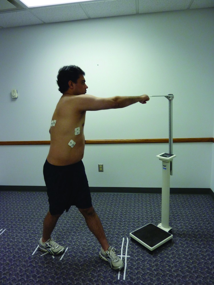
Abstract
Background:
Poor activation of the serratus anterior (SA) muscle may result in abnormal shoulder rhythm, and secondarily contribute to impingement and rotator cuff tears. Sequential activation of the trunk, pelvis, and lower extremity (LE) muscles is required to facilitate the transfer of appropriate forces from these body segments to the upper extremity. Myofascial connections that exist in the body, and LE and trunk muscles (TM) activity may influence scapular and upper limb activity. The purpose of this study was to investigate the effect of simultaneous recruitment of the LE muscles and TM on the SA muscle activation when performing a forward punch plus (FPP) and six variations of the FPP exercise.
Study Design:
Experimental, within‐subject repeated measures.
Methods:
Surface electromyographic (EMG) activity of the SA, latissimus dorsi, and external oblique muscles on the dominant side, bilateral gluteus maximus muscles, and contra‐lateral femoral adductor muscles were analyzed in forward punch plus (FPP) movement and six variations in twenty one healthy male adults. The percentage of maximum voluntary isometric contraction (%MVIC) for each muscle was compared across various exercises using a 1‐way repeated –measures analysis of variance with Sidak pair wise comparison as post‐hoc test (p < 0.05).
Results:
Pairwise comparisons found that the EMG activity of the serratus anterior (SA) during the FPP with contralateral closed chain leg extension (CCLE), FPP with ipsilateral closed chain leg extension (ICLE), FPP with closed chain serape effect (CS), and FPP with open chain serape effect (OS) showed significantly higher EMG activity than the FPP.
Conclusions:
Simultaneous recruitment of the lower extremity and trunk muscles increases the activation of the SA muscle during the FPP exercise.
Clinical Relevance:
Rehabilitation clinicians should have understanding of the kinetic chain relationships between the LE, the trunk, and the upper extremity while prescribing exercises. The results of this study may improve clinicians' ability to integrate the kinetic chain model in a shoulder rehabilitation program.
Level of Evidence:
2b
Keywords: Electromyography, kinetic chain, myofascial connections, serratus anterior
INTRODUCTION
Shoulder pain is the most common complaint in overhead throwing athletes.1,2 Overhead throwing motions place incredibly high demand on the shoulder complex requiring high muscular activation around the joint.3,4 Researchers have reported that abnormal biomechanics of the shoulder girdle and repeated overhead movements could lead to injuries in overhead throwing athletes.2,5 Muscular imbalances around the shoulder complex could lead to diminished scapular control and dyskinesis resulting in glenohumeral joint injuries like instability and impingement.6,7
Upper extremity (UE) injuries occurring in sports are also due to alterations in the function of the muscles that control the scapula.6,7 The scapula provides a link between the arm and trunk. It acts as a stable base for the humeral head during overhead movements of the arm by providing a congruent socket.8 The serratus anterior (SA) is a prime mover of the scapula, contributing to the maintenance of normal scapulohumeral rhythm and motion.9 Due to its insertion into the inferior medial border and inferior angle of the scapula, the SA has large moment arms to produce upward rotation and posterior tipping.9 It contributes to the three‐dimensional movement of scapula by producing upward rotation, posterior tilting, and external rotation of the scapula during arm elevation.10 Poor activation of the SA muscle may result in reduced scapular rotation and protraction, resulting in anterior‐superior translation of the humeral head, causing secondary impingement and rotator cuff tears.11
The ultimate ability to produce forces necessary for performance of overhead sports is not solely due to the UE contributions. 3,12 Efficient distal segment motions occurring in such functional motions as overhead throwing and striking involve proximal core muscle activation patterns. More than half of the force production required by a tennis player in an overhead tennis serve is produced from trunk muscles (TM) and lower extremity (LE) muscles.4,12 Weakness or limited trunk and hip mobility can alter the normal activation pattern required in overhead throwing athletes, producing distal joint dysfunction.13 The core musculature acts as a connecting link between the upper and the lower extremity limbs in overhead athletic endeavors.14
The shoulder complex does not function in isolation. Synchronized sequential rotation from the LE through the trunk needs to occur in order for the shoulder joint to act efficiently in overhead sports.15 The shoulder is a part of the kinetic chain and the body is considered as a linked system of articulated segments.12 Each segment (LE, trunk, pelvis and UE) in the kinetic chain has a specific role in ensuring that the UE performs efficiently in athletic endeavors.3,12 This coordinated sequencing of the segments is known as the kinetic chain. Sequential activation of the LE, pelvis and trunk muscles is required to facilitate the transfer of appropriate forces from these body segments to the UE.16 Such forces result in a harmonized movement at the UE needed in throwing activities in various sports. Momentum generated by the larger segments in the kinetic chain is transferred to the adjacent distal segments.12,17 This mechanism results in summation of individual speeds and forces at each segment. Putnam12 demonstrated that the forces acting at a joint are influenced by the motions at the adjacent segments in the chain. The ultimate speed and forces achieved by the distal segment are the product of all the individual segments in the kinetic link.12 Thus the timely flow of the energy from the lower to the upper part of the body is vital for effective sporting motions to occur at the shoulder joint.18–20
Current shoulder rehabilitation programs integrate the kinetic chain model in order to mimic normal UE motor patterns that are utilized during sporting movements and daily activities.21,22 Kinetic chain rehabilitation programs are considered functional and address the shoulder function in a proximal to distal manner.17,22 Proximal TM and the LE are used to initiate scapular and arm activations. Such force‐dependent integrated muscle activation patterns are used to coordinate the motions of connecting segments, resulting in better gains during strength training programs.23,24 Inclusion of multiple body segments in a diagonal manner during exercises can facilitate activation of the involved muscles in order to develop functionally appropriate use of the shoulder complex.25,26 Therefore, clinicians incorporate the TM and LE muscles in shoulder rehabilitation to mimic the normal upper extremity motor patterns during sporting movements and daily activities.17,21,22,27,28
Several authors have illustrated myofascial connections by which the LE and the TM activity may influence the scapular and the upper limb activity.29–31 Clinically such connections become evident when dysfunction in one area of the body can be related to a body region away from the primary site.32 Alteration of the knee flexion angle in tennis players decreased the contribution by the hip and trunk, leading to increased loads and injuries at the shoulder and elbow.18 Posterior‐superior glenoid labral tears were arthroscopically proven in athletes with weakness or tightness at the hip joint.33 The following myofascial linkages resulting in overlapping among the muscles of the shoulder complex and the trunk have been reported:29,30
Latissimus dorsi (LatD) and ipsilateral SA.
SA, ipsilateral rhomboid and external oblique (ExOb) muscle, and contralateral internal oblique, and femoral adductor muscle (FAd). The SA courses anteriorly around the rib cage to attach to the ribs and interdigitates with the ExOb. The fiber line of ExOb then becomes continuous with the internal oblique and FAd on the contralateral side. The orientation of these muscles, anatomically linking the UE, trunk, and the LE across the front of the body is referred as the “serape effect”.
LatD and contralateral gluteus maximus (cGMax) via thoracolumbar fascia.
Such crossing relationships among the neighboring muscles have been reported to influence each other and could increase stability and strength in that specific region.21,29–31,34,35 Maenhout et al21 recruited individuals to determine the influence of the kinetic chain on the scapular muscle activity in knee push up plus exercises. They concluded that ipsilateral leg extension increased the SA activity whereas contralateral leg extension decreased the SA activity. Similar results were reported by Kim et al35 as they compared the shoulder and the TM activation during the ipsilateral leg extension with the knee push up plus exercises.
Several authors have explored the SA muscle EMG activation in closed‐chain exercises.21,34–37 Closed kinetic chain exercises are safe, enhance co‐contractions in early stages of rehabilitation, and provide a foundation for more functional open kinetic chain exercises.17,22 No studies have been published to investigate the importance of the mysofascial connections between the LE, TM, and the UE muscles in closed kinetic chain exercises using diagonal pattern muscle recruitment. The purpose of this study was to investigate the effect of simultaneous recruitment of the LE muscles and TM on the SA muscle activation when performing a forward punch plus (FPP) and six variations of the FPP exercise. The research hypothesis was that the SA activation would be significantly higher when LE muscles and TMs are recruited simultaneously during the SA muscle training than when LE muscles and TMs are not actively recruited.
METHODS
Subjects
Twenty one healthy males with fair to very lean body composition (% body fat), as reported in ACSM's Guidelines for Exercise Testing and Prescription38 completed the study. The mean anthropometric characteristics ± standard deviation (SD) of the males were age, 22.36 ± 3.26 years; height, 176.02 ± 7.42cm; weight, 77.78 ± 8.58 kg, %body fat, 9.64 ± 3.84%.
The Institutional Review Board of Rocky Mountain University of Health Professions (RMUoHP), Provo, Utah, and Saginaw Valley State University (SVSU), approved the study. Sample size of 17 subjects was needed using conventional values for a medium effect size (f= 0.25); degrees of freedom = 6, power = 0.80; alpha = 0.05.39 Twenty one subjects actually completed the study.
Procedures
The subjects were given the informed consent and then were screened for inclusion and exclusion criteria (Table 1). Skin fold measurements were taken using the guidelines provided in ACSM's Guidelines for Exercise Testing and Prescription.38 Lange® skin fold calipers (model # 68902, Fitness Mart®, division of Country Technology, Inc.) and three site formula regression equations for men (chest, abdomen, and thigh) were used to assess body composition.38 Leg length was measured by the PI (N.K.) to in order to standardize the step length during exercises to allow for accurate comparisons of performance among participants.40 The leg length was measured from the anterior superior iliac spine (ASIS) to the end of the medial malleolus with the subject lying supine while not wearing shoes.
TABLE 1.
Describes inclusion/exclusion criteria of the study.
| Inclusion Criteria | Exclusion Criteria |
|---|---|
|
|
The Biopac MP 36 System (Biopac Systems Inc, Santa Barbara, CA) was used to collect all EMG data. Skin impedance of less than 20 KΩ was accepted. 41 The skin was prepared before the electrode placement using vigorous cleaning of the area with SKIN‐PREP protective wipes (Smith & Nephew plc, London, UK). The subject shaved the area if body hair was present. EMG electrodes were applied to the muscle fibers of the SA, LatD, and ExOb muscles on the dominant side, GMax bilaterally, and FAd of the contralateral side of the subjects according to the procedure described by Cram et al.41 Surface EMG data was collected using 10‐mm‐contact‐area Ag‐AgCl disposable electrodes (Trace Rite® Bio‐Detek Inc, Pawtucket, RI). As a warm up, the subjects then performed jumping jacks for 30 seconds.
For normalization of the EMG data, of maximum voluntary isometric contraction (MVICs) were established for each muscle. Test positions were consistent with those described by Kendall 42 for SA, LatD, GMax and Fad muscles and with previous research for ExOb muscle.43 The MVICs were performed over a five second period using a metronome involving a gradual build up to maximum muscle activity. Each muscle test was repeated three times, with a five‐second rest between contractions.21 The MVIC value for each muscle was calculated as the average of the three trials. Between MVIC measurements of different muscles, a two minute of rest period was provided. Verbal feedback was provided for each subject for the MVIC procedures.
Exercises
The exercises under investigation are either based on the previous recommendations34,44 or myofascial connections reported in the literature.21,29,30 Exercises were performed without any footwear. The exercises were performed in a random order using a computerized random sequence generator as follows.
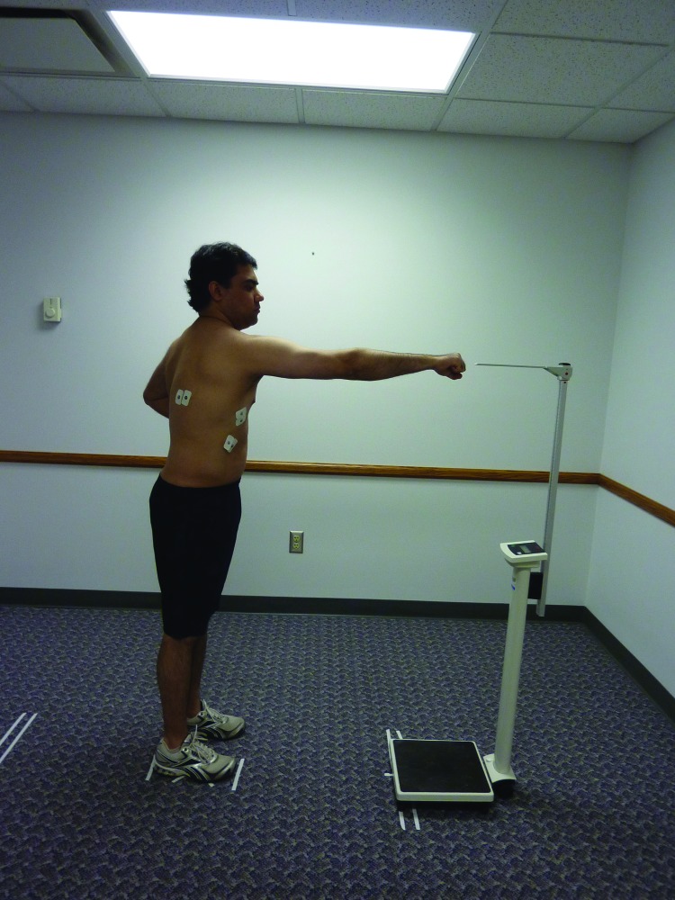
Forward punch plus (FPP) (Figure 1) was performed while the subject stood with feet shoulder‐width apart in a parallel stance. The exercise was started with subject's dominant arm at the side of the body with elbow flexed to 90° and neutral position. The subject flexed the shoulder to 90° and fully extended the elbow, the humerus internally rotated 45°, and the scapula was protracted. The subject then returned to the initial position by extending the shoulder, flexing the elbow in neutral position, and standing in parallel stance.
Figure 1.
Forward Punch Plus (FPP). The subject has engaged serratus anterior during the punching action.
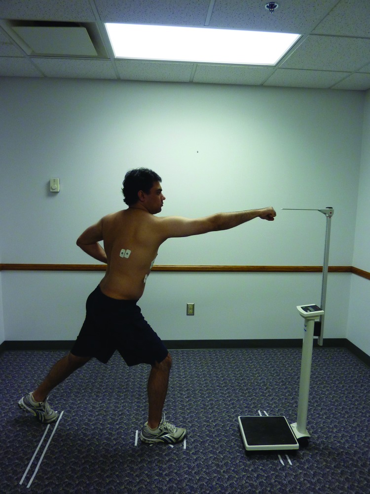
For FPP with contralateral closed chain leg extension (CCLE) (Figure 2) the subject performed a FPP with dominant arm and lunged straight back with the contralateral leg in closed chain. The subject then returned to the initial position by extending the shoulder and flexing the elbow in neutral position and standing in parallel stance.
Figure 2.
Forward Punch Plus with Contralateral Closed Chain Leg Extension (FPP – CCLE). The subject has engaged all the muscles contributing to the kinetic chain. Serratus anterior was engaged during the punching action, gluteus maximus of the ipsilateral side using eccentric hip flexion, gluteus maximus of the contralateral side using leg extension.
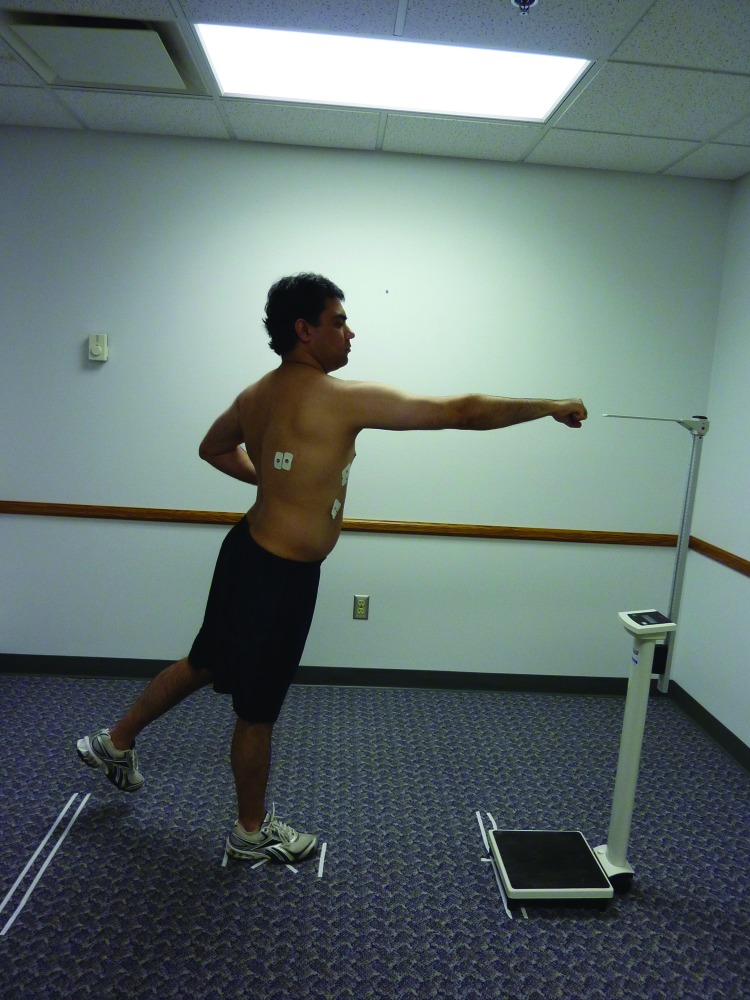
For FPP with contralateral open chain leg extension (COLE) (Figure 3), the subject performed FPP with dominant arm and swung their contralateral leg back in extension. The subject was instructed to keep the trunk straight and not to bend the contralateral knee or lean forward while swinging the leg backwards in open chain. The subject then returned to the initial position by extending the shoulder, flexing the elbow in neutral position and standing in parallel stance.
Figure 3.
Forward Punch Plus with Contralateral Open Chain Leg Extension (FPP – COLE). The subject has engaged all the muscles contributing to the kinetic chain. Serratus anterior was engaged during punching action, gluteus maximus of the ipsilateral side using single leg stance, gluteus maximus of the contralateral side using leg extension.
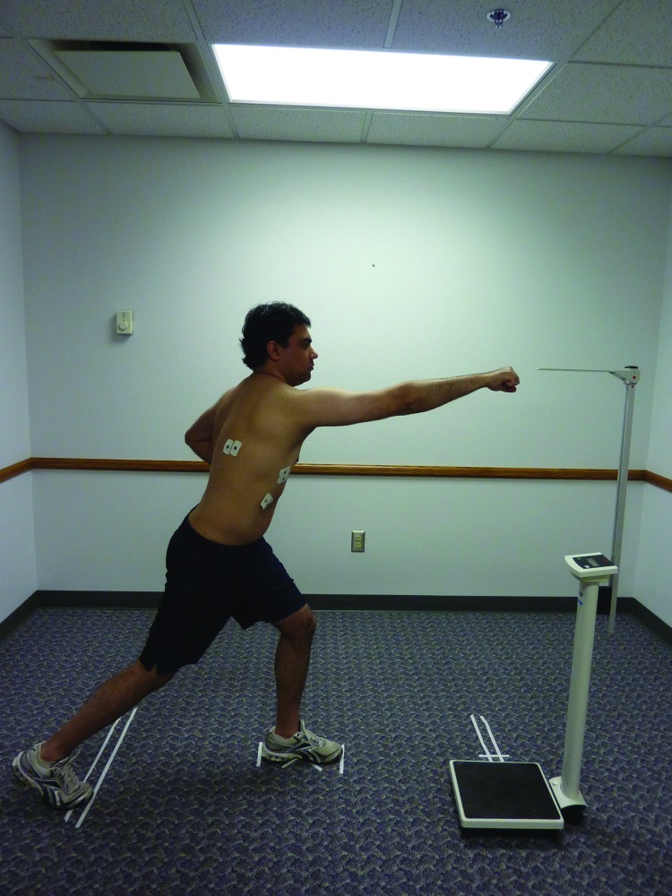
For FPP with ipsilateral closed chain leg extension (ICLE) (Figure 4), the subject performed FPP with dominant arm and lunged straight back with the ipsilateral leg in closed chain. The subject then returned to the initial position by extending the shoulder and flexing the elbow in neutral position and standing in parallel stance.
Figure 4.
Forward Punch Plus with Ipsilateral Closed Chain Leg Extension (FPP – ICLE). The subject has engaged all the muscles contributing to the kinetic chain. Serratus anterior was engaged during punching action, gluteus maximus of the ipsilateral side using leg extension, gluteus maximus of the contralateral side using eccentric hip flexion.
For FPP with ipsilateral open chain leg extension (IOLE) (Figure 5) the subject performed FPP with dominant arm and swung their ipsilateral leg back in extension. Subject was instructed to keep the trunk straight and not to bend the ipsilateral knee or lean forward while swinging the leg backwards in open chain. The subject then returned to the initial position by extending the shoulder, flexing the elbow in neutral position and standing in parallel stance.
Figure 5.
Forward Punch Plus with Ipsilateral Open Chain Leg Extension (FPP – IOLE). The subject has engaged all the muscles contributing to the kinetic chain. Serratus anterior was engaged during punching action, gluteus maximus of the ipsilateral side using leg extension, gluteus maximus of the contralateral side using single leg stance.
For FPP with closed chain serape effect (CS) (Figure 6) the subject performed FPP with dominant arm as he rotated and flexed the trunk to the contralateral hip and performed contralateral leg flexion and adduction in closed chain. The subject stepped forward and stopped at the midline of the trunk as marked by the white tape on the ground. The subject then returned to the initial position by extending the shoulder, flexing the elbow in neutral position and standing in parallel stance.
Figure 6.
Forward Punch Plus with Closed Chain Serape Effect (FPP – CS). The subject has engaged all the muscles contributing to the serape effect. Serratus anterior was engaged during punching action, external oblique and internal oblique were engaged during trunk rotation, and hip flexors and hip adductors were engaged with contralateral leg flexion and adduction.
For FPP with open chain serape effect (OS) (Figure 7) the subject performed FPP with dominant arm as the trunk was rotated and flexed to the contralateral hip and performed contralateral leg flexion and in an open chain. The subject swung the contralateral leg in front and stopped at the midline of the trunk as he maintained his balance. The subject then returned to the initial position by extending the shoulder, flexing the elbow in neutral position and standing in parallel stance.
Figure 7.
Forward Punch Plus with Open Chain Serape Effect (FPP – OS). The subject has engaged all the muscles contributing to the serape effect. Serratus anterior was engaged during punching action, external oblique and internal oblique were engaged during trunk rotation, and hip flexors and hip adductors were engaged with contralateral leg flexion and adduction.
For closed chain exercises, the step length for leg flexion and extension was standardized for each subject by measuring the floor distance from the mid‐foot to 75% of their leg length. Each subject was required to step with their toes between the two pieces of tape placed 3 cm on either side of the 75% of their leg length measurements. To ensure adequate protraction of scapula with FPP, a stand with a visual marker was placed at the maximum reach distance. The subject was verbally instructed to punch as hard as possible with maximum force to reach the stand with a visual marker placed at the maximum reach distance and the subject was required to bring their fist close to the marker with each exercise trial.
Practice trials were provided to the subjects and five trials of the exercises were performed with five seconds rest between repetitions. The speed of the trials were regulated by a metronome set to 50 beats per minute, where each phase (starting position, maximum reach, and ending position) was performed during one beat.21,44 The subjects were given verbal commands to begin and end each exercise trial for proper technique during the training and data collection. A minimum of two minutes of rest was provided between different exercises to prevent the influence of fatigue on muscle activation.21
Data Processing
All collected signals were subsequently band pass filtered (between 10 and 500 Hz), then rectified and finally smoothed by using a root‐mean‐square (RMS) calculation with a 50‐millisecond sliding window. The electrocardiac contributions to the SA, LatD and ExOb muscles (being close to heart) were removed using high pass digital filtering (finite impulse response (FIR) using a Hamming window, and fourth‐order Butterworth (BW) filter) at 30 Hz cutoff frequency. This method has been reported to provide optimal balance between levels for the EMG.45 For all subjects, MVIC was averaged across the three intermediate seconds for each muscle to calculate the mean of the peak RMS value of the three trials. The mean RMS EMG activity of each muscle was calculated across the best three trials of every exercise, for all subjects. The mean RMS value of the three trials for each muscle was normalized to its respective MVIC value and represented as a percentage of MVIC (%MVIC) using the following equation:21
Data Analysis
The Statistical Package for the Social Sciences (SPSS Inc, v18.0, Chicago, IL, USA) was used for analysis. A separate one‐way repeated measures analysis of variance (ANOVA) was performed on %MVIC EMG activity for each of the six muscles (SA, LatD, ExOb, GMax, FAd) across the seven exercises (within‐subject factor) with Sidak pair wise comparison was used for post‐hoc comparisons. A separate analysis of each muscle helped to determine if the change in the SA activation was due to the recruitment of TM and LE muscles, and any significant difference among the seven exercises. The level of significance was set at 0.05 for all analysis and 95% confidence intervals (CIs) were reported around the %MVIC for each exercise.
RESULTS
Statistically significant main effects existed among all the exercises for the SA (p < .001), ExOb (p < .001), FAd (p < .001), cGMax (p = .001), and ipsilateral gluteus maximus (iGMax) (p < .001). Results of pairwise comparisons found that the EMG activity (% MVIC) of the SA during CCLE (p = .001), (ICLE (p = .002), CS (p = .001), and OS (p = .001) was significantly higher than the EMG activity of the FPP (Table 2). The ExOb was significantly more active during the performance of IOLE (p = .002), CS (p = .003), and OS (p = .024) when compared to FPP, and CS produced significantly higher ExOb activation than IOLE (p = .048) (Table 4). The FAd activity was significantly higher in CCLE (p = .005), ICLE (p <.001), IOLE (p= .040), CS (p < .001), and OS (p < .001) as compared to FPP exercise (Table 5). In addition, FAd activity was also significantly higher in OS compared to ICLE (p = .002) and in CS and OS in comparison to IOLE (p = .001). The cGMax was also found to be significantly higher during the performance of COLE (p = .018), ICLE (p = .002), IOLE (p = .037), CS (p = .036), and OS (p = .012) when compared to FPP (Table 6). No other pair wise comparisons for cGMax were significantly different from each other. The iGMax was significantly more active in CCLE (p < .001), COLE (p = .002), ICLE (p = .001), IOLE (p < .001), CS (p = .002), and OS (p < .001) when compared to FPP (Table 7). Pair wise comparisons revealed that IOLE produced significantly higher iGMax activity as compared to all the other exercises. There was no statistically significant effect for the activation of LatD (p = .09) among the seven exercises (Table 3).
TABLE 2.
Mean EMG activation of serratus anterior (SA) muscle expressed as a percentage of maximum voluntary isometric contraction (MVIC) for 7 different exercises.
| Exercise | Mean (%MVIC) | SD | 95% CIs | |
|---|---|---|---|---|
| Lower bound | Upper bound | |||
| FPP | 44.56 | 18.46 | 36.16 | 52.97 |
| CCLE | 63.86* | 26.29 | 51.90 | 75.83 |
| COLE | 46.09 | 20.88 | 36.59 | 55.60 |
| ICLE | 59.77* | 19.63 | 50.84 | 68.71 |
| IOLE | 54.95 | 22.31 | 44.80 | 65.11 |
| CS | 71.30* | 26.71 | 59.14 | 83.45 |
| OS | 70.82* | 29.88 | 57.22 | 84.42 |
The 1‐way repeated‐measures ANOVA indicated a significant main effect across exercises; p≤0.05; SD= standard deviation; FPP=forward punch plus; CCLE= FPP with contralateral closed chain leg extension; COLE= FPP with contralateral open chain leg extension; ICLE=FPP with ipislateral closed chain leg extension; IOLE=FPP with ipsilateral open chain leg extension; CS= FPP with closed chain serape effect; OS= FPP with open chain serape effect.
TABLE 4.
Mean EMG activation of external oblique (ExOb) muscle expressed as a percentage of maximum voluntary isometric contraction (MVIC) for 7 different exercises.
| Exercise | Mean (%MVIC) | SD | 95% CIs | |
|---|---|---|---|---|
| Lower bound | Upper bound | |||
| FPP | 27.92 | 2.80 | 22.07 | 33.76 |
| CCLE | 39.36 | 6.54 | 25.71 | 53.01 |
| COLE | 39.66 | 7.90 | 23.17 | 56.15 |
| ICLE | 32.01 | 3.72 | 24.24 | 39.78 |
| IOLE | 44.08* | 4.26 | 35.19 | 52.98 |
| CS | 75.40* | 11.64 | 51.12 | 99.67 |
| OS | 109.55* | 22.94 | 61.71 | 157.40 |
The 1‐way repeated‐measures ANOVA indicated a significant main effect across exercises; p≤0.05; SD= standard deviation. FPP= forward punch plus; CCLE= FPP with contralateral closed chain leg extension; COLE= FPP with contralateral open chain leg extension; ICLE= FPP with ipislateral closed chain leg extension; IOLE= FPP with ipsilateral open chain leg extension; CS= FPP with closed chain serape effect; OS= FPP with open chain serape effect.
TABLE 5.
Mean EMG activation of femoral adductors (FAd) muscle expressed as a percentage of maximum voluntary isometric contraction (MVIC) for 7 different exercises.
| Exercise | Mean (%MVIC) | SD | 95% CIs | |
|---|---|---|---|---|
| Lower bound | Upper bound | |||
| FPP | 11.10 | 3.30 | 4.24 | 17.97 |
| CCLE | 38.01* | 7.51 | 22.35 | 53.67 |
| COLE | 25.96 | 6.15 | 13.13 | 38.79 |
| ICLE | 34.74* | 3.85 | 26.72 | 42.76 |
| IOLE | 27.19* | 6.21 | 14.24 | 40.136 |
| CS | 56.99* | 9.00 | 38.21 | 75.77 |
| OS | 61.95* | 6.32 | 48.77 | 75.12 |
The 1‐way repeated‐measures ANOVA indicated a significant main effect across exercises; p≤0.05; SD= standard deviation; FPP= forward punch plus; CCLE= FPP with contralateral closed chain leg extension; COLE= FPP with contralateral open chain leg extension; ICLE= FPP with ipislateral closed chain leg extension; IOLE= FPP with ipsilateral open chain leg extension; CS= FPP with closed chain serape effect; OS= FPP with open chain serape effect.
TABLE 6.
Mean EMG activation of contralateral gluteus maximus (cGmax) muscle expressed as a percentage of maximum voluntary isometric contraction (MVIC) for 7 different exercises.
| Exercise | Mean (%MVIC) | SD | 95% CIs | |
|---|---|---|---|---|
| Lower bound | Upper bound | |||
| FPP | 13.35 | 4.115 | 4.76 | 21.93 |
| CCLE | 22.86 | 4.396 | 13.69 | 32.03 |
| COLE | 54.94* | 10.656 | 32.71 | 77.16 |
| ICLE | 39.32* | 6.787 | 25.16 | 53.47 |
| IOLE | 27.63* | 3.853 | 19.59 | 35.67 |
| CS | 28.10* | 7.173 | 13.13 | 43.06 |
| OS | 27.60* | 5.101 | 16.96 | 38.24 |
The 1‐way repeated‐measures ANOVA indicated a significant main effect across exercises; p≤0.05; SD= standard deviation. FPP= forward punch plus, CCLE= FPP with contralateral closed chain leg extension, COLE= FPP with contralateral open chain leg extension, ICLE= FPP with ipislateral closed chain leg extension, IOLE= FPP with ipsilateral open chain leg extension, CS= FPP with closed chain serape effect, OS= FPP with open chain serape effect.
TABLE 7.
Mean EMG activation of ipsilateral gluteus maximus (iGmax) muscle expressed as a percentage of maximum voluntary isometric contraction (MVIC) for 7 different exercises.
| Exercise | Mean (%MVIC) | SD | 95% CIs | |
|---|---|---|---|---|
| Lower bound | Upper bound | |||
| FPP | 9.92 | 2.247 | 5.23 | 14.61 |
| CCLE | 45.41* | 5.656 | 33.61 | 57.20 |
| COLE | 27.10* | 4.317 | 18.10 | 36.11 |
| ICLE | 36.95* | 5.617 | 25.23 | 48.66 |
| IOLE | 83.60* | 9.331 | 64.13 | 103.06 |
| CS | 28.72* | 4.617 | 19.09 | 38.35 |
| OS | 46.24* | 6.254 | 33.20 | 59.29 |
The 1‐way repeated‐measures ANOVA indicated a significant main effect across exercises; p≤0.05; SD= standard deviation; FPP= forward punch plus; CCLE= FPP with contralateral closed chain leg extension; COLE= FPP with contralateral open chain leg extension; ICLE= FPP with ipsilateral closed chain leg extension; IOLE= FPP with ipsilateral open chain leg extension; CS= FPP with closed chain serape effect; OS= FPP with open chain serape effect.
TABLE 3.
Mean EMG activation of latissimus dorsi (LatD) muscle expressed as a percentage of maximum voluntary isometric contraction (MVIC) for 7 different exercises.
| Exercise | Mean (%MVIC) | SD | 95% CIs | |
|---|---|---|---|---|
| Lower bound | Upper bound | |||
| FPP | 9.61 | 1.169 | 7.17 | 12.04 |
| CCLE | 13.97 | 2.793 | 8.15 | 19.80 |
| COLE | 12.63 | 1.640 | 9.21 | 16.05 |
| ICLE | 11.89 | 1.805 | 8.11 | 15.64 |
| IOLE | 13.92 | 2.105 | 9.53 | 18.31 |
| CS | 12.93 | 1.699 | 9.39 | 16.48 |
| OS | 13.74 | 2.44 | 8.65 | 18.83 |
*The 1‐way repeated‐measures ANOVA indicated a significant main effect across exercises; p≤0.05; SD= standard deviation; FPP= forward punch plus; CCLE= FPP with contralateral closed chain leg extension; COLE= FPP with contralateral open chain leg extension; ICLE= FPP with ipislateral closed chain leg extension; IOLE= FPP with ipsilateral open chain leg extension; CS= FPP with closed chain serape effect; OS= FPP with open chain serape effect.
DISCUSSION
The SA activation significantly increased during CCLE, ICLE, CS, and OS but did not during COLE and IOLE even though all these exercises simultaneously recruited LE and TMs. The results support the present theoretical rationale that the myofascial connections between the LE and the TM activity may influence the scapular and the upper limb activity. Such interconnections between the UE and LE through trunk muscles form the basis for the kinetic chain theory and have been documented to play a vital role in various core related activities.3,4,14
Forward Punch Plus (FPP) Versus Contralateral Closed Chain Leg Extension (CCLE)
When comparing FPP (Figure 1) and CCLE (Figure 2), there was a significant difference in the activation of the iGMax between the two exercises. As the subject steps back with the contralateral leg and lowers the body down, the iGMax bears most of the body weight and contracts eccentrically.40 This increased activation of the iGMax likely tightens the thoracolumbar fascia, transferring the energy to the LatD, facilitating the SA muscle activation.31 There was no significant increase in the activation of the cGMax possibly due to the fact that most of the subjects were right side dominant, stimulating the iGMax more than the cGMax. Research has shown that such dominancy affects the muscle activation patterns.46 Maenhout et al21 found decreased activation of the SA muscle as their subjects extended the contralateral leg in the knee push up plus (KPP) exercise. They argued that as the contralateral leg was extended, the flexors on that side were inhibited resulting in inhibition of the serape effect and decreased the SA activation as compared to a standard KPP. This study included open chain exercises for the UE and a closed chain activity for the contralateral leg as the subject stepped back with that leg. In contrast, the previous study21 compared a closed chain exercise for the UE and an open chain activity for the contralateral leg as the subject swung the leg back. Such differences between the exercises could be the reason for differences in the SA activation patterns between the studies.
Forward Punch Plus (FPP) Versus Ipsilateral Closed Chain Leg Extension (ICCLE)
When comparing FPP (Figure 1) with ICLE (Figure 4), the iGMax and the cGMax activity was also significantly increased. Stepping back with ipsilateral leg and lowering the body toward the ground results in the cGMax bearing more body weight and contracting eccentrically.40 Also, as the subject stepped back with the ipsilateral leg, there was a significant increase in the activation of the iGMax. This increased activation of the GMax bilaterally may have increased the SA activity by tightening the thoracolumbar fascia, transferring the energy to the SA through the LatD and myofascial connections between the GMax and the contralateral LatD.31 Maenhout et al21 measured the EMG activity of the trapezius and the SA muscles and concluded that, theoretically, the ipsilateral leg extension during KPP exercise increased the SA activity due to stimulation of serape effect, but they did not measure the EMG activity of all the muscles involved in the myofascial chain (serape). Kim et al35 also supported the increase in the SA activation with ipsilateral leg extension in KPP due to the stimulation of the GMax, tightening the thoracolumbar fascia, leading to increased ExOb activation, resulting in the higher SA activity due to the myofascial connections between the ExOb and the SA. The subjects in both the studies21,35 performed a closed chain exercise for the UE as opposed to open chain exercise in the present study, leading to involvement of serape muscles with ipsilateral leg extension. Authors in the present study did not find any significant change in the stimulation of all the muscles involved in serape chain as the participant did not engage those muscles during the performance of ICLE. This study is in agreement with Maenhout at al.21 and Kim et al35 that the SA activation increases with ipsilateral leg extension but there are different myofascial connections that are being activated in these studies due to difference in the nature of the exercises performed.
Forward Punch Plus (FPP) Versus Closed Chain Serape (CS) and Open Chain Serape (OS)
Increased activation of the SA muscle during CS (Figure 6) and OS (Figure 7) could be explained due to various myofascial connections working together: the serape effect and the connections between the iGMax and cGMax with the thoracolumbar fascia and the LatD, connecting to the SA.29–31 Statistically significant differences were found between the muscles (ExOb, bilateral GMax, FAd) in these mysofacial chains as authors compared FPP to CS and OS, in agreement with the present hypothesis that simultaneous recruitment of LE and TMs increased the SA activation during the SA muscle training.
Forward Punch Plus (FPP) Versus Contralateral Open Chain Leg Extension (COLE) and Ipsilateral Open Chain Leg Extension (IOLE)
The lack of difference in the SA muscle activation between FPP as compared to COLE (Figure 3) and IOLE (Figure 5), even when there is a statistically significant difference between the GMax muscles on both sides, could be due to the difference in the nature of open versus closed chain exercises. During COLE and IOLE, as the subject swung one leg back while standing on the other leg, it reduced the base of support as compared to stepping back in a closed chain exercise (CCLE and ICLE). As the subject swung the leg back to activate the GMax during COLE and IOLE, there was significant increase in the activation of the GMax bilaterally. Thus, as the subject stands on one leg to swing the opposite leg back, the body stabilizes itself by increasing the muscle activation of the bilateral GMax which acts as a core muscle. Since most of the muscle activation from the bilateral GMax could have been used to maintain the pelvis and spine in neutral position and prevent loss of balance, there might not be enough transfer of energy to the thoracolumbar fascia, the LatD and finally to the SA to increase its activation between these exercises. This explanation has been supported by other authors21,35 as they found that addition of unstable surfaces during the performance of KPP exercise did not increase the SA activation because introduction of unstable surfaces may have influenced the recruitment patterns differently. Kim et al35 further explained that addition of unstable surfaces may have increased the demands on the muscles responsible for balance and proprioception. This resulted in increased activity from core muscles (external and internal oblique) to provide lumbar stabilization. Similar findings have been documented in the literature reporting that to maintain the stability under unstable conditions, core muscles compensate by increasing the level and altering the pattern of activation.47,48
Latissimus Dorsi (LatD) Activation Pattern
The LatD muscle did not show any significant differences across the exercises. It has been reported49 that muscle activity of less than 25%MVIC may indicate that the muscle is functioning as a stabilizer. There was some activation of the LatD with all the exercises (<14%MVIC), which indicates that the LatD was working to maintain the stability of the scapula for the SA to act efficiently. It could be also due to the fact that LatD was not stimulated separately for its primary action during all the exercises and therefore it operated as a connection in the myofascial chain to transfer the energy from the core muscles below to the SA above.
Limitations and Future Scope
Despite the sound methodology used in this study, surface EMG does have its limitations including electrode positioning, variations in motor unit recruitment by subjects, variations in electrode placement, crosstalk between electrodes, and the possibility of sub maximal effort being given by the subjects.41 Incorrect conclusions could also be drawn if results of this study are generalized to a population with any pathology. Subjects in this study were healthy males between the ages of 18 to 40 and overhead throwing athletes were excluded from the study. It would be interesting to replicate this study in different populations such as additional age groups, in subjects with varying baseline activity levels, and people with musculoskeletal disorders.
Every attempt was made to standardize the exercises across all subjects but there may have been some differences in joint kinetics and kinematics during performance of the exercises due to subject variability that could influence muscle activation patterns. However, the objective was to perform the exercises in a manner similar to a clinical setting. Authors in this study could not measure all the muscles in the kinetic chain due to inability of surface EMG to collect data on deeper muscles like the hip flexors and the internal obliques. Future studies using fine wire EMG could provide more insight into the concept of kinetic chain theory and to be able to measure EMG activity in all the superficial and deep muscles in the muscles involved in a particular kinetic chain. Future studies could be performed to provide more information about the flow of muscle activation patterns in a kinetic chain and to see if there was a distal to proximal muscle activation. Since the change in tension of the thoracolumbar fascia was not measured in the current study, authors cannot be certain if increased SA muscle activity occurred due to increase in tension in the thoracolumbar fascia because of the recruitment of the LE and TM. Future studies could also monitor any changes in the tension of the thoracolumbar fascia in UE exercises involving LE and TM. This study introduced the variables of normal base of support and reduced base of support by varying the point of contact on the ground. More studies could be performed to check the effects of various surfaces on these exercises.
CONCLUSION
The results of the present study demonstrate that the use of the muscles that are a part of a kinetic chain, activation of the SA muscle can be increased. Based on the results of the mean normalized amplitudes of EMG activation from lower to higher recruitment of the SA, the following pattern of exercise development emerges:
This could serve as a guideline for progression of these exercises in a clinic as the patient progresses from easier to more advanced levels of exercise training with normal movement patterns. These exercises could also be used to strengthen the SA muscle in healthy adults that could help prevent injury as they perform UE activities that demand higher SA activation such as overhead lifting and throwing motions. Choices regarding the use of the exercises in the above suggested continuum depends upon the educated choices made by the clinician as well as patients/clients needs and limitations. Muscle activity patterns revealed in this study may help clinicians choose the exercises that best fit their patients/clients objectives.
REFERENCES
- 1.Marshall SW Hamstra‐Wright KL Dick R Grove KA Agel J Descriptive epidemiology of collegiate women's softball injuries: National Collegiate Athletic Association injury surveillance system, 1988‐1989 through 2003‐2004. J. Athl. Train. 2007;42(2):286‐294. [PMC free article] [PubMed] [Google Scholar]
- 2.Hootman JM Dick R Agel J Epidemiology of collegiate injuries for 15 sports: Summary and recommendations for injury prevention initiatives. J. Athl. Train. 2007;42(2):311‐319. [PMC free article] [PubMed] [Google Scholar]
- 3.Aguinaldo AL Buttermore J Chambers H Effects of upper trunk rotation on shoulder joint torque among baseball pitchers of various levels. J. Appl. Biomech. 2007;23(1):42‐51. [DOI] [PubMed] [Google Scholar]
- 4.Kibler WB Biomechanical analysis of the shoulder during tennis activities. Clin. Sports Med. 1995;14(1):79‐85. [PubMed] [Google Scholar]
- 5.McFarland EG Wasik M Epidemiology of collegiate baseball injuries. Clin. J. Sport Med. 1998;8(1):10‐13. [DOI] [PubMed] [Google Scholar]
- 6.Ludewig PM Cook TM Alterations in shoulder kinematics and associated muscle activity in people with symptoms of shoulder impingement. Phys. Ther. 2000;80(3):276‐291. [PubMed] [Google Scholar]
- 7.Warner JJP Micheli LJ Arslanian LE Kennedy J Kennedy R Scapulothoracic motion in normal shoulders and shoulders with glenohumeral instability and impingement syndrome: A study using Moire topographic analysis. Clin. Orthop. 1992(285):191‐199. [PubMed] [Google Scholar]
- 8.Doukas WC Speer KP Anatomy, pathophysiology, and biomechanics of shoulder instability. Oper. Tech. Sports Med. 2000;8(3):179‐187. [DOI] [PubMed] [Google Scholar]
- 9.Dvir Z Berme N The shoulder complex in elevation of the arm: a mechanism approach. J. Biomech. 1978;11(5):219‐225. [DOI] [PubMed] [Google Scholar]
- 10.Ludewig PM Cook TM Nawoczenski DA Three‐dimensional scapular orientation and muscle activity at selected positions of humeral elevation. J. Orthop. Sports Phys. Ther. 1996;24(2):57‐65. [DOI] [PubMed] [Google Scholar]
- 11.Allegrucci M Whitney SL Irrgang JJ Clinical implications of secondary impingement of the shoulder in freestyle swimmers. J. Orthop. Sports Phys. Ther. 1994;20(6):307‐318. [DOI] [PubMed] [Google Scholar]
- 12.Putnam CA Sequential motions of body segments in striking and throwing skills: Descriptions and explanations. J. Biomech. 1993;26(SUPPL. 1): 125‐135. [DOI] [PubMed] [Google Scholar]
- 13.Burkhart SS Morgan CD Ben Kibler W The disabled throwing shoulder: Spectrum of pathology Part I: Pathoanatomy and biomechanics. Arthroscopy. 2003;19(4):404‐420. [DOI] [PubMed] [Google Scholar]
- 14.Hirashima M Kadota H Sakurai S Kudo K Ohtsuki T Sequential muscle activity and its functional role in the upper extremity and trunk during overarm throwing. J. Sports Sci. 2002;20(4):301‐310. [DOI] [PubMed] [Google Scholar]
- 15.Bahamonde RE Knudson D Kinetics of the upper extremity in the open and square stance tennis forehand. J. Sci. Med. Sport. 2003;6(1):88‐101. [DOI] [PubMed] [Google Scholar]
- 16.Halder AM Itoi E An KN Anatomy and biomechanics of the shoulder. Orthop. Clin. North Am. 2000;31(2):159‐176. [DOI] [PubMed] [Google Scholar]
- 17.McMullen J Uhl TL A Kinetic Chain Approach for Shoulder Rehabilitation. J. Athl. Train. 2000;35(3):329‐337. [PMC free article] [PubMed] [Google Scholar]
- 18.Elliott B Fleisig G Nicholls R Escamilia R Technique effects on upper limb loading in the tennis serve. J. Sci. Med. Sport. 2003;6(1):76‐87. [DOI] [PubMed] [Google Scholar]
- 19.Elliott B RM Crespo M Biomechanics of Advanced Tennis. London, England: The International Tennis Federation; 2003. [Google Scholar]
- 20.Roetert EP Ellenbecker TS Reid M Biomechanics of the tennis serve: Implications for strength training. Strength Cond. J. 2009;31(4):35‐40. [Google Scholar]
- 21.Maenhout A Van Praet K Pizzi L Van Herzeele M Cools A Electromyographic analysis of knee push up plus variations: What is the influence of the kinetic chain on scapular muscle activity? Br. J. Sports Med. 2010;44(14):1010‐1015. [DOI] [PubMed] [Google Scholar]
- 22.Wilk KE Meister K Andrews JR Current concepts in the rehabilitation of the overhead throwing athlete. Am. J. Sports Med. 2002;30(1):136‐151. [DOI] [PubMed] [Google Scholar]
- 23.Kraemer WJ Adams K Cafarelli E, et al. Progression models in resistance training for healthy adults. Med. Sci. Sports Exerc. 2002;34(2):364‐380. [DOI] [PubMed] [Google Scholar]
- 24.Nichols TR A biomechanical perspective on spinal mechanisms of coordinated muscular action: An architecture principle. Acta Anat. (Basel). 1994;151(1):1‐13. [DOI] [PubMed] [Google Scholar]
- 25.Knott M, Voss, D.E. Proprioceptive Neuromuscular Facilitation Patterns and Techniques. 2nd ed. Philadelphia, PA: Harper Row; 1968. [Google Scholar]
- 26.Voss DE Proprioceptive neuromuscular facilitation. Am. J. Phys. Med. 1967;46(1):838‐899. [PubMed] [Google Scholar]
- 27.Ben Kibler W Sciascia AD Uhl TL Tambay N Cunningham T Electromyographic analysis of specific exercises for scapular control in early phases of shoulder rehabilitation. Am. J. Sports Med. 2008;36(9):1789‐1798. [DOI] [PubMed] [Google Scholar]
- 28.Wilk KE Arrigo C Current concepts in the rehabilitation of the athletic shoulder. J. Orthop. Sports Phys. Ther. 1993;18(1):365‐378. [DOI] [PubMed] [Google Scholar]
- 29.Myers TW Anatomy Trains. Myofascial Meridians for Manual An Movement Therapists. Edinburgh: Churchill Livingstone; 2001. [Google Scholar]
- 30.Porterfield JA DeRosa C Mechanical Shoulder Disorders. Perspectives in Functional Anatomy. St Louis, Missouri: USA: Elsevier Science; 2004. [Google Scholar]
- 31.Porterfield JA DeRosa C Mechanical Low Back Pain. Perspectives in Functional Anatomy. 2nd ed. Philadelphia: W.B.Saunders; 1998. [Google Scholar]
- 32.Wainner RS Whitman JM Cleland JA Flynn TW Regional interdependence: A musculoskeletal examination model whose time has come. J. Orthop. Sports Phys. Ther. 2007;37(11):658‐660. [DOI] [PubMed] [Google Scholar]
- 33.Burkhart SS Morgan CD Kibler WB Shoulder injuries in overhead athletes: The ‘dead arm’ revisited. Clin. Sports Med. 2000;19(1):125‐158. [DOI] [PubMed] [Google Scholar]
- 34.Park SY Yoo WG Differential activation of parts of the serratus anterior muscle during push‐up variations on stable and unstable bases of support. J. Electromyogr. Kinesiol. 2011;21(5):861‐867. [DOI] [PubMed] [Google Scholar]
- 35.Kim JB Choi IR Yoo WG A comparison of scapulothoracic and trunk muscle activities among three variations of knee push‐upplus exercises. J. Phys. Ther. Sci. 2011;23(3):365‐367. [Google Scholar]
- 36.Tucker WS Bruenger AJ Doster CM Hoffmeyer DR Scapular muscle activity in overhead and nonoverhead athletes during closed chain exercises. Clin. J. Sport Med. 2011;21(5):405‐410. [DOI] [PubMed] [Google Scholar]
- 37.Moseley Jr JB Jobe FW Pink M Perry J Tibone J EMG analysis of the scapular muscles during a shoulder rehabilitation program. Am. J. Sports Med. 1992;20(2):128‐134. [DOI] [PubMed] [Google Scholar]
- 38.Thompson WR Gordon NF Pescatello LS ACSM's Guidelines for Exercise Testing and Prescription. 8th ed. Baltimore, MD: Lippincott Williams & Wilkins; 2009. [Google Scholar]
- 39.Portney LG Watkins MP Foundations of Clinical Research: Applications to Practice 3rd ed. NJ: Prentice Hall; 2008. [Google Scholar]
- 40.Farrokhi S Pollard CD Souza RB Chen YJ Reischl S Powers CM Trunk position influences the kinematics, kinetics, and muscle activity of the lead lower extremity during the forward lunge exercise. J. Orthop. Sports Phys. Ther. 2008;38(7):403‐409. [DOI] [PubMed] [Google Scholar]
- 41.Cram JR E C Cram's Introduction to Surface Electromyography. 2nd ed. Sudbury MA: Jones & Bartlett Publishers; 2010. [Google Scholar]
- 42.Kendall FP ME Provance PG Rodgers MM WA R Muscles: Testing and Function, with Posture and Pain. 5th ed. Baltimore MD: Lippincott Williams & Wilkins; 2005. [Google Scholar]
- 43.Souza GM Baker LL Powers CM Electromyographic activity of selected trunk muscles during dynamic spine stabilization exercises. Arch. Phys. Med. Rehabil. 2001;82(11):1551‐1557. [DOI] [PubMed] [Google Scholar]
- 44.Decker MJ Hintermeister RA Faber KJ Hawkins RJ Serratus anterior muscle activity during selected rehabilitation exercises. Am. J. Sports Med. 1999;27(6):784‐791. [DOI] [PubMed] [Google Scholar]
- 45.Drake JDM Callaghan JP Elimination of electrocardiogram contamination from electromyogram signals: An evaluation of currently used removal techniques. J. Electromyogr. Kinesiol. 2006;16(2):175‐187. [DOI] [PubMed] [Google Scholar]
- 46.Newton RU Gerber A Nimphius S, et al. Determination of functional strength imbalance of the lower extremities. J. Strength Cond. Res. 2006;20(4):971‐977. [DOI] [PubMed] [Google Scholar]
- 47.Lehman GJ MacMillan B MacIntyre I Chivers M Fluter M Shoulder muscle EMG activity during push up variations on and off a Swiss ball. Dyn. Med. 2006;5. [DOI] [PMC free article] [PubMed] [Google Scholar]
- 48.Marshall PW Murphy BA Core stability exercises on and off a Swiss ball. Arch. Phys. Med. Rehabil. 2005;86(2):242‐249. [DOI] [PubMed] [Google Scholar]
- 49.Vezina MJ Hubley‐Kozey CL Muscle activation in therapeutic exercises to improve trunk stability. Arch. Phys. Med. Rehabil. 2000;81(10):1370‐1379. [DOI] [PubMed] [Google Scholar]


