Abstract
A retired man in his 60s was referred to the on call orthopaedic team by his general practitioner following several attempts to extricate a human botfly larva from his forearm. While on holiday in Belize with his daughter 8 weeks previously they both were bitten by some insects. She developed an infestation which was treated locally. Once back in the UK, he subsequently reported of localised itching and discomfort. A botfly larva was successfully removed in the emergency department following local anaesthetic infiltration.
Background
Myiasis, a cutaneous infestation of larvae, caused by the human botfly is rarely seen in the UK. Dermatobia hominis, the human botfly, is native to Central and South America and cases of infestation are only seen in travellers to these areas.1 We present a case of human botfly larval removal in the emergency department under local anaesthetic and review the literature on the management of human infestation by the botfly, with an aim to raise awareness of this diagnosis.
Case presentation
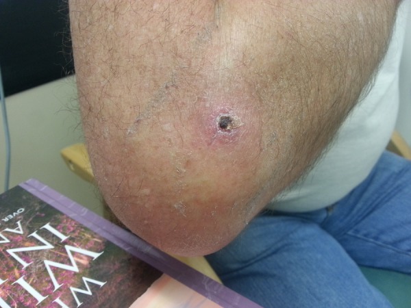
A 67-year-old man had visited the Central American country of Belize 8 weeks prior to presentation, where he was bitten by a mosquito on the posterolateral aspect of his right proximal forearm. Initially there was only a mild itching sensation, but this persisted with a circular erythematous area developing. Later there was a necrotic centre with skin breakdown, within which he could intermittently see a mobile white thread-like protrusion (figure 1).
Figure 1.
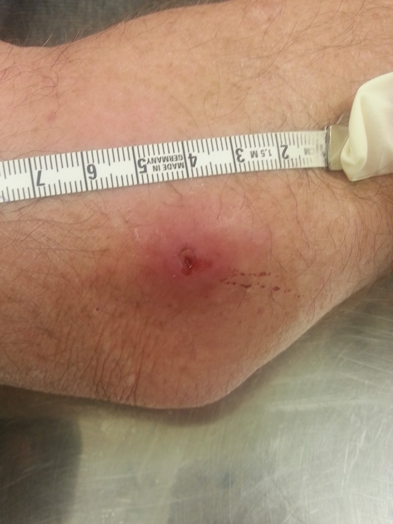
Myiasis with Dermatobia hominis, showing central necrotic puntum with visible breathing apparatus of larva.
He was systemically well with no fever. His daughter who had accompanied him on the trip to Belize had a similar cutaneous infestation which was treated locally, and he was concerned that he might have a similar organism under his skin.
Following attempts by his general practitioner to remove the larva he was referred to the orthopaedic on call team, and seen in the emergency department.
There was a raised erythematous swelling measuring 5 cm in diameter, and a central punctum area with the tip of the larva visible. There was no neurovascular deficit, and he had no axillary or cervical lymphadenopathy.
Investigations
Blood tests revealed a mild eosinophilia, otherwise no other significant findings noted.
Differential diagnosis
Differential diagnosis would include myiasis caused by the larva stage of other flies for example, blowfly, screw fly etc.
Treatment
After verbal consent, 10 mL of 1% lidocaine was infiltrated in the skin and surrounding subcutaneous tissues.
Under aseptic conditions, a 3 cm incision was made along the skin crease lines and a single botfly larva was removed easily using a pair of dissecting forceps (figures 2 and 3). No further larvae were seen. The central necrotic area was debrided (figure 4), then betadine wound dressing applied.
Figure 2.
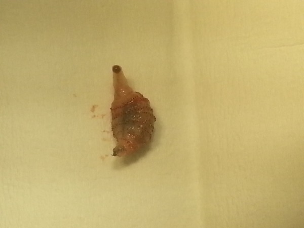
Wholly extracted larva of Dermatobia hominis following mini surgical incision under local anaesthetic.
Figure 3.
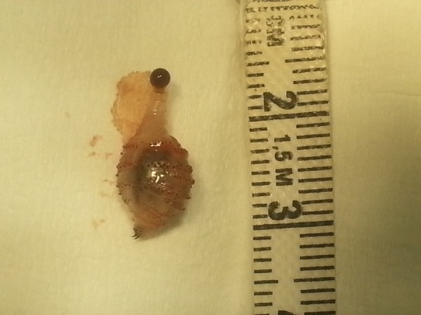
Larva of Dermatobia hominis measuring 3 cm in length.
Figure 4.
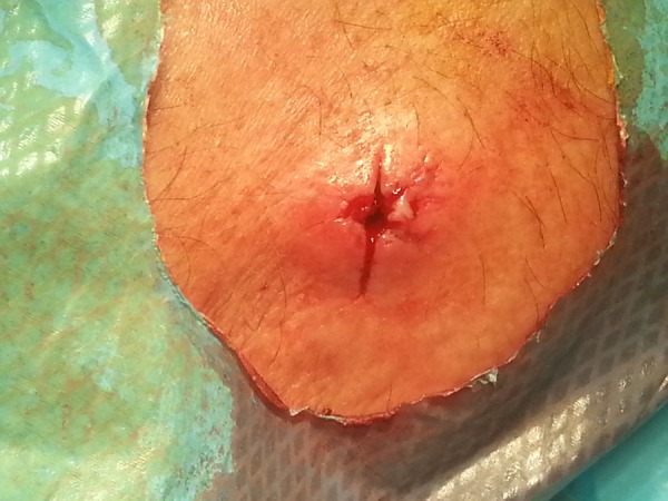
Post larva extraction and debridement of myiasis with Dermatobia hominis.
Outcome and follow-up
The patient was seen 2 weeks postprocedure, the wound was healing well and there were no complications (figure 5). The larva was sent to Liverpool Tropical Medicine Hospital for dissecting microscopy and has been confirmed to be the larva stage of the D. hominis.
Figure 5.
Follow-up 2 weeks post up, satisfactory wound healing.
Discussion
Overview: introduction
The human Botfly (D. hominis), which is native to Central and South America,2 causes skin furuncular myiasis at the site of egg penetration. Myiasis is the invasion of subcutaneous tissues by the larvae of the dipterous fly. The adult form of the human botfly is rarely seen, ranges between 1 and 3 cm long and has been described as sharing semblance with a bumble bee.
Epidemiology
The human Botfly species is native to Central and South America.
Pathogenesis
The female human Botfly lays her eggs on the body of an intermediate host, usually a mosquito, or fly, which acts as a vector onto the human skin when it feeds.3 The heat of the skin causes the eggs to hatch into larvae where they rapidly burrow themselves. The larval stage in the skin tissue can last between 27 and 128 days before the adult larva drops to the ground where it pupates for between 27 and 78 days before maturing into the adult botfly. The whole life cycle lasts between 3 and 4 months.4 The larvae breathe through a central punctum. Unlike other subfamily of the botfly, the larva of the human botfly does not migrate far into the skin from its point of entrance.5–7
The presence of the larva in the skin often triggers a local inflammatory response with the migration and proliferation of inflammatory cells. These include neutrophils, mast cells, eosinophils, fibroblasts and endothelial cells8–10 and can cause irritation, itching and local tissue damage. The majority of cases affect the limbs, though presentation on the genitals, scalp, breast and eye have been reported.11–14 Palpebral infestation was seen in a Danish zoologist who had travelled to Brazil15 and fatal cerebral Myiasis was reported in two babies where the larva was thought to have penetrated through the fontanelles.16 17 Multiple skin infestation is uncommon and it is usual to see only one larva in a skin burrow. Respiratory allergic symptoms in humans have been reported, precipitated by nasal invasion of affected patients by the sheep Botfly specie.18
Secondary bacteria infection is rare as the larvae secrete bacteriostatic and bacteriocidal substance which ingestion of bacterial organisms.
Clinical presentation
There is a localised, itchy, eryhtematous raised skin lesion with a central punctum through which the larva may occasionally be seen and through which it breathes. A serous, bloody and rarely purulent discharge can occur. Pain is unusual and should raise the possibility of a secondary bacterial infection. Travel history to the Americas is most significant, particularly where treatment of a simple bacterial furuncle with antibiotics has not led to the expected improvement in symptoms.
Investigation
Clinical diagnosis is usually possible in most cases, though atypical presentation in unusual anatomical sites may require imaging techniques.19
Treatment
Treatment options vary with different degree of success. It is important to extract whole larva completely.
Non-surgical options include suffocation, with topical application of vaseline, petroleum jelly, beeswax or pork fat. The use of the sap from the Matatorsalo tree found in Costa Rica has being reported to kill larva but does not extrude it.20 However, the dead larva may then need a surgical extraction.
Manually squeezing the larva out through the centre punctum is not advised as it can lead to the rupture of larva with release of its fluid and subsequent risk of anaphylactic reaction21 and is more likely to lead to incomplete extraction and secondary infection. This may also happen with the use of adhesive tape.
Venom extractor has also been employed with or without immobilising the larva that exerts a suction pressure and theoretically causes its extrusion.22
Antiparasitic agent like Ivermectin has shown effectiveness in treating both Myiasis and alimentary forms of Botfly infestation.
Surgical excision using local anaesthesia is an effective treatment option, allowing complete removal of the larvae and debridement of the necrotic cavity.
Learning points.
Travel history to the Americas in a patient with prolonged itchy skin lesion with a central punctum should draw one's mind to human botfly myiasis.
Surgical excision allows complete removal of the larva.
Send extracted larva to an appropriate institution for confirmation of larval species.
Footnotes
Contributors: JCN attended to patient and wrote up case report. RM contributed to review of literature.
Competing interests: None.
Patient consent: Obtained.
Provenance and peer review: Not commissioned; externally peer reviewed.
References
- 1.US Army Center for Health Promotion and Preventive Medicine. Human Bot Fly Myiasis. August 2007. Retrieved 2008–10–09.
- 2.Piper R. “Human botfly”. Extraordinary animals: an encyclopedia of curious and unusual animals. Westport, Connecticut: Greenwood Publishing Group: 192–4. ISBN: 0-313-33922-8. OCLC 191846476. Retrieved 2009–02–13. [Google Scholar]
- 3.Dunleavy S. Life In The Undergrowth: Intimate Relations (Programme synopses). BBC. Retrieved 2008–12–17.
- 4.Powers NR, Yorgensen ML, Rumm PD et al. Myiasis in humans: an overview and a report of two cases in the Republic of Panama. Mil Med 1996;161:495–7. [PubMed] [Google Scholar]
- 5.Catts EP. Biology of NewWorld bot flies: Cuterebridae. Annu Rev Entomol 1982;27:313–38. 10.1146/annurev.en.27.010182.001525 [DOI] [Google Scholar]
- 6.Catts EP, Mullen GR. Myiasis (Muscoidea, Oestroidea). See Ref. 64, 2002:314–48.
- 7.Colwell DD. Bot flies and warble flies (order Diptera: family Oestridae). In: Parasitic diseases of wild mammals. Samuel WM, Pybus MJ, Kocan AA, eds. Ames: Iowa State University Press, 2001:46–71. [Google Scholar]
- 8.Bennett GF. Studies on Cuterebra emasculator Fitch 1856 (Diptera: Cuterebridae) and a discussion of the status of the genus Cephenemyia Ltr. 1818. Can J Zool 1955;33:75–98. 10.1139/z55-004 [DOI] [Google Scholar]
- 9.Cogley TP. Warble development by the rodent bot Cuterebra fontinella (Diptera: Cuterebridae) in the deer mouse. Vet Parasitol 1991;38:276–88. 10.1016/0304-4017(91)90140-Q [DOI] [PubMed] [Google Scholar]
- 10.Payne JA, Cosgrove GE. Tissue changes following Cuterebra infestation in rodents. Am Midl Nat 1966;75:205–13. 10.2307/2423491 [DOI] [Google Scholar]
- 11.Parkinson RJ, Robinson S, Lessells R et al. Fly caught in foreskin: an usual case of preputial myiasis. Ann R Coll Surg Engl 2008;90:7–8. 10.1308/147870808X303010 [DOI] [PMC free article] [PubMed] [Google Scholar]
- 12.Harbin LJ, Khan M, Thompson EM et al. A sebaceous cyst with a difference: Dermatobia hominis. J Clin Pathol 2002;55:798–9. 10.1136/jcp.55.10.798 [DOI] [PMC free article] [PubMed] [Google Scholar]
- 13.Khan DG. Myiasis secondary to Sermatobia hominis (human botfly) presenting as a long-standing breast mass. Arch Pathol Lab Med 1999;123:829–31. [DOI] [PubMed] [Google Scholar]
- 14.Goodman RL, Montalvo MA, Reed JB et al. Photo essay: anterior orbital myiasis caused by human botfly (Dermatobia hominis). Arch Opthal 2000;118:1002–3. [PubMed] [Google Scholar]
- 15.Bangsgaard R, Holst B, Krogh E et al. Palpebral myiasis in a Danish traveler caused by the human bot-fly (Dermatobia hominis). Acta Ophthalmol Scand 2000;78:487–9. 10.1034/j.1600-0420.2000.078004487.x [DOI] [PubMed] [Google Scholar]
- 16.Rossi MA, Zucoloto S. Fatal cerebralmyiasis caused by the tropical warble fly. Dermatobia hominis. Am J Trop Med Hyg 1973;22:267–9. [DOI] [PubMed] [Google Scholar]
- 17.Dunn LH. Prevalence and importanceof the tropical warble fly Dermatobia hominisLinn, in Panama. J Parasitol 1934;20:219–26. 10.2307/3272463 [DOI] [Google Scholar]
- 18.Masoodi M, Hosseini K. The respiratory and allergic manifestations of human myiasis caused by larvae of the sheep bot fly (Oestrus ovis): a report of 33 pharyngeal cases from southern Iran. Ann Trop Med Parasitol 2003;97:75–81. 10.1179/000349802125000000 [DOI] [PubMed] [Google Scholar]
- 19.Bowry R, Cottingham RL. Use of ultrasound to aid management of late presentation of Dermatobia hominis larva infestation. J Accid Emerg Med 1997;14:177–8. 10.1136/emj.14.3.177 [DOI] [PMC free article] [PubMed] [Google Scholar]
- 20.Pariser HS. Explore Costa Rica. Manatee Press, 2006. ISBN: 1-893643-55-7. [Google Scholar]
- 21.Companion animal parasite council. CAPC recommendation 2012. http://www.capcvet.org/capc-recommendations/cuterebriasis
- 22.Boggild AK, Keystone JS, Kain KC. Furuncular myiasis: a simple and rapid method for extraction of intact Dermatobia hominis larvae. Clin Infect Dis 2002;35:336–8. 10.1086/341493 [DOI] [PubMed] [Google Scholar]


