Abstract
A 17-month-old girl with no medical history presented at our emergency room with abnormal vaginal bleeding and vaginal tissue loss with a “grape bunch” appearance. Physical examination showed no abnormalities, but gynaecological examination showed abnormal vaginal tissue protruding through the vagina introitus. Given the typical clinical presentation, the age of the girl and the location and aspect of the lesion, there was a high suspicion of the botryoid variant of embryonal rhabdomyosarcoma of the vagina. Histology of a biopsy of the lesion was consistent with embryonal rhabdomyosarcoma. As no metastases were detected, the girl received chemotherapy. This case report describes the importance of early recognition of the typical clinical symptoms of sarcoma botryoides, since a rapid diagnosis followed by treatment is necessary to prevent death.
Background
Sarcoma botryoides is a diagnosis not to be missed in an infant since it can be extremely malignant. This case report presents a classic textbook presentation of ‘grape-like’ vaginal tissue loss. This classical presentation should alarm every doctor since a rapid diagnosis followed by treatment is necessary to prevent death. This case report may help to achieve awareness for this rare disease with a typical presentation.
Case presentation
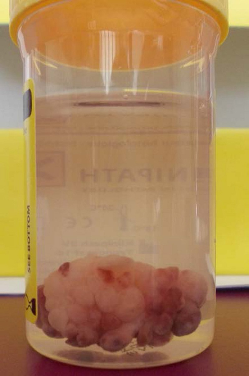
A 17-month-old girl presented at our emergency room with abnormal vaginal bleeding and vaginal tissue loss with a “grape bunch” appearance (figure 1). Two days before presentation, her parents noticed abnormal tissue protruding out of the girl’s vagina during diaper change. The tissue was found in her diaper and consisted of multiple polypoid soft tissue fragments of 3×4 cm. The girl had no medical history and no symptoms.
Figure 1.

Vaginal tissue with a ‘grape bunch’ appearance.
Management
Physical examination showed no abnormalities, but gynaecological examination showed abnormal vaginal tissue protruding through the vagina introitus.
MRI of the abdominal region showed an inhomogeneous, polycyclic, grape-like solid lesion (6.9×3.7×4.1 cm in dimension) arising from the vagina, which was not connected to the uterus. Staging showed no distant metastases. Microscopic examination of the tumour showed botryoid rhabdomyosarcoma: a polypoid lesion consisting of primitive mesenchymal cells with, in parts, rhabdomyoblastic differentiation showing bright eosinophilic cytoplasm. There was immunohistochemical evidence for skeletal-muscle differentiation with focal positivity for desmin and myogenin (figure 2).
Figure 2.

Typical features of embryonal rhabdomyosarcoma with primitive cells and rhabdomyoblasts showing prominent eosinophil cytoplasm. There is nuclear expression of myogenin (inset). Mucosal epithelium is seen in the upper left.
Outcome
Given the typical clinical presentation, the age of the girl, and the location and aspect of the lesion, there was a high suspicion of embryonal rhabdomyosarcoma of the vagina. The ‘grape bunch’ appearance matches the subtype rhabdomyosarcoma botryoides. Another cause such as a germ cell tumour was excluded as the laboratory results showed normal levels of β-human chorionic gonadotropin and α-fetoprotein. Histology of a biopsy of the lesion was consistent with embryonal rhabdomyosarcoma. The patient was treated with nine cycles of chemotherapy (ifosfamide, vincristin, actinomycin, protocol EpSSG RMS 2005, group C). Owing to the location, the type of tumour and the good response on chemotherapy, radical surgery or radiotherapy was omitted.1 At the end of treatment MRI showed a small rest lesion. Within 6 months she had a local relapse, and was treated again with chemotherapy and brachytherapy. She has now been in complete remission for almost 1 year.
Discussion
Rhabdomyosarcoma is the most common soft tissue sarcoma in childhood and adolescence. It is a malignant tumour that arises from embryonic muscle cells and accounts for approximately 5% of all malignancies in children.1 2 The botryoid variant presents as a submucosal lesion with a typical ‘grape bunch’ (botryoid in Greek) appearance. Sarcoma botryoides arise from the vagina or urinary bladder and rarely occur in the uterine fundus or cervix. In the latter case it is extremely malignant.2 Early recognition of this clinical presentation is therefore important.
Learning points.
A vaginal protruding lesion with a ‘grape bunch’ appearance is a red flag for rhabdomyosarcoma botryoides.
Early recognition of the typical clinical presentation of rhabdomyosarcoma botryoides is extremely important since it can be life saving.
Footnotes
Competing interests: None.
Patient consent: Obtained.
Provenance and peer review: Not commissioned; externally peer reviewed.
References
- 1.Maharaj NR, Nimako D, Hadley GP. Multimodal therapy for the initial management of genital embryonal rhabdomyosarcoma in childhood. Int J Gynaecol Cancer 2008;18:190–200. [DOI] [PubMed] [Google Scholar]
- 2.Koukourakis GV, Kouloulias V, Zacharias G et al. . Embryonal rhabdomyosarcoma of the uterine cervix. Clin Transl Oncol 2009;11:399–402. [DOI] [PubMed] [Google Scholar]


