

Abstract
We describe a case of chronic sclerosing osteomyelitis of Garré in a 50-year-old woman occurring in her right femur and presenting with uncontrolled pain. The patient was initially treated with intramedullary reaming of the femur, but 3 years later re-presented with similar symptoms. This required further reaming and intramedullary nailing, achieving good clinical outcomes and lasting pain relief at 8-year follow-up.
Background
Primary chronic sclerosing osteomyelitis was first described by Garré in 1893 with thickening of the cortices and loss of the medullary canal, but no features of acute infection such as suppuration, bony sequestra or drainage tracts.1 2
Chronic sclerosing osteomyelitis (CSO) is a rare condition that affects young children and adults. It is also known as Garré's sclerosing osteomyelitis (GSO), chronic osteomyelitis with proliferative periostitis, ossifying periostitis or non-suppurative chronic sclerosing osteomyelitis.3 4
It most commonly affects males and has a preponderance for the tibia.5 It usually presents to clinicians after a period of localised pain around the site of bone involvement in the absence of constitutional symptoms. CSO has an insidious onset with symptoms recurring at any time. A secondary lesion can occur at a distant site many years after the initial onset.
The radiographic changes include obliteration of the marrow cavity with widening of the cortex associated with increased bone density.2 5 Involvement can be multifocal and in the active phase inflammatory markers, including C reactive protein (CRP) or erythrocyte sedimentation rate (ESR), may be raised,2 but not predictably. Histological findings show chronic, non-specific osteomyelitis with negative blood and tissue cultures.
A set of related disorders include SAPHO syndrome (characterised by synovitis, acne, pustulosis of the hands and feet, hysterostosis and osteitis) and chronic recurrent multifocal osteomyelitis (CRMO).6 7 The aetiology of these related disorders remains unclear, however some studies suggest an autoimmune or genetic cause.8–10
Treatment is mainly symptomatic with some patients responding temporarily to analgesia. Antibiotic therapy can cause temporary improvement although the exact mechanism remains unclear. Few other treatments have been shown to have a beneficial impact on disease progression. Surgical options include debridement of the bone with exposure of the medulla, resection of the area of chronic osteomyelitis, excision with bone grafting and wire fixation.5 11–13 However, following any treatment the patients may again become symptomatic after a number of years or be found to have other lesions, for example osteoid osteomas or other bone tumours.2 14
Case presentation
We present the case of a 50-year-old woman with CSO of her right femur, since the age of 9. Previous local debridement of the distal femur had been performed during her adolescence with management thereafter based on recurrent courses of empirical antibiotics. In adulthood, the patient presented to the rheumatology service at the age of 39 with a 5-year history of pain in her right femur, surgical scars remained healed with no sinuses. There was no history of acnes or pustulosis of her hands or feet.
Routine laboratory testing at presentation were normal (flood blood count, liver function tests, bone profile, immunoglobulins, clotting, vitamin D and parathyroid hormone). ESR and CRP were minimally raised at 34 and 29, respectively (normal ranges 1–10 mm/h and <6 mg/L, respectively). The results of blood and urine cultures were negative. The patient was not on bisphosphonate therapy and was not postmenopausal at presentation.
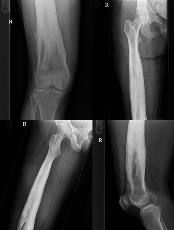
Radiographs and CT confirmed a pancortical, sclerosing osteomyelitis in the middle two-thirds of the right femur (figure 1). There was no evidence of sequestration. An isotope bone scan showed abnormal uptake within the diaphysis with an increase in blood pool activity. An MRI showed heterogenous low signal intensity.
Figure 1.

Anteroposterior full-length plain radiographs of right femur at initial presentation.
The patient was initially treated with antibiotics without significant improvement and orthopaedic opinion sought. Intramedullary (IM) debridement and biopsy was advocated to confirm the diagnosis. A classical Lautenbach compartmental IM reaming was performed through a proximal piriformis fossa entry and distal lateral cortical window.15 A combination of hand and power reamers were used to debride and recanalise the IM cavity. The open medulla was lavaged with 3 L of normal saline under power to remove reaming debris. No pus was found on exploration. Wound swabs, tissue and bone samples were taken. All results were negative and no anaerobic organisms were grown. Histology confirmed no malignancy and microscopic examination revealed chronic inflammatory changes with periosteal reaction.
A diagnosis of CSO of Garré was confirmed based on the clinical and radiological features, with non-specific histology findings. Inflammatory makers normalised postoperatively and she was treated empirically with broad spectrum oral antibiotics for 6 weeks. Postoperative imaging confirmed opening of the medullary cavity (figure 2). At 1 year follow-up she remained asymptomatic with no further episodes of pain.
Figure 2.
Plain radiographs showing the femoral canal open.
However, 3 years later, due to debilitating and worsening pain the patient was re-referred. Further radiological investigation confirmed recurrent sclerosis of the femoral canal with complete obliteration of the medulla (figure 3). ESR and CRP at that point were raised at 29 mm/h and 18 mg/L respectively.
Figure 3.

Plain radiographs showing the femoral canal closed.
After thought and discussion with the patient, repeat IM debridement and to recanalise the medullary canal was proposed. A Lautenbach compartmental debridement was performed as described above.15 To reduce the risk of recurrent obliteration of the medullary cavity and with it the preoperative symptoms the decision was made presurgery to introduce an IM femoral nail (figure 4). The IM nail was unlocked distally as there was no acute fracture and therefore no risk of shortening, malrotation or nail migration. The theoretical risk of distal migration is overcome by inserting the extraction device before removing the proximal locking screw. Postoperatively a 6-week course of broad spectrum oral antibiotics were used on an empirical basis.
Figure 4.

Postoperative radiographs with intramedullary femoral nail in situ.
Eight years following femoral nailing the patient continued to have complete resolution of her symptoms, is pain free with no restriction on activity.
Discussion
The diagnosis of CSO should be made only after a thorough workup and exclusion of more aggressive conditions (eg, Ewing sarcoma).16 Although there are no definite diagnostic criteria found in the literature, it has been suggested that the diagnosis may be made on the basis of a prolonged disease course (more than 3 months), evidence of chronic inflammation on biopsy and lack of organism growth.17
Tissue cultures are typically negative in this condition, but there have been reports showing the anaerobic bacterium Propionibacterium acnes and fungal infections Actinomycosis naeslundii and A. israelii as causative organisms.18 19
Being a rare entity, there remains controversy regarding the treatment of this chronic condition. Intermittent courses of antibiotics may bring short-term resolution of symptoms in some patients.17 20 IM reaming of long bones has been the most successful treatment with the largest case series of 8,14 showing good long-term survival outcomes regardless of previous failed treatments.17 It is however, associated with an increased risk of pulmonary embolism.20 These outcomes have since been replicated in a further smaller case series.21
A more recent approach for long bone involvement in children is with bone resection and transport using a circular external fixator.22 This has also achieved a good functional result in a single case report, although is clearly a more demanding procedure for the patient. Involvement of smaller bones can be managed using the same surgical principles. Intramedullary K-wiring of a fifth metacarpal has resulted in resolution of symptoms in one patient with proven chronic osteomyelitis.12 Metatarsal involvement has also been treated successfully using fenestration techniques.13 Other examples of the use of intramedullary nailing for sclerosing conditions include the rare Ribbing disease or sclerosing bone dysplasia.23
In our case although there was an improvement in symptoms following the original reaming, the patient re-presented 3 years later with recurrent symptoms and progressive pain. Her radiographs showed re-sclerosis and closure of the medullary canal and long-term resolution was only achieved through reaming and IM nailing. To the best of our knowledge, this is the first description of using IM reaming and nailing to manage recurrent, CSO in an adult long bone. Lautenbach compartmental reaming and immediate IM nailing have the advantages of being less destructive and debilitating than excision and bone transport and allow immediate full weight bearing and rehabilitation. Additionally, IM nailing keeps ‘open’ the recanalised medulla and changes the mechanical loading of the long bone which possibly helps in reducing pain.
Insertion of an IM nail in the potential presence of infection may seem a controversial technique in the management of this condition. However, once the surgical clearance of infection has been performed using the Lautenbach technique,15 using an IM nail is no more controversial than performing one-stage revision for an infected total knee replacement.24
The use of postoperative broad spectrum antibiotics is open to discussion and may not be necessary other than as perioperative prophylaxis.
CSO of Garré is a rare condition and its symptomatic management is difficult. IM reaming is generally recommended but the role of IM nailing should also be considered in weight bearing long bones to prevent recurrence of symptoms.
Learning points.
Chronic sclerosing osteomyelitis is a rare condition that affects young children and adults.
It most commonly affects males and has a preponderance for the tibia.
It usually presents to clinicians after a period of moderate pain around the site of bone involvement and has an insidious onset with symptoms recurring at any time.
Treatment is mainly symptomatic with some patients responding temporarily to analgesia.
Surgical options include: debridement of the bone with exposure of the medulla, resection of the area of chronic osteomyelitis, excision with bone grafting and wire fixation or intramedullary reaming and nailing.
Footnotes
Contributors: NBV performed the literature review and wrote the manuscript. HLMW was a major contributor in writing the manuscript. BH assessed the patient throughout the course of treatment. RM-J assessed the patient throughout the course of treatment, performed the operation and reviewed in follow-up clinic. All authors read and approved the final manuscript.
Competing interests: None.
Patient consent: Obtained.
Provenance and peer review: Not commissioned; externally peer reviewed.
References
- 1.Garré C. Ueber besondre Formen und Folgezustande d. akuten infekt. Osteomyelitis Beitr z klin Chir 1893;10:257. [Google Scholar]
- 2.Macnicol MF, Watts AC. Haematogenous osteomyelitis (SURGERY 23:1). The Medicine Publishing Company Ltd, 2005:25–30. [Google Scholar]
- 3.Wood RE, Nortjé CJ, Grotepass F et al. Periostitis ossificans versus Garrè's osteomyelitis. Part I. What did Garrè really say? Oral Surg Oral Med Oral Pathol 1988;65:773–7. 10.1016/0030-4220(88)90028-X [DOI] [PubMed] [Google Scholar]
- 4.Vienne P, Exner GU. Garré sclerosing osteomyelitis. Orthopade 1997;26:902–7. 10.1007/PL00003340 [DOI] [PubMed] [Google Scholar]
- 5.Jensen DR. Chronic sclerosing osteomyelitis: Garré. Am J Surg 1941;2:377–83. 10.1016/S0002-9610(41)90384-7 [DOI] [Google Scholar]
- 6.Demharter J, Bohndorf K, Michl W et al. Chronic recurrent multifocal osteomyelitis: a radiological and clinical investigation of five cases. Skeletal Radiol 1997;26:579–88. 10.1007/s002560050290 [DOI] [PubMed] [Google Scholar]
- 7.Jurik AG. Chronic recurrent multifocal osteomyelitis. Semin Musculoskeletal Radiol 2004;8:243–53. 10.1055/s-2004-835364 [DOI] [PubMed] [Google Scholar]
- 8.Letts M, Davidson D, Birdi N et al. The SAPHO syndrome in children: a rare cause of hyperostosis and osteitis. J Pediatr Orthop 1999;19:297–300. 10.1097/01241398-199903000-00004 [DOI] [PubMed] [Google Scholar]
- 9.El-Shanti HI, Ferguson PJ. Chronic recurrent multifocal osteomyelitis: a concise review and genetic update. Clin Orthop 2007;462:11–19. 10.1097/BLO.0b013e3180986d73 [DOI] [PubMed] [Google Scholar]
- 10.Ferguson PJ, Bing X, Vasef MA et al. A missense mutation in pstpip2 is associated with the murine autoinflammatory disorder chronic multifocal osteomyelitis. Bone 2006;38:41–7. 10.1016/j.bone.2005.07.009 [DOI] [PMC free article] [PubMed] [Google Scholar]
- 11.Viejo-Fuertes D, Rossillon R, Mousny M et al. Primary chronic sclerosing osteomyelitis—a case report. Joint Bone Spine 2005;72:73–5. 10.1016/j.jbspin.2004.02.007 [DOI] [PubMed] [Google Scholar]
- 12.Kelkar AS, Malshikare VA. Chronic sclerosing osteomyelitis of a metacarpal. J Hand Surg Eur Vol 2005;3:298–301. 10.1016/j.jhsb.2005.01.009 [DOI] [PubMed] [Google Scholar]
- 13.Sharma H, Taylor GR. Chronic sclerosing osteomyelitis of Garré affecting fifth metatarsal bone of the foot. The Foot 2003;13:209–11. 10.1016/j.foot.2003.08.005 [DOI] [Google Scholar]
- 14.Collert S, Isacson J. Chronic sclerosing osteomyelitis (Garré). Clin Orthop 1982;164:136–40. [PubMed] [Google Scholar]
- 15.Caesar BC, Morgan-Jones RL, Warren RE et al. Closed double-lumen suction irrigation in the management of chronic diaphyseal osteomyelitis: long-term follow-up. Bone Joint Surg Br 2009;91:1243–8. 10.1302/0301-620X.91B9.21768 [DOI] [PubMed] [Google Scholar]
- 16.Carbanela ME, Sim FH, Beabout JW et al. Osteomyelitis appearing as neoplasms. A diagnostic problem. Arch Surg 1974;109:68–72. 10.1001/archsurg.1974.01360010050012 [DOI] [PubMed] [Google Scholar]
- 17.Schultz C, Holterhus PM, Seidel A et al. Chronic recurrent multifocal osteomyelitis in children. Pediatr Infect Dis J 1999;18:1008–13. 10.1097/00006454-199911000-00015 [DOI] [PubMed] [Google Scholar]
- 18.Phillon P, Pajon A, Juvin R et al. Tibial hyperostosis and Proiobacterium acnes. Rev Rheum Mal Osteartic 1992;59:349–51. [PubMed] [Google Scholar]
- 19.Kadish LJ, Muller CJ, Mezger H. Chronic sclerosing osteomyelitis in a long bone caused by actinomycosis. A case report. S Afr Med J 1982;62:658–9. [PubMed] [Google Scholar]
- 20.Pape HC, Zwipp H, Regel G et al. Chronic treatment refractory osteomyelitis of long tubular bones—possibilities and risks of intramedullary boring. [Article in German] Unfallchirurg 1995;98:139–44. [PubMed] [Google Scholar]
- 21.Fery A. Chronic sclerosing osteomyelitis. A propos of 4 cases and the value of the treatment by closed intramedullary reaming. J Chir Paris 1990;127:157–63. [PubMed] [Google Scholar]
- 22.Nikomarov D, Zaidman M, Katzman A et al. New treatment option for sclerosiing osteomyelitis of Garre. J Pediatr Orthop B 2013;22:577–82. 10.1097/BPB.0b013e32836330a6 [DOI] [PubMed] [Google Scholar]
- 23.Matas M, Torrededia L, Via-Duresne O. Symptomatic Ribbing's disease—case report. Rev Esp Cir Ortop Traumatol 2008;52:322–5. [Google Scholar]
- 24.Vanhegan IS, Morgan-Jones R, Barrett DS et al. Developing a strategy to treat established infection in total knee replacement: a review of the latest evidence and clinical practice. J Bone Joint Surg Br 2012;94:875–81. 10.1302/0301-620X.94B7.28710 [DOI] [PubMed] [Google Scholar]