Abstract
Background
We have developed a novel head-holding device for behaving non-human primates that affords stability suitable for reliable chronic electrophysiological recording experiments. The device is completely non-invasive, and thus avoids the risk of infection and other complications that can occur with the use of conventional, surgically implanted head-fixation devices.
New method
The device consists of a novel non-invasive head mold and bar clamp holder, and is customized to the shape of each monkey’s head. The head-holding device that we introduce, combined with our recording system and reflection-based eye-tracking system, allows for chronic behavioral experiments and single-electrode or multi-electrode recording, as well as manipulation of brain activity.
Results and comparison with existing methods
With electrodes implanted chronically in multiple brain regions, we could record neural activity from cortical and subcortical structures with stability equal to that recorded with conventional head-post fixation. Consistent with the non-invasive nature of the device, we could record neural signals for more than two years with a single implant. Importantly, the monkeys were able to hold stable eye fixation positions while held by this device, demonstrating the possibility of analyzing eye movement data with only the gentle restraint imposed by the non-invasive head-holding device.
Conclusions
We show that the head-holding device introduced here can be extended to the head holding of smaller animals, and note that it could readily be adapted for magnetic resonance brain imaging over extended periods of time.
Keywords: chronic recording, primate, head holding, electrophysiology, eye-tracking
1. Introduction
Single-electrode and multi-electrode recordings have been successfully applied in conscious monkeys to explore neural substrates of behavior, providing valuable information about the functions of the brain. Most of these experiments have been performed in monkeys that were head-fixed, as most recording systems depend critically on adequate fixation of the animal’s head so that electrical activity from single and multiple units can be recorded without noise artifact and with a constant relationship of the subject’s head position to experimental stimuli and monitoring equipment. For this purpose, series of methods for head holding have been developed (Adams et al., 2007; Davis et al., 2009; Evarts, 1968; Isoda et al., 2005; Pigarev et al., 2009; Srihasam et al., 2010), most of which, following the pioneering work of Evarts, require head-restraint bolts to be implanted into the skull (Adams et al., 2007; Davis et al., 2009; Evarts, 1968). These methods all allowed excellent stabilization of the subject’s head, but with prolonged times following implantation, head-bolt systems become increasingly at risk for intracranial infection or necrosis and softening of bone around the head post, where the principal torques are applied during experimental sessions. This situation can lead to instability and ultimately failure of the implanted devices. In an attempt to solve such problems, less invasive methods without bolts screwed on the skull have been developed in some studies (Isoda et al., 2005; Pigarev et al., 2009). They achieved rigid head fixation, but even with those methods, small incisions of the skin are still necessary at the time of installation.
In this study, we developed a completely non-invasive head-holding device to prevent head movement during experimental sessions. The device is constructed with a head mold, bar clamp and metal brackets for fixation. The head mold is made with a flexible plastic that readily conforms to the contours of each monkey’s head, and, as a result, the device achieves a strong but gentle securing of the head. A bar clamp holds the head mold on both sides and can be attached to a primate chair by metal brackets. This head-holding device has allowed us to record from the neocortex and deeper subcortical structures in the forebrain and midbrain both with conventional single-electrode recording methods (Evarts, 1968) and with a multi-electrode recording method developed in-house (Feingold et al., 2012) as monkeys perform tasks that require eye movements and ocular fixation.
Here we present the procedure for making the head-holding device, demonstrate the precision of eye-tracking possible with this system, and illustrate the reliability of both acute and chronic recordings of neuronal activity from the neocortex and from subcortical structures performed with the head-holding device as the sole source of head restraint. The completely non-invasive character of this device should help researchers to maintain non-human primates in a robustly healthy condition even with multiple electrodes chronically implanted in multiple sites in the brain.
2. Materials and methods
2.1. Design of the head-holding device
2.1.1. Description of the head-holding device
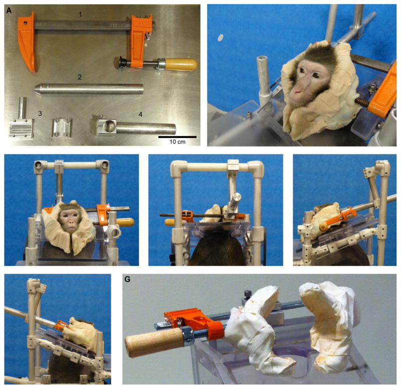
Each part of this head-holding device and its overall configuration are shown in Fig. 1. The device is made of five parts: (1) a bar clamp (21 cm long) with a left arm and an adjustable right arm with a large screw (Jorgensen #3908, Chicago, IL, labeled 1 in Fig. 1A), (2) a custom-made extender (2 in Fig. 1A), (3) a custom-made clamp attachment with screws (3 in Fig. 1A), (4) a holder for the extender (4 in Fig. 1A), and (5) three sheets of “Aquaplast” moldable plastic (Patterson Medical, Bolingbrook, IL, not shown). One sheet is about 23 cm × 36 cm, and the other two are about 15 cm × 20 cm.
Fig. 1.
Design of the head-holding device. (A) Individual components of the device: adjustable 21-cm-long bar clamp (1), extender (2), attachment bracket (3) and extender holder (4). (B) Head mold made with flexible plastic. (C–F) Front (C), rear (D), left side (E) and right side (F) views of the head-holding device attached to a primate chair. (G) Head-holding device designed for squirrel monkey.
Fig. 1B–F illustrates the assembled device. The tips of the left arm of the bar clamp and the large screw on the right arm of the bar clamp (1 in Fig. 1A) are covered by the smaller Aquaplast sheets (Fig. 1B–F). The covered left arm and right screw are connected to the left and right sides of the head mold, which was formed with the larger sheet of Aquaplast and touches the monkey’s head (Fig. 1B–F). The head molds are customized to each monkey’s head (Fig. 1B). The bar clamp is attached to the clamp attachment (3 in Fig. 1A), which is inserted into the extender (2 in Fig. 1A). This extender is held on the primate chair by means of the holder (4 in Fig. 1A). All of the attachments and the extender are secured by tightening screws.
2.1.2. Manufacture of the head-holding device
The entire procedure for making the head-holding device takes only a few hours. During the procedure, the monkey sits in a primate chair under mild sedation with ketaset (5 mg/kg i.m.). Important points are (1) preventing the hot Aquaplast from touching directly to the monkey’s head; (2) molding the device to fit the monkey’s neck as well as the monkey’s occipital, parietal, temporal, zygomatic and mandible bones for secure holding of the head; (3) shaping the mold quickly before the Aquaplast hardens; and (4) preventing the mold from covering the face, including the regions around the mouth, nose, eyes and forehead, to allow comfortable task performance.
During the procedure, we cover the monkey’s face, neck, chin, and ears with a thin and soft underpad (<1 mm thickness, McKesson #477563, Waltham, MA) that is waterproofed outside and has fluffy filler inside to prevent heat from the Aquaplast from reaching the monkey’s skin. We attach the bar clamp to the primate chair for preparation. We boil water in a flat pan and put a 23 cm × 36 cm sheet of Aquaplast into the hot water. At >70°C, the Aquaplast becomes soft and transparent. We then wrap the softened Aquaplast sheet around the monkey’s head as shown in Fig. 1B. We mold and cover the monkey’s neck as well as the monkey’s occipital, parietal, temporal, zygomatic and mandible bones precisely with gloved hands. This step is essential for secure fixation of monkey’s head. The Aquaplast hardens in about 15 min. Just before this time, the head mold is separated into left and right halves by carefully cutting the center of the mold with heated or non-heated scissors. After the head mold becomes hard, we then make a joint with the bar clamp. We cover and wrap the tip of the left arm and the tip of the large screw on the right arm of the bar clamp with the smaller sheets of the Aqaplast, which have been heated in the hot water bath. While these smaller Aquaplast sheets are still transparent and flexible, we slide the right arm of the adjustable bar clamp and tighten the large screw into the head mold. We push the head mold tightly from both sides and make sure that it attaches to the bar clamp, and we then allow the Aquaplast to harden. Finally, we wrap the underpad to the head mold with tape to make a cushion between the monkey’s head and the device.
2.1.3. Attachment procedures
We first place an attachment bracket (3 in Fig. 1A) on a horizontal metal bar of the bar clamp (1 in Fig. 1A). We then insert the projecting part of the bracket (3 in Fig. 1A) into the holding pocket of the extender (2 in Fig. 1A). The extender is held by a holder (4 in Fig. 1A) which is attached to the primate chair. We tighten the screws to make all parts of the head-holding device stable. In each session, with the monkey in the primate chair, the head-holding device is carefully attached to the chair from the back. After the head mold is put on the sides of the monkey’s face, we first slide the right arm of the clamp (1 in Fig. 1A) to adjust roughly the distance, and we then tighten the large screw. When we remove the device, we loosen the large screw, and then slide the right arm to release holding.
2.1.4. Adaptation of the head-holding device for squirrel monkeys
We also made a smaller version of the macaque head-holding device to fit to squirrel monkeys (see Fig. 1G). We used a 10-cm-long bar clamp from the same company (Jorgensen 3904-LD, Chicago, IL). This head-holding device has the same design as the device for macaques and achieved successful stabilization of the squirrel monkey’s head.
2.2. Experimental application of the head-holding device
We collected behavioral and neuronal data while two macaque monkeys, fitted with the head-holding device, performed cognitive tasks involving hand and eye movements as well as licking movements to obtain reward.
2.2.1. Subjects
Recording experiments with the head-holding device in place were performed on two female Macaca mulatta monkeys (S: 7.5 kg and P: 6.3 kg), with the approval from the Committee on Animal Care of the Massachusetts Institute of Technology. Multi-electrode recordings were performed with methods described in detail previously (Feingold et al., 2012). In monkey S, we recorded units in the frontal neocortex and caudate nucleus. In monkey P, we acquired eye and head movement data during single-electrode recording in the midbrain.
2.2.2. Behavioral tasks
Monkey S was trained to perform an approach-approach decision task, in which the monkey was required to choose which of a pair of targets yielded a larger reward by making a joystick movement with the right hand in response to visual cues presented on a computer screen in front of the animal (Amemori and Graybiel, 2012). Ocular fixation was not required. Monkey P was trained to perform an active/passive reward task in which eye movements and ocular fixation were required. At the start of the passive version of the task, a fixation cue appeared at the center of a black screen in front of the monkey. After a 0.3–1.8 s fixation period during which the monkey was required to direct its gaze to within a fixation window of 8°, a target cue replaced the fixation cue, and the monkey was required to fixate this target for 500 ms. In the active task-version, after the fixation period, a target cue appeared 10° to the right or left of the fixation point, with randomized locations from trial to trial. The monkey was required to hold its gaze on target for 500 ms, and then, according to the target-cue colors, reward was either delivered or not delivered. During the task, eye position was recorded by an infrared eye-movement camera system (Eyelink 1000, SR Research, Ontario, Canada) that uses coronal reflection to obtain eye movements. The Eyelink 1000 system allows accurate eye-position monitoring with tolerance of head movements within a range of ±25 mm horizontally or vertically and ±10 mm in depth. Because the head-fixation device restrained head movements within the range acceptable for the Eyelink system, the residual movements allowed by the device did not degrade the precision of the measurement. Eye positions were converted to digital signals at 1 kHz. If the monkey lost fixation, the trial was terminated, and a beep sound was delivered.
2.2.3. Unit recording and measurement of head movements
After behavioral training, a plastic recording chamber was implanted on the skull of each monkey. After recovery, over 60 platinum-iridium electrodes (impedance, 0.8–1.5 MΩ; FHC Inc., Bowdoin, ME) were implanted into the prefrontal cortex and the caudate nucleus of monkey S. All electrodes were held by custom-made micromanipulators affixed to the grid (Amemori and Graybiel, 2012; Feingold et al., 2012). Single-electrode recording was performed in monkey P with glass-coated tungsten electrodes (impedance, 1.5–2.5 MΩ; Alpha Omega Co. USA Inc., Alpharetta, GA) that were advanced by an oil-driven micromanipulator (MO-97A, Narishige) affixed at 5° off-vertical to a grid with openings spaced at 1 mm, each containing stainless steel guide tubes to pierce the dura mater. Signals were amplified with a 0.2–5 kHz band-pass filter (Lynx-8, Neuralynx, Bozeman, MT) and were collected at 1 kHz via a custom-made window discriminator. Head movements for monkey P were measured in 3D with analog gyroscopes that were fitted on top of the recording chamber and that have a full scale of ±300°/s (LPY430AL and LPR430AL, STMicroelectronics, Geneva, Switzerland).
3. Results
3.1. Eye and head movements during performance with the head-holding device
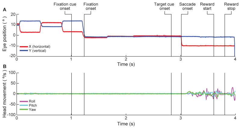
Fig. 2A shows an example of horizontal (x-axis) and vertical (y-axis) eye positions recorded while monkey P was performing the active task with the head-holding device in place. The monkey fixated the central fixation cue for 1.5 s, then made a leftward saccade, and then held her gaze at that target position for 500 ms. Fig. 2B shows the angular velocity of head movement for the same trial. Despite the fact that the monkey’s head was held only by the head-holding device, and not by a head-post implanted into the skull, the head was steady until the monkey licked the spout to receive the liquid reward. Moreover, the eye-tracking camera successfully tracked the monkey’s eye position and eye movements throughout the task, even when the monkey’s head was moving at reward-delivery time (Fig. 2A), because eye position data were obtained based on the corneal reflection.
Fig. 2.
Negligible effect of head movements on stability of eye tracking. (A) Example of horizontal (red) and vertical (blue) eye-position traces during the active task. (B) Angular velocity of head movement monitored during the same trial as Fig. 2A. Magenta, cyan, and green lines indicate, respectively, roll, pitch, and yaw movements. Head movements were minimal except during reward-licking period.
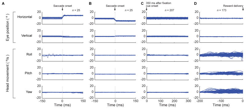
Fig. 3A and B illustrates the eye-tracker data (upper two rows) and gyroscope records (lower three rows) from multiple trials for the time-window of 150 ms before and after saccade onset in the active task in which the monkey had to make 10° saccades to the peripheral targets. The monkey successfully made rightward (Fig. 3A) or leftward (Fig. 3B) saccades following the onset of the targets. During the saccades, the monkey occasionally moved its head, but the tracker camera detected the eye movements. Thus the saccades were tracked accurately with or without head movements.
Fig. 3.
Eye position and head movement traces in the active and passive tasks. (A and B) Horizontal (top row) and vertical (second row) eye position, relative to the fixation cue position, and roll (third row), pitch (fourth row) and yaw (bottom row) angular velocity of head movements recorded during rightward saccades (A) and leftward saccades (B) made during performance of the active task. Zero on x-axis indicates saccade onset time. (C and D) Traces showing eye position and head movements during fixations for the 300 ms starting at 350 ms after fixation cue onset (C), and during fixations for 200 ms before the reward delivery (D) by a monkey performing the passive task. Traces from multiple trials are overlaid.
Fig. 3C shows the eye positions (upper two rows) and head movements (lower three rows) for 300 ms of the fixation period starting at 350 ms after fixation onset in the passive task in which the monkey had to fixate the central fixation cue and the following target cue for more than 800 ms. The standard deviation for horizontal eye position was 0.92°, and that for vertical eye position was 0.45°. During fixations, eye position was well maintained within the 8° window. Mean values of angular velocity of the head movement during the 300 ms fixations were not different from those during the 300 ms rightward and leftward saccade periods (two tailed t-test, p > 0.05 for both rightward and leftward saccades). Thus the amplitude of the head movements was similar during saccades and fixations.
Fig. 3D shows the positions of the eyes (upper two rows) and head (lower three rows) for the 200 ms period before reward delivery in the passive task, during which the monkey had to fixate at the central target in order to receive reward. The standard deviation of eye position was 0.22° horizontally and 0.18° vertically. The mean angular velocities of the head movement during this pre-reward period were significantly greater than those during the fixation period (two tailed t-test, p < 0.05). Although there were head movements caused by anticipatory licking toward the reward delivery, the monkey maintained fixation on the target. These head movements were within the tolerated movement-range of the eye position tracker system. Thus, even though there were small head movements, usually on the order of 20°/s, during saccades and fixations, the monkey could perform the task correctly, and the tracker system could successfully track eye position during the entire time. Thus the head-holding device could be also used to track eye movements when combined with a reflection-based eye tracker that can compensate for small head movements.
3.2. Neuronal recordings with the head-holding device
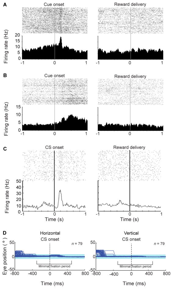
For both multi-electrode and conventional single-electrode recordings that we performed, the manipulator holding the electrodes was affixed to a grid inserted into the recording chamber. Stable recordings thus could be made in the two monkeys despite the small head movements allowed by the head-holding device (Fig. 4). Fig. 4A shows the cue-related activity of a unit recorded in the dorsolateral prefrontal cortex with our chronically implanted multi-electrode system as monkey S performed the approach-approach task in which the monkey had to choose the target indicating larger reward by moving a joystick to control a cursor on the screen. An example of a simultaneously recorded subcortical unit in the caudate nucleus is shown in Fig. 4B. As shown in the right panel of Fig. 4A and B, the units were stably monitored during the session lasting over 3 h without noise related to head movements made during licking. Fig. 4C illustrates subcortical recordings made in monkey P by conventional single-unit recording methods with a single electrode implanted in the substantia nigra pars compacta while the monkey was performing the passive task requiring ocular fixation. Again, the recorded neuronal activity was stable even in the reward period when licking occurred (right panel of Fig. 4C). Fig. 4D illustrates records of eye movements during the same recording session. The monkey was required to start fixation 300–500 ms before the target cue appeared and to keep fixation within a fixation window of 8° for 500–800 ms, when reward delivery occurred. The duration of fixation was randomly determined within this range. We found that during this fixation period, the monkey could maintain its gaze within the fixation window of 8°. Thus the head-holding device can be used for recording of the neuronal activity with or without head movements while monkeys perform behavioral tasks requiring eye movements.
Fig. 4.
Cue-related spike activity recorded during use of the head-holding device. (A and B) Raster plots (above) and spike histograms (below) of spike activity recorded during cue (left) and reward (right) periods in the dorsolateral prefrontal cortex (A) and caudate nucleus (B) in a monkey performing the approach-approach task. Activity was recorded with the multi-electrode recording method. (C) Spike activity recorded from the substantia nigra pars compacta with conventional single-electrode recording methods during performance of the passive task. Spike histograms are aligned to cue onset (left) and reward delivery (right). (D) Eye position trace from the same recording session as shown in Fig. 4C. Horizontal (left) and vertical (right) eye positions, relative to the fixation cue position, are shown for the ±800 ms period around the onset of the target cue. Shading indicates the range within which the monkey was required to maintain eye position (8° square window).
4. Discussion
The major advantage of the head-holding device described here is that it is completely non-invasive. By making surgical intervention unnecessary, both before and during recording, the fixation device avoids problems related to infection associated with implants that are subjected to high torques, and thus improves conditions for prolonged chronic recording. By holding the head with a conformable padded mold, the device allows behavioral monitoring and low-noise neuronal recordings to be made from superficial and deep regions of the brain with both conventional single-electrode and multi-electrode recording methods. The eye position data and electrophysiological recording data that we obtained with electrode manipulators affixed to the recording chamber grid are comparable in quality and stability to those obtained with traditional head-post fixation methods. To date, we have used the head-holding device for work with five macaque monkeys. None has shown discomfort when performing tasks while held with the head-holding device.
Implanting a head-post or comparable device by means of screws or bolts inserted into the bony calvarium can, over time, produce softening of the bone and eventual necrosis, and can add to the risk of infection produced by recording chamber implantation. We have used the head-holding device in place of such conventional head-posts, and also to replace a failed head-post; we found that the head-holding device allowed the infection to clear and the monkey to perform experimental tasks daily for over two years. Further, the head-holding device is suitable for experiments combined with aversive stimuli such as air-puffs to face. Even if a monkey attempts to avoid the air-puff stimulus, which usually produces heavy strain on conventional head-posts, the holding device that we describe can safely and securely hold the head without imposing any strain on the monkey. Further advantages of the device include its ease of fabrication, low price (<$100 US), and ready adaptability to individual research needs.
We emphasize that the head-holding device does allow some movement of a head, particularly, in our experience, when the monkey licks at reward delivery. We did not observe detectable effects of these head movements on the eye-tracking data obtained with the Eyelink 1000 eye-tracking system, which measures corneal reflection and center of the pupil as parameters with which to derive eye position. Even when we used the eye-tracking system without head movement compensation, the monkey could still perform the task using eye movements, though the preciseness of the eye tracking was decreased especially during reward periods in which licks frequently occurred. Further, the head movements that the head-holding device allowed did not affect the quality of neural recording. Because the recording chamber in the system that we describe here was separated from the head-holding device, and the manipulators holding microelectrodes were attached to a grid fixed within the recording chamber implanted on the skull (Feingold et al., 2012), the recording configuration allowed for stable recording even with the small head movements that did occur.
There are limitations to the current head-holding device that could be overcome by small changes in design. One is that the device covers the cheeks, neck and posterior parts of the head. This design precludes neuronal recording from far posterior and temporal parts of the brain, but redesign could open suitable recording windows as needed. Further, because the device covers the ears, it is not in its present form suitable for examination of the auditory system; but again, adaptation of the design would allow microphones to be placed at the ears. Finally, the device as we have made it in our prototypes is not compatible with the magnetic resonance imaging (MRI), but MRI-compatible material could be substituted in the design. Despite these drawbacks, the relative stability afforded by the device and the non-invasive nature of the device are strong positive features that recommend it for many types of experiments.
HIGHLIGHTS.
We developed a completely non-invasive head-holding device for behaving monkeys.
The device is made from a head mold that is customized for each monkey’s head.
Accurate monitoring of ocular movements was possible during use of the device.
Stable chronic recording of neuronal activity during behavior was also possible.
Acknowledgments
We thank S. Hong, P.L. Tierney, H. Shimazu, H.F. Hall and J. Gill for their technical support and advice. We also thank Y. Kubota and A. McWhinnie for their insightful comments on our manuscript and figures. This work was funded by National Institutes of Health (R01 NS025529), Office of Naval Research (N00014-07-1-0903) and CHDI Foundation (A-5552).
Abbreviations
- MRI
magnetic resonance imaging
Footnotes
Publisher's Disclaimer: This is a PDF file of an unedited manuscript that has been accepted for publication. As a service to our customers we are providing this early version of the manuscript. The manuscript will undergo copyediting, typesetting, and review of the resulting proof before it is published in its final citable form. Please note that during the production process errors may be discovered which could affect the content, and all legal disclaimers that apply to the journal pertain.
References
- Adams DL, Economides JR, Jocson CM, Horton JC. A biocompatible titanium headpost for stabilizing behaving monkeys. J Neurophysiol. 2007;98:993–1001. doi: 10.1152/jn.00102.2007. [DOI] [PubMed] [Google Scholar]
- Amemori K, Graybiel AM. Localized microstimulation of primate pregenual cingulate cortex induces negative decision-making. Nat Neurosci. 2012;15:776–85. doi: 10.1038/nn.3088. [DOI] [PMC free article] [PubMed] [Google Scholar]
- Davis TS, Torab K, House P, Greger B. A minimally invasive approach to long-term head fixation in behaving nonhuman primates. J Neurosci Methods. 2009;181:106–10. doi: 10.1016/j.jneumeth.2009.04.012. [DOI] [PMC free article] [PubMed] [Google Scholar]
- Evarts EV. A technique for recording activity of subcortical neurons in moving animals. Electroencephalogr Clin Neurophysiol. 1968;24:83–6. doi: 10.1016/0013-4694(68)90070-9. [DOI] [PubMed] [Google Scholar]
- Feingold J, Desrochers TM, Fujii N, Harlan R, Tierney PL, Shimazu H, Amemori K, Graybiel AM. A system for recording neural activity chronically and simultaneously from multiple cortical and subcortical regions in nonhuman primates. J Neurophysiol. 2012;107:1979–95. doi: 10.1152/jn.00625.2011. [DOI] [PMC free article] [PubMed] [Google Scholar]
- Isoda M, Tsutsui K, Katsuyama N, Naganuma T, Saito N, Furusawa Y, Mushiake H, Taira M, Tanji J. Design of a head fixation device for experiments in behaving monkeys. J Neurosci Methods. 2005;141:277–82. doi: 10.1016/j.jneumeth.2004.07.003. [DOI] [PubMed] [Google Scholar]
- Pigarev IN, Saalmann YB, Vidyasagar TR. A minimally invasive and reversible system for chronic recordings from multiple brain sites in macaque monkeys. J Neurosci Methods. 2009;181:151–8. doi: 10.1016/j.jneumeth.2009.04.024. [DOI] [PubMed] [Google Scholar]
- Srihasam K, Sullivan K, Savage T, Livingstone MS. Noninvasive functional MRI in alert monkeys. Neuroimage. 2010;51:267–73. doi: 10.1016/j.neuroimage.2010.01.082. [DOI] [PMC free article] [PubMed] [Google Scholar]


