Fig. 5.


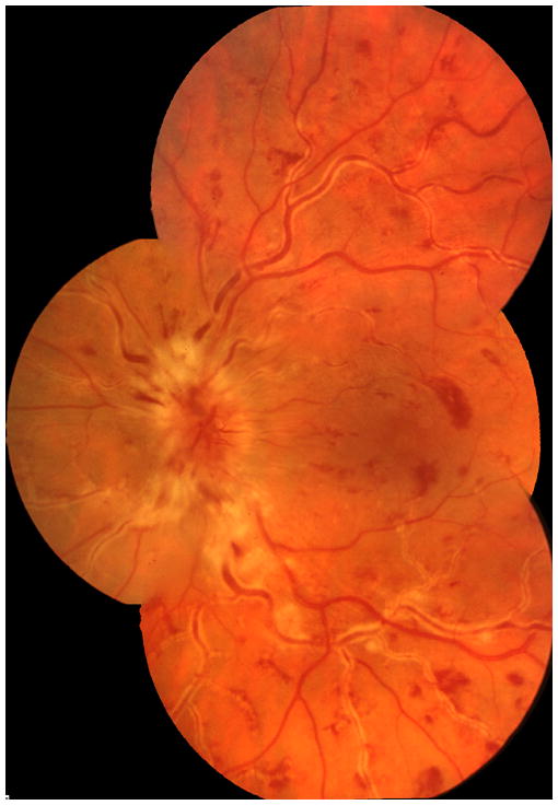
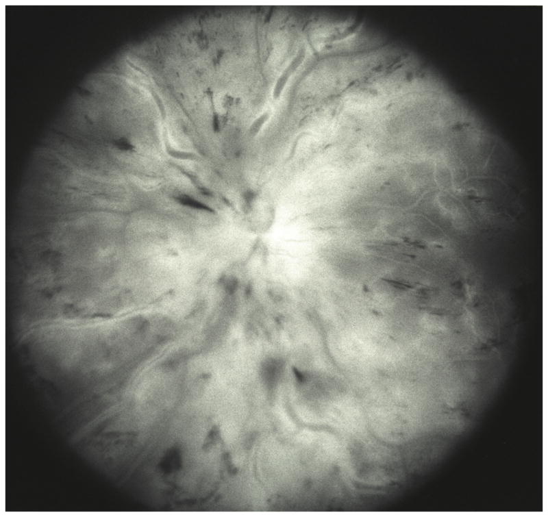
Fundus photograph (A) and fluorescein angiogram (B) of left with non-ischemic CRVO 4 days after onset of visual complaint.
A. This compost fundus photograph shows extensive perivenous sheathing, optic disc edema, macular edema and scattered retinal hemorrhages. .
B. Late phase of fluorescein angiography showing marked perivenous staining due to fluorescein leakage.
