Abstract
Objective
Acute kidney injury (AKI) in patients hospitalized for acute heart failure (AHF) is usually type 1 of the cardiorenal syndrome (CRS) and has been associated with increased morbidity and mortality. Early recognition of AKI is critical. This study was to determine if the new KDIGO criteria (Kidney Disease: Improving Global Outcomes) for identification and short-term prognosis of early CRS type 1 was superior to the previous RIFLE and AKIN criteria.
Methods
The association between AKI diagnosed by KDIGO but not by RIFLE or AKIN and in-hospital mortality was retrospectively evaluated in 1005 Chinese adult patients with AHF between July 2008 and May 2012. AKI was defined as RIFLE, AKIN and KDIGO criteria, respectively. Cox regression was used for multivariate analysis of in-hospital mortality.
Results
Within 7 days on admission, the incidence of CRS type 1 was 38.9% by KDIGO criteria, 34.7% by AKIN, and 32.1% by RIFLE. A total of 110 (10.9%) cases were additional diagnosed by KDIGO criteria but not by RIFLE or AKIN. 89.1% of them were in Stage 1 (AKIN) or Stage Risk (RIFLE). They accounted for 18.4% (25 cases) of the overall death. After adjustment, this proportion remained an independent risk factor for in-hospital mortality [odds ratios (OR)3.24, 95% confidence interval(95%CI) 1.97–5.35]. Kaplan-Meier curve showed AKI patients by RIFLE, AKIN, KDIGO and [K(+)R(−)+K(+)A(−)] had lower hospital survival than non-AKI patients (Log Rank P<0.001).
Conclusion
KDIGO criteria identified significantly more CRS type 1 episodes than RIFLE or AKIN. AKI missed diagnosed by RIFLE or AKIN criteria was an independent risk factor for in-hospital mortality, indicating the new KDIGO criteria was superior to RIFLE and AKIN in predicting short-term outcomes in early CRS type 1.
Introduction
Acute kidney injury (AKI) is common and one of the most powerful determinants of outcome in acute heart failure (AHF) [1]–[3]. According to a recently published classification, AKI after hospitalization for AHF is usually characteristic of the acute (Type 1) cardiorenal syndrome (CRS) [4]–[6]. Early recognition of AKI is critical in AHF [7]. Indeed, worsening renal function after hospitalization for AHF is frequently observed and has been a predictor of longer hospital stay and increased mortality [1]–[3].
The definition of AKI was recently revised. The first consensus classification of AKI, known as the RIFLE criteria, was defined based on a ≥50% increase in serum creatinine (SCr) level occurring over 1–7 days or the presence of oliguria for more than 6 hours [8]. The RIFLE criteria subsequently were modified by the AKI Network (AKIN) in 2007, by the addition of an absolute increase in SCr level of 0.3 mg/dL and reduced the timeframe for the increase in SCr level to 48 hours [9]. The diagnosis of AKI may be missed when using one or the other classification schemes [10]. Thus combining the two criteria ensures that the diagnosis is capture. The most recent consensus definition proposed by the Kidney Disease Improving Global Outcomes (KDIGO) Work Group in 2012 [11], harmonizing RIFLE and AKIN definitions, contains those individuals diagnosed as AKI but not by RIFLE or AKIN. However, the new KDIGO criterion was not yet widely validated. More importantly, it remains unclear whether the proportion of AKI diagnosed by KDIGO criteria but missed by RIFLE or AKIN is associated with an increased risk of death during hospitalization. This study was to evaluate the incidence of unidentified AKI by RIFLE or AKIN criteria and their prognostic impact in AHF patients. We hypothesize that KDIGO is superior to RIFLE and AKIN criteria in predicting in-hospital mortality in the setting of early CRS type 1 (within 7 days on admission).
Patients and Methods
Study Cohort
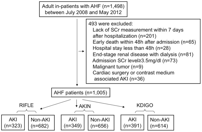
This retrospective cohort study was conducted at Guangdong General Hospital and the First Affiliated Hospital of Sun Yat-sen University in Guangzhou, China. We collected 1,498 adult patients (aged ≥18 years) hospitalized with acute heart failure (AHF) between July 2008 and July 2012. AHF was defined as either now-onset HF or decompensation of chronic HF with symptoms sufficient to warrant hospitalization. The diagnosis of AHF was based on European Society of Cardiology Criteria [12]. All patients had a New York Heart Association (NYHA) functional class of either Class III or IV. Only the first hospital admission was considered if a patient had more than one hospitalization for AHF during the study period. Patients were excluded if they met the following exclusion criteria: lack of SCr measurement during the first 7 days of hospitalization, early death within 48 h after admission, hospital stay <48 h, end-stage renal disease with dialysis, admission SCr level ≥3.5 mg/dl, malignant tumor, cardiac surgery- or contrast medium-associated AKI.
The Ethics Research Committee at Guangdong General Hospital and the First Affiliated Hospital of Sun Yat-sen University approved the study and agreed that informed consent was not necessary because of the purely observational nature of this study. Information of all patients was anonymized and de-identified prior to analysis.
Definition and Staging of AKI
We defined and staged AKI according to SCr-based criteria per the RIFLE, AKIN and KDIGO criteria within the first 7 days of hospitalization (Table 1). Here the RIFLE classification only included the three categories of severity but not the two categories of clinical outcome. AKI diagnosed by KDIGO criteria but missed by RIFLE or AKIN was defined as [K(+)R(−)+K(+)A(−)]. Urine output data were not available in this study. Baseline SCr was estimated from either the admission value (if this was within the normal range) or if available [13]–[14], from another value within 3 months, whichever was lowest [15]–[17]. Patients with SCr elevation at the time of admission but without SCr record in the previous 3 months before admission were considered having AKI, according to the use of estimation of SCr by backward calculation from the Modification of Diet in Renal Disease (MDRD) equation as recommended [11]. In this study, baseline SCr within 3 months before admission accounted for 27%. SCr was measured by the same means of a kinetic Jaffé method in the 2 hospitals.
Table 1. Diagnosis and staging criteria for AKI of RIFLE, AKIN, KDIGO definitions based on SCr.
| Classification | Definition for AKI | Stage | Serum Creatinine criteria for AKI staginga |
| RIFLE | Increase in SCr ≥50% within 7 d | Risk | To ≥1.5 times baseline |
| Injury | To ≥2 times baseline | ||
| Failure | To ≥3 times baseline or ≥44 µmol/L increase to at least 354 µmol/L | ||
| AKIN | Increase in SCr ≥26.5 µmol/L or ≥50% within 48 h | 1 | Increase of ≥26.5 µmol/L or to 1.5–2 times baseline |
| 2 | To 2–3 times baseline | ||
| 3 | To ≥3 times baseline or ≥26.5 µmol/L increase to at least 354 µmol/L or initiation of RRT | ||
| KDIGO | Increase in SCr ≥26.5 µmol/L within 48 h or ≥50% within 7 d | 1 | Increase in SCr ≥26.5 µmol/L within 48 h or to 1.5–2 times baseline |
| 2 | To 2–3 times baseline | ||
| 3 | To ≥3 times baseline or to at least 354 µmol/L or initiation of RRT |
For patients meeting diagnosis criteria for AKI according to RIFLE, AKIN or KDIGO, the stage based on percentage increase were determined by the ratio of peak SCr value obtained during hospitalization to baseline. AKI: Acute Kidney Injury; RIFLE: Risk Injury Failure Loss ESRD; AKIN: Acute Kidney Injury Network; KDIGO: Kidney Disease Global Outcomes; SCr: serum creatinine; RRT: renal replacement therapy.
Urine output was not used, because records of hourly urine output were not available in the majority of patients.
Data Collection
Data including the patients' demographics, medical history (smoking, diabetes, hypertension, coronary heart disease and previous AHF), physical examination, etiology of AHF, medication use and biochemical parameters (including SCr on admission, or if available, SCr within 3 months prior to admission), length of stay (LOS) in the ICU and in the hospital, requirement for renal replacement therapy (RRT), and date of death were obtained from the hospitals' computerized database. On admission, baseline estimated glomerular filtration rate (eGFR) was calculated using MDRD equation [18]. Left ventricular ejection fraction (LVEF) was evaluated by Doppler echocardiography during hospitalization.
Statistical analysis
Analyses were performed using the Statistical Package for Social Sciences software (SPSS, version 16.0). Continuous variables were expressed as means ± SDs or medians (with 25th and 75th percentiles) values, and were tested by the t test, Mann-Whitney U test or ANOVA, as appropriate. Categorical variables were described as numbers and percentages, and were compared using the chi-squared test. In-hospital mortality was estimated by the Kaplan-Meier method, and curves were compared with the log-rank test. Univariate and multivariate Cox proportional regression analyses were performed to assess the relationship between AKI and in-hospital mortality. Two-tailed P values <0.05 were considered significant.
Results
Study population characteristics
Of the total of 1,498 patients, 493 patients were excluded from analysis (Fig. 1.). The final studied cohort was composed of 1005 subjects. The mean age was (68.5±15.0) years, and 439(43.7%) were women. The commonest etiology of AHF was ischemic heart disease (IHD) (43.8%), followed by valvular disease (22.6%) and cardiomyopathy (12.8%). 32.7% had diabetes and 27.5% had previous IHD. The mean eGFR was (59.1±24.9)ml/min/1.73 m2 at baseline, and mean LVEF, (49.3±12.9)%. The median LOS for hospitalization was 12(IQR: 8–20) days. 23.9% patients were admitted to ICU during hospitalization. A total of 64 patients (6.4%) were treated with RRT for AKI during hospitalization. Baseline characteristics stratified by AKI according to RIFLE, AKIN and KDIGO classification was described in Table 2. AKI group had more diabetic patients, lower eGFR, hemoglobin, serum albumin, higher C-reactive protein (CRP), less ACEI/ARB medication, more diuretic use and longer LOS in ICU.
Figure 1. Schematic of study sample with exclusions from analysis.
Table 2. Demographic and clinical characteristics of patients with AHF according to the development of AKI by RIFLE, AKIN, KDIGO definitions during hospitalization.
| Characteristic | RIFLE | AKIN | KDIGO | |||
| No AKI (n = 682) | AKI (n = 323) | No AKI (n = 656) | AKI (n = 349) | No AKI (n = 614) | AKI (n = 391) | |
| Age(year) | 68.6±15.0 | 68.3±15.1 | 68.4±15.2 | 68.8±14.8 | 68.4±15.3 | 68.8±14.7 |
| Gender(Male) —n (%) | 408(59.8) | 158(48.9) * | 381(58.1) | 185(53.0) | 360(58.6) | 206(52.7) |
| Smoker—n (%) | 173(25.4) | 64(19.8) | 164(25.0) | 73(20.9) | 154(25.1) | 83(21.2) |
| Systolic BP (mmHg) | 136±28 | 134±30 | 135±27 | 135±32 | 135±28 | 135±31 |
| Diastolic BP (mmHg) | 78±16 | 76±18 | 78±16 | 76±18 | 77±16 | 77±18 |
| HR(beat/min) | 90±21 | 93±21* | 90±21 | 93±22 | 90±21 | 93±21 |
| LVEF(%) | 49.7±13.2 | 48.5±12.3 | 49.5±13.0 | 49.1±12.7 | 49.5±13.0 | 49.0±12.7 |
| Hypertension—n (%) | 446(65.4) | 203(62.8) | 422(64.3) | 227(65.0) | 395(64.3) | 254(65.0) |
| Diabetes—n (%) | 208(30.5) | 121(37.5) * | 194(29.6) | 135(38.7) * | 182(29.6) | 147(38.4) * |
| Ischemic heart disease—n (%) | 185(27.1) | 91(28.2) | 184(28.0) | 92(26.4) | 172(27.7) | 104(27.2) |
| Causes of AHF | ||||||
| Coronary artery disease | 296(43.4) | 144(44.6) | 296(45.1) | 144(41.3) | 270(44.0) | 170(43.5) |
| Valvular heart disease | 161(23.6) | 66(20.4) | 147(22.4) | 80(22.9) | 142(23.1) | 85(21.7) |
| Cardiomyopathy. | 89(13.0) | 40(12.4) | 83(13.1) | 46(13.2) | 79(12.9) | 50(12.8) |
| Hypertension | 81(11.9) | 43(13.3) | 75(11.4) | 49(14.0) | 72(11.7) | 52(13.3) |
| Congenital heart disease | 37(5.4) | 20(6.2) | 35(5.3) | 22(6.3) | 35(5.7) | 22(5.6) |
| Others | 18(2.6) | 10(3.1) | 18(2.7) | 8(2.3) | 16(2.6) | 12(3.1) |
| Admission eGFR (ml/min/1.73 m2) | 58.8±24.1 | 59.9±26.5 | 61.3±23.1 | 55.2±27.5* | 60.9±23.3 | 56.4±27.0* |
| Hemoglobin(g/L) | 121±25 | 112±26* | 123±24 | 110±27* | 122±24 | 112±26* |
| Serum albumin(g/L) | 32.7±5.9 | 31.7±6.1* | 32.8±5.8 | 31.5±6.1* | 32.8±5.9 | 31.6±6.1* |
| C-reactive protein(mg/L) | 16.0 (7.9–46.0) | 25.5* (11.8–67.0) | 15.5 (7.9–43.8) | 27.2* (12.0–72.1) | 15.5 (7.5–43.6) | 25.8* (11.8–68.3) |
| LDL-cholesterol(mmol/L) | 2.5±1.1 | 2.4±1.1 | 2.4±1.1 | 2.5±1.1 | 2.4±1.1 | 2.5±1.1 |
| ACEI/ARB medication—n (%) | 477(69.9) | 200(61.9) * | 469(71.5) | 208(59.6) * | 435(70.8) | 242(61.9) * |
| Diuretic medication—n (%) | 527(77.3) | 281(87.0) * | 510(77.7) | 298(85.4) * | 473(77.0) | 335(85.7) * |
| RRT—n (%) | 14(2.1) | 50(15.5) * | 11(1.7) | 53(15.2) * | 10(1.6) | 54(13.8) * |
| LOS in hospital (days) | 13(9–21) | 14(8–25) | 14(9–21) | 14(8–24) | 12(8–20) | 12(7–21) |
| LOS in ICU (days) | 7(3–11) | 6(3–12) * | 7(3–11) | 6(3–12) * | 7(3–11) | 6(3–12)* |
| ICU stay—n (%) | 106(15.5) | 134(41.5) * | 105(16.0) | 135(41.8)* | 88(14.3) | 152(38.9)* |
AHF: acute heart failure; AKI: acute kidney injury; BP: blood pressure; eGFR: estimated glomerular filtration rate; LVEF: left ventricular eject fraction; ACEI: angiotensin converting enzyme inhibitor; ARB: angiotensin receptor blocker; RRT: renal replacement therapy; LOS: length of stay; ICU: intensive care unit.
*:compared with NO-AKI group, P<0.05.
Identification of early CRS type 1
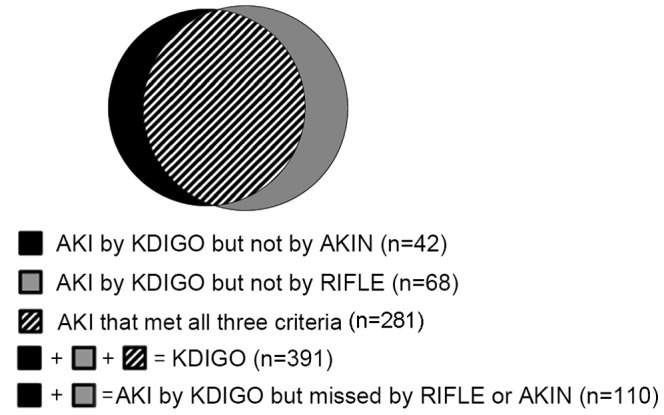
Within 7 days, CRS type 1 that could be diagnosed by all three criteria occurred in 28.0% (281/1005). Incidence of CRS type 1 according to RIFLE, AKIN and KDIGO criteria was 32.1%, 34.7% and 38.9%, respectively. A total of 110 (10.9%) cases were diagnosed by KDIGO but missed by RIFLE or AKIN criteria (Table 3). KDIGO classification identified 68 more cases (17.4% in KDIGO) than RIFLE did and 42 more cases (10.7% in KDIGO) than AKIN did. 89.1% (98/110) of them were in either Stage 1 of AKIN or Stage Risk of RIFLE. The distribution of AKI patients among RIFLE, AKIN and KDIGO criteria was depicted in Fig. 2. and Table 4. Compared with patients in No-AKI group (by any criteria), the missed diagnosed AKI patients had lower hemoglobin, higher level of serum uric acid, low density lipoprotein (LDL), CRP and longer LOS in ICU. 87.2% patients developed CRS type 1 within two days of hospitalization.
Table 3. Incidence of CRS type 1 according to the RIFLE, AKIN, KDIGO and K(+)R(−)+K(+)A(−) definitions.
| Classification | Incidence of AKI by all stages | Stage | Incidence of AKI by each stage |
| RIFLE | 323(32.1%) | Risk | 141(14.0%) |
| Injury | 116(11.5%) | ||
| Failure | 66(6.6%) | ||
| AKIN | 349(34.7%) | 1 | 157(15.6%) |
| 2 | 101(10.0%) | ||
| 3 | 91(9.1%) | ||
| KDIGO | 391(38.9%) | 1 | 194(19.3%) |
| 2 | 104(10.3%) | ||
| 3 | 93(9.3%) | ||
| K(+)R(−) + K(+)A(−)a | 110(10.9%) | 1 | 98(9.8%) |
| 2 | 3(0.3%) | ||
| 3 | 9(0.9%) |
CRS: cardiorenal syndrome;
: AKI diagnosed by KDIGO criteria but missed by RIFLE or AKIN criteria. AKI: Acute Kidney Injury.
Figure 2. AKI distribution among RIFLE, AKIN and KDIGO classification.

Table 4. Concordance of AKI designation.
| Definition | Comparison | No AKI by RIFLE or AKIN | AKI stage by RIFLE or AKIN | ||
| Risk/Stage 1 | Injury/Stage 2 | Failure/Stage 3 | |||
| KDIGO | RIFLE | ||||
| NO AKI | 614(61.1%) | 0 | 0 | 0 | |
| Stage 1 | 61(6.1%)a | 133(13.2%) | 0 | 0 | |
| Stage 2 | 0a | 0 | 104(10.3%) | 0 | |
| Stage 3 | 7(0.7%)a | 8(0.8%) | 12(1.2%) | 66(6.6%) | |
| KDIGO | AKIN | ||||
| NO AKI | 614(61.1%) | 0 | 0 | 0 | |
| Stage 1 | 37(3.7%)b | 157(15.6%) | 0 | 0 | |
| Stage 2 | 3(0.3%)b | 0 | 101(10.0%) | 0 | |
| Stage 3 | 2(0.2%)b | 0 | 0 | 91(9.1%) | |
AKI: acute kidney injury;
: K(+)R(−), AKI diagnosed by KDIGO criteria but not by RIFLE criteria.
: K(+)A(−), AKI diagnosed by KDIGO criteria but not by AKIN criteria.
AKI and in-hospital mortality
The in-hospital mortality rate was 13.5% (136 patients). Hospital mortality in AKI patients was 23.5% by RIFLE, 23.8% by AKIN, and 23.5% by KDIGO criteria. A total of 18.4% deaths (25/136) occurred in the additional AKI group, of which 16 cases (11.8% of the overall death) were in K(+)R(−) group and 9 cases (6.6% of the overall death) in K(+)A(−) group. Those deaths in added AKI group by KDIGO criteria were mostly in Stage 1.AKI patients by four classifications, either by all stages or by each stage, showed higher mortality than patients without AKI (P<0.05). Hospital mortality stratified by four definitions showed increasing trends with stage progression. However, only AKIN and KDIGO criteria showed significance when compared Stage 1 to Stage 3 or Stage 2 to Stage 3 (Table 5).
Table 5. In-hospital mortality stratified by AKI or No-AKI.
| Classification | AKI | No AKI |
| RIFLE(all stages) | 76(23.5%)* | 60(8.8%) |
| Risk | 27(19.1%)* | - |
| Injury | 28(24.1%)* | - |
| Failure | 21(31.8%)* # | - |
| AKIN(all stages) | 83(23.8%)* | 53(8.1%) |
| Stage 1 | 28(17.8%)* | - |
| Stage 2 | 22(21.8%)* | - |
| Stage 3 | 33(36.3%)* #& | - |
| KDIGO(all stages) | 92(23.5%)* | 44(7.2%) |
| Stage 1 | 36(18.6%)* | - |
| Stage 2 | 23(22.1%)* | - |
| Stage 3 | 33(35.5%)* #& | - |
| K(+)R(−) + K(+)A(−) | 25(22.7%)* | 44(7.2%) |
| Stage 1 | 20(20.4%)* | - |
| Stage 2 | 1(33.3%) | - |
| Stage 3 | 4(44.4%)* | - |
K(+)R(−)+K(+)A(−): AKI diagnosed by DIGO criteria but missed by RIFLE or AKIN.
*: compared with No-AKI group, P<0.05;
: compared with Stage 1 or Stage Risk, P<0.05;
: compared with Stage 2, P<0.05.
Prognostic value of different AKI definitions on in-hospital mortality
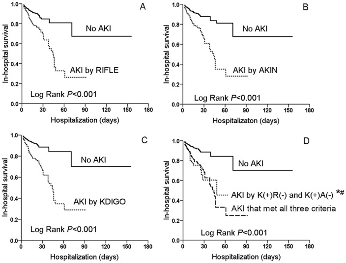
Kaplan-Meier curve showed AKI patients by RIFLE, AKIN, KDIGO and [K(+)R(−)+K(+)A(−)] definitions had lower hospital survival than non-AKI patients (Fig. 3., Log Rank P<0.001). After adjustment, the development of AKI by any definitions remained independently associated with in-hospital mortality. Odds ratio (OR) for RIFLE, AKIN, KDIGO and [K(+)R(−)+K(+)A(−)] was 2.56, 2,68, 4.00 and 3.24, respectively (P<0.05). By multivariable analysis, hospital mortality was also associated with stage progression by RIFLE, AKIN and KDIGO criteria (Table 6).
Figure 3. In-hospital survival of CRS type 1 according to RIFLE(A), AKIN(B), KDIGO(C) and K(+)R(−)+K(+)A(−) definitions(D).
*: vs No-AKI, P<0.001;#:vs AKI by all three criteria, P = 0.061.
Table 6. Cox proportional analysis for in-hospital mortality in AHF patients.
| Unadjusted OR (95%CI) | P Value | Adjusted ORc (95%CI) | P Value | |
| RIFLE by all stagesa | 2.66(1.89–3.73) | <0.001 | 2.56 (1.79–3.65) | <0.001 |
| RIFLE by each stage | ||||
| No AKI | reference | reference | ||
| Risk | 2.21(1.40–3.48) | 0.001 | 2.24(1.41–3.56) | 0.001 |
| Injury | 2.80(1.78–4.38) | <0.001 | 2.70(1.69–4.31) | <0.001 |
| Failure | 3.31(2.01–5.44) | <0.001 | 2.98(1.75–5.06) | <0.001 |
| AKIN by all stagesa | 3.01(2.13–4.26) | <0.001 | 2.68(1.86–3.84) | <0.001 |
| AKIN by each stage | ||||
| No AKI | reference | reference | ||
| 1 | 2.39(1.51–3.78) | <0.001 | 2.09(1.31–3.33) | 0.002 |
| 2 | 2.85(1.73–4.69) | <0.001 | 2.72(1.62–4.55) | <0.001 |
| 3 | 4.07(2.63–6.30) | <0.001 | 3.69(2.31–5.89) | <0.001 |
| KDIGO by all stagesa | 3.38(2.36–4.84) | <0.001 | 3.22 (2.24–4.62) | <0.001 |
| KDIGO by each stage | ||||
| No AKI | reference | reference | ||
| 1 | 2.80(1.80–4.35) | <0.001 | 2.68(1.72–4.16) | <0.001 |
| 2 | 3.26(1.96–5.40) | <0.001 | 3.05(1.82–5.09) | <0.001 |
| 3 | 4.52(2.87–7.12) | <0.001 | 4.33(2.75–6.82) | <0.001 |
| K(+)R(−) and K(+)A(−)b | 3.47 (2.12–5.68) | <0.001 | 3.24 (1.97–5.35) | <0.001 |
AHF: acute heart failure; K(+)R(−)/K(+)A(−): AKI diagnosed by KDIGO criteria but missed by RIFLE and AKIN; OR: odds ratio; AKI: acute kidney injury;
: including 1005 AHF patients;
: including 724 AHF patients (614 without AKI and 110 with AKI diagnosed by KDIGO criteria but missed by RIFLE and AKIN criteria);
: adjusted by gender, age, hemoglobin, serum albumin, medication with ACEI or ARB and diuretic and AKI.
Discussion
The reported incidence of AKI after AHF (usually referred to as CRS type 1) varies widely depending on the definition used as well as the etiologies. Several studies have examined AKI in the context of AHF before the first consensus RIFLE criteria was proposed. Using the term ‘worsening renal failure (WRF)’ to describe the acute and/or subacute changes in kidney function that occurs following AHF, incidence of WRF ranged from 23–45% in acute decompensated heart failure (ADHF), and 9–19% in acute coronary syndrome [2], [19]–[23]. This broad incidence is mainly attributed to disparities in the definition used for WRF, difference in the observed time at risk and heterogeneity of selected populations [24]. Besides, there is no consensus on how best to define WRF, with some studies utilizing the absolute and others using relative changes in SCr values.
Data investigating the incidence of CRS type 1 was scarce after recommendation of RIFLE criteria. Most epidemiologic AKI data were from the subset of critically ill patients, sepsis patients or patients after cardiac surgery [25]–[27]. Only three studies evaluated the incidence of early CRS type 1.One retrospective study reported the incidence of AKI was 33% (125/376) in ADHF patients upon admission [28], and the other was 68.8% (344/500) in AHF patients admitted to ICU [29]. Both of these studies evaluated AKI by RIFLE criteria. In the very recent prospective study including 637 AHF admissions, AKI by four definitions (RIFLE, AKIN, KDIGO and WRF) was assessed and compared. AKI occurred in 25.6% by RIFLE, 27.9% by AKIN, 36.7% patients by KDIGO, and 33.0% by WRF [30]. In the present study, 28.0% patients met all three AKI criteria, and incidence of AKI was 32.1%, 34.7% and 38.9% by RIFLE, AKIN and KDIGO criteria, respectively. Our results showed the consistent epidemiology of early CRS type 1 with previous data in which AHF patients were in general ward but not in ICU [28]–[30]. Furthermore, 10.9% patients in our study were classified as AKI in KDIGO but not in RIFLE or AKIN criteria. Most of them were predominantly staged in the lowest AKI severity (Stage 1). This can be explained by the principles of AKI in KDIGO criteria.
In KDIGO criteria, only an absolute SCr increase of 0.3 mg/dl within 48 hs is sufficient for an AKI diagnosis, whereas in RIFLE, a SCr increase of ≥50% from baseline is necessary [11]. Compared with AKIN, KDIGO have an extended time frame to diagnose AKI. The combined use of small absolute increases in SCr and enough observation time in the KDIGO criteria may potentially make it more sensitive than RIFLE or AKIN, which therefore undoubtedly identified additional early CRS type 1 cases. However, those added cases may be less severe with lower mortality risk. Identification of more AKI cases may on one hand reduce misclassification, but on the other hand, increase risk dilution.
An important strength of our study was that we separated the additional diagnosed AKI by KDIGO to determine if the expanded proportion could predict short-term outcome of AHF. The mortality of missed diagnosed AKI by RIFLE or AKIN accounted for 18.4% in the total death and had a significantly longer stay in ICU, indicating a large number of patients with CRS type 1 and elevated risk for mortality would be missed by the RIFLE or AKIN criteria.
Interestingly, although the mortality of patients with AKI classified by RIFLE, AKIN and KDIGO criteria in our study was similar, the 3 classifications classified individual patients differently. The percentage of patients who were identified as non-AKI by the AKIN classification but fulfilled the RIFLE criteria for AKI was 4.2% (42/1005), among which 21.4% died (9/42). By contrast, 6.8% (68/1005) of patients were classified as non-AKI according to the RIFLE criteria but fulfilled the AKIN criteria for AKI, among which 23.5% died (16/68). From this standpoint, AKIN criteria missed less risk patients than RIFLE. KDIGO criteria detected all those high risk patients additionally. Mortality of the combined AKI group was nearly three times that of patients who did not have AKI by any criteria (22.7% vs 7.2%). Even after adjustment for covariables, the additional diagnosed AKI remained an independent risk factor for in-hospital mortality. These results suggest that KDIGO is a useful tool in clinical practice to identify “true AKI” because those diagnosed by this classification but missed by RIFLE or AKIN increased in-hospital mortality. Our results were consistent with Rodrigues's prospective study enrolling 1050 patients with AMI [31]. They compared incidence and mortality of AKI after AMI between KDIGO and RIFLE criteria and found that KDIGO criteria detected substantially more AKI patients than RIFLE. Patients diagnosed as AKI by KDIGO but not RIFLE criteria had a significantly higher early and late mortality. The author suggested KDIGO criteria were more suitable for AKI diagnosis in AMI patients than RIFLE. Using 3 criteria to compare their prognostic power, studies on cardiac surgery-related to AKI got the same results [32]. However, Roy et al's results demonstrate that the RIFLE and KDIGO classification systems have only marginally superior prognostic ability when compared to WRF and AKIN to predict the composite outcomes (death, re-hospitalization and RRT) [29]. One potential explanations for this discrepancy maybe they present no analysis of those additional diagnosed cased by KDIGO criteria. Altogether, there is little evidence assessing and comparing the three criteria so far.
Similar to other studies, our study found those missed diagnosed AKI patients who died during hospitalization were mostly in Stage 1, demonstrating even slight changes in SCr would have impact on prognosis in CRS type 1 patients. Small changes in SCr have also been associated with early and long-term mortality in cardiothoracic surgery patients, cohorts of AMI patients as well as other hospitalized individuals [33]–[35].
Although our findings confirmed the helpful utilization of KDIGO criteria, there still remain specificity limitations [36]. An important one is how to determine the baseline kidney function in which this baseline is not known. Whereas RIFLE and KDIGO suggest the use of back calculation, AKIN recommends using the evolution of SCr relative to the first observed value in that episode. The lack of a uniform approach to estimate this baseline has been recently shown to compound risk for AKI misclassification, hindering effective comparisons of this disease between settings [37]–[39]. In our study, patients with elevated SCr on admission but without previous SCr data were considered AKI according to a presumed ‘standard GFR’ of 75 ml/min/1.73 m2 as the KDIGO guideline recommended [11]. Another explanation was that SCr often increased at severe AHF presentation because of the hypoinfusion of kidney. However, those patients may have chronic kidney disease or acute-on-chronic kidney injury, which may overestimate the incidence of AKI.
There are several potential limitations in our study. Firstly, only the SCr criteria of AKI classification was evaluated, because urine output was difficult to collect in the general wards, and on the other hand, urine output was influenced by the diuretic therapy administered to the majority of AHF patients. Secondly, this was a retrospective study and only short-term prognosis was analyzed. Although a retrospective study had revealed relationship between the long-term prognosis of AHF and AKI [35], a prospective study with long-term follow-up will better demonstrate the implication by different AKI criteria. However, our study was so far the largest cohort to investigate the epidemiology and prognosis of CRS type 1 by KDIGO criteria.
In conclusion, the present study provided further insight into the epidemiology of early CRS type 1 using the new criteria KDIGO and revealed the association between AKI diagnosed by KDIGO but not RIFLE or AKIN and short-come prognosis. KDIGO criteria identified more episode CRS type 1 and predicted hospital survival. The clinical application of the KDIGO AKI definition helped to increase the early recognition of AKI, allowing more individuals at high risk of AKI for intervention, indicating the new KDIGO criteria were superior to RIFLE and AKIN criteria in predicting short-term outcomes in CRS type 1.
Supporting Information
Information of enrolled 1,005 AHF patients.
(XLS)
Data Availability
The authors confirm that all data underlying the findings are fully available without restriction. Our data are available from the Guangdong General Hosptial and the First Affiliated Hospital of Sun Yat-sen University Data Access/Ethics Committee for researchers who meet the criteria for access to confidential data.
Funding Statement
This study was supported by grants from the National Natural Science Foundation (81170683 and 81200544), the Science and Technique Project of Guangzhou City (2013J4100064), the National Key Technology R&D Program (2011BAI10B08), and the National Clinical Key Specialty Construction Preparatory Projects, China. The funders had no role in study design, data collection and analysis, decision to publish, or preparation of the manuscript.
References
- 1. McAlister FA, Ezekowitz J, Tonelli M, Armstrong PW (2004) Renal insufficiency and heart failure: prognostic and therapeutic implications from a prospective cohort study. Circulation. 109:1004–1009. [DOI] [PubMed] [Google Scholar]
- 2. Cowie MR, Komajda M, Murray-Thomas T, Underwood J, Ticho B, et al. (2006) Prevalence and impact of worsening renal function in patients hospitalized with decompensated heart failure: results of the prospective outcomes study in heart failure (POSH). Eur Heart J. 27:1216–1222. [DOI] [PubMed] [Google Scholar]
- 3. Shirakabe A, Hata N, Kobayashi N, Shinada T, Tomita K, et al. (2012) Long-term prognostic impact after acute kidney injury in patients with acute heart failure. Int Heart J. 53:313–319. [DOI] [PubMed] [Google Scholar]
- 4. Ronco C, Cicoira M, McCullough PA (2012) Cardiorenal syndrome type 1: pathophysiological crosstalk leading to combined heart and kidney dysfunction in the setting of acutely decompensated heart failure. J Am Coll Cardiol. 60:1031–1042. [DOI] [PubMed] [Google Scholar]
- 5. Haase M, Müller C, Damman K, Murray PT, Kellum JA, et al. (2013) Pathogenesis of cardiorenal syndrome type 1 in acute decompensated heart failure: workgroup statements from the eleventh consensus conference of the Acute Dialysis Quality Initiative (ADQI). Contrib Nephrol. 182:99–116. [DOI] [PubMed] [Google Scholar]
- 6. Ismail Y, Kasmikha Z, Green HL, McCullough PA (2012) Cardio-renal syndrome type 1: epidemiology, pathophysiology, and treatment. Semin Nephrol. 32:18–25. [DOI] [PubMed] [Google Scholar]
- 7. Ronco C, McCullough PA, Chawla LS (2013) Kidney attack versus heart attack: evolution of classification and diagnostic criteria. Lancet. 382:939–940. [DOI] [PubMed] [Google Scholar]
- 8. Bellomo R, Ronco C, Kellum JA, Mehta RL, Palevsky P, et al. (2004) Acute renal failure - definition, outcome measures, animal models, fluid therapy and information technology needs: the Second International Consensus Conference of the Acute Dialysis Quality Initiative (ADQI) Group. Crit Care. 8:R204–212. [DOI] [PMC free article] [PubMed] [Google Scholar]
- 9. Mehta RL, Kellum JA, Shah SV, Molitoris BA, Ronco C, et al. (2007) Acute Kidney Injury Network: report of an initiative to improve outcomes in acute kidney injury. Crit Care. 11:R31. [DOI] [PMC free article] [PubMed] [Google Scholar]
- 10. Rodrigues FB, Bruetto RG, Torres US, Otaviano AP, Zanetta DM, et al. (2013) Incidence and mortality of acute kidney injury after myocardial infarction: a comparison between KDIGO and RIFLE criteria. PLoS One. 2013 8(7):e69998. [DOI] [PMC free article] [PubMed] [Google Scholar]
- 11.Kidney Disease: Improving Global Outcomes (KDIGO) Acute Kidney Injury Work Group (2012) KDIGO clinical practice guideline for acute kidney injury. Kidney Int Suppl 2: 1–138.
- 12. McMurray JJ, Adamopoulos S, Anker SD, Auricchio A, Böhm M, et al. (2012) ESC Guidelines for the diagnosis and treatment of acute and chronic heart failure 2012: The Task Force for the Diagnosis and Treatment of Acute and Chronic Heart Failure 2012 of the European Society of Cardiology. Developed in collaboration with the Heart Failure Association (HFA) of the ESC. Eur Heart J. 33:1787–1847. [DOI] [PubMed] [Google Scholar]
- 13. Joannidis M, Metnitz B, Bauer P, Schusterschitz N, Moreno R, et al. (2009) Acute kidney injury in critically ill patients classified by AKIN versus RIFLE using SAPS 3 database. Intensive Care Med. 35:1692–1702. [DOI] [PubMed] [Google Scholar]
- 14. Hsu CY, McCulloch CE, Fan D, Ordoñez JD, Chertow GM, et al. (2007) Community-based incidence of acute renal failure. Kidney Int. 72:208–212. [DOI] [PMC free article] [PubMed] [Google Scholar]
- 15. Zhou Q, Zhao C, Xie D, Xu D, Bin J, et al. (2012) Acute and acute-on-chronic kidney injury of patients with decompensated heart failure: impact on outcomes. BMC Nephrol. 13:51. [DOI] [PMC free article] [PubMed] [Google Scholar]
- 16. Siew ED, Matheny ME, Ikizler TA, Lewis JB, Miller RA, et al. (2010) Commonly used surrogates for baseline renal function affect the classification and prognosis of acute kidney injury. Kidney Int. 77:536–542. [DOI] [PMC free article] [PubMed] [Google Scholar]
- 17. Siew ED, Ikizler TA, Matheny ME, Shi Y, Schildcrout JS, et al. (2012) Estimating baseline kidney function in hospitalized patients with impaired kidney function. Clin J Am Soc Nephrol. 7:712–719. [DOI] [PMC free article] [PubMed] [Google Scholar]
- 18. Levey AS, Bosch JP, Lewis JB, Greene T, Rogers N, et al. (1999) A more accurate method to estimate glomerular filtration rate from serum creatinine: a new prediction equation. Modification of Diet in Renal Disease Study Group. Ann Intern Med. 130:461–470. [DOI] [PubMed] [Google Scholar]
- 19. Belziti CA, Bagnati R, Ledesma P, Vulcano N, Fernández S (2010) Worsening renal function in patients admitted with acute decompensated heart failure: incidence, risk factors and prognostic implications. Rev Esp Cardiol. 63:294–302. [DOI] [PubMed] [Google Scholar]
- 20. Metra M, Nodari S, Parrinello G, Bordonaili T, Bugatti S, et al. (2008) Worsening renal function in patients hospitalised for acute heart failure: clinical implications and prognostic significance. Eur J Heart Fail. 10:188–195. [DOI] [PubMed] [Google Scholar]
- 21. Goldberg A, Hammerman H, Petcherski S, Zdorovyak A, Yalonetsky S, et al. (2005) In-hospital and 1-year mortality of patients who develop worsening renal function following acute ST-elevation myocardial infarction. Am Heart J. 150:330–337. [DOI] [PubMed] [Google Scholar]
- 22. Parikh CR, Coca SG, Wang Y, Masoudi FA, Krumholz HM (2008) Long-term prognosis of acute kidney injury after acute myocardial infarction. Arch Intern Med. 168:987–995. [DOI] [PubMed] [Google Scholar]
- 23. Nohria A, Hasselblad V, Stebbins A, Pauly DF, Fonarow GC, et al. (2008) Cardiorenal interactions: insights from the ESCAPE trial. J Am Coll Cardiol. 51:1268–1274. [DOI] [PubMed] [Google Scholar]
- 24. Ostermann M, Chang RW (2011) Challenges of defining acute kidney injury. QJM 104:237–243. [DOI] [PubMed] [Google Scholar]
- 25. Lopes JA, Fernandes P, Jorge S, Goncalves S, Alvarez A, et al. (2008) Acute kidney injury in intensive care unit patients: a comparison between the RIFLE and the Acute Kidney Injury Network classifications. Crit Care 12:R110. [DOI] [PMC free article] [PubMed] [Google Scholar]
- 26. Chen YC, Jenq CC, Tian YC, Chang MY, Lin CY, et al. (2009) Rifle classification for predicting in-hospital mortality in critically ill sepsis patients. Shock. 31:139–145. [DOI] [PubMed] [Google Scholar]
- 27. Haase M, Bellomo R, Matalanis G, Calzavacca P, Dragun D, et al. (2009) A comparison of the RIFLE and Acute Kidney Injury Network classifications for cardiac surgery-associated acute kidney injury: a prospective cohort study. J Thorac Cardiovasc Surg. 138:1370–1376. [DOI] [PubMed] [Google Scholar]
- 28. Hata N, Yokoyama S, Shinada T, Kobayashi N, Shirakabe A, et al. (2010) Acute kidney injury and outcomes in acute decompensated heart failure: evaluation of the RIFLE criteria in an acutely ill heart failure population. Eur J Heart Fail. 12:32–37. [DOI] [PubMed] [Google Scholar]
- 29. Shirakabe A, Hata N, Kobayashi N, Shinada T, Tomita K, et al. (2012) Long-term prognostic impact after acute kidney injury in patients with acute heart failure. Int Heart J. 53:313–319. [DOI] [PubMed] [Google Scholar]
- 30. Roy AK, Mc Gorrian C, Treacy C, Kavanaugh E, Brennan A, et al. (2013) A Comparison of Traditional and Novel Definitions (RIFLE, AKIN, and KDIGO) of Acute Kidney Injury for the Prediction of Outcomes in Acute Decompensated Heart Failure. Cardiorenal Med. 3:26–37. [DOI] [PMC free article] [PubMed] [Google Scholar]
- 31. Rodrigues FB, Bruetto RG, Torres US, Otaviano AP, Zanetta DM, et al. (2013) Incidence and mortality of acute kidney injury after myocardial infarction: a comparison between KDIGO and RIFLE criteria. PLoS One. 8:e69998. [DOI] [PMC free article] [PubMed] [Google Scholar]
- 32. Sampaio MC, Máximo CA, Montenegro CM, Mota DM, Fernandes TR, et al. (2013) Comparison of diagnostic criteria for acute kidney injury in cardiac surgery. Arq Bras Cardiol. 101:18–25. [DOI] [PMC free article] [PubMed] [Google Scholar]
- 33. Zeng X, McMahon GM, Brunelli SM, Bates DW, Waikar SS (2014) Incidence, Outcomes, and Comparisons across Definitions of AKI in Hospitalized Individuals. Clin J Am Soc Nephrol. 9:12–20. [DOI] [PMC free article] [PubMed] [Google Scholar]
- 34. Lassnigg A, Schmidlin D, Mouhieddine M, Bachmann LM, Druml W, et al. (2004) Minimal changes of serum creatinine predict prognosis in patients after cardiothoracic surgery: a prospective cohort study. J Am Soc Nephrol. 15:1597–605. [DOI] [PubMed] [Google Scholar]
- 35. Praught ML, Shlipak MG (2005) Are small changes in serum creatinine an important risk factor? Curr Opin Nephrol Hypertens. 14:265–270. [DOI] [PubMed] [Google Scholar]
- 36. Lameire N (2013) The definitions and staging systems of acute kidney injury and their limitations in practice. Arab J Nephrol Transplant. 6:145–152. [PubMed] [Google Scholar]
- 37. Bagshaw SM, Uchino S, Cruz D, Bellomo R, Morimatsu H, et al. (2009) A comparison of observed versus estimated baseline creatinine for determination of RIFLE class in patients with acute kidney injury. Nephrol Dial Transplant. 24:2739–2744. [DOI] [PubMed] [Google Scholar]
- 38. Lafrance JP, Miller DR (2010) Defining acute kidney injury in database studies: the effects of varying the baseline kidney function assessment period and considering CKD status. Am J Kidney Dis. 56:651–660. [DOI] [PubMed] [Google Scholar]
- 39. Siew ED, Matheny ME, Ikizler TA, Lewis JB, Miller RA, et al. (2010) Commonly used surrogates for baseline renal function affect the classification and prognosis of acute kidney injury. Kidney Int. 77:536–542. [DOI] [PMC free article] [PubMed] [Google Scholar]
Associated Data
This section collects any data citations, data availability statements, or supplementary materials included in this article.
Supplementary Materials
Information of enrolled 1,005 AHF patients.
(XLS)
Data Availability Statement
The authors confirm that all data underlying the findings are fully available without restriction. Our data are available from the Guangdong General Hosptial and the First Affiliated Hospital of Sun Yat-sen University Data Access/Ethics Committee for researchers who meet the criteria for access to confidential data.


