Abstract
Background
The objective of this study was to test the effect of removal of a ureteral obstruction (renal calculus) from anesthetized patients on the perfusion index (PI), as measured by a pulse oximeter, and on the estimated glomerular filtration rate (eGFR).
Patients and Methods
This prospective study enrolled 113 patients with unilateral ureteral obstructions (kidney stones) who were scheduled for ureteroscopy (URS) laser lithotripsy. One urologist graded patient hydronephrosis before surgery. A pulse oximeter was affixed to each patient's index finger ipsilateral to the intravenous catheter, and a non-invasive blood pressure cuff was placed on the contralateral side. Ipsilateral double J stents and Foley catheters were inserted and left indwelling for 24 h. PI and mean arterial pressure (MAP) were determined at baseline, 5 min after anesthesia, and 10 min after surgery; eGFR was determined at admission, 1 day after surgery, and 14 days after surgery.
Results
Patients with different grades of hydronephrosis had similar age, eGFR, PI, mean arterial pressure (MAP), and heart rate (HR). PI increased significantly in each hydronephrosis group after ureteral stone disintegration. None of the groups had significant post-URS changes in eGFR, although eGFR increased in the grade I hydronephrosis group after 14 days. The percent change of PI correlates significantly with the percent change of MAP, but not with that of eGFR.
Conclusion
Our results demonstrate that release of a ureteral obstruction leads to a concurrent increase of PI during anesthesia. Measurement of PI may be a valuable tool to monitor the successful release of ureteral obstructions and changes of microcirculation during surgery. There were also increases in eGFR after 14 days, but not immediately after surgery.
Introduction
Modern anesthetic practice places increasing emphasis on changes in microcirculation following the initiation of newly developed devices, and this has led to improved organ perfusion and reduced post-operative morbidity [1]. The oximeter probe (Masimo Corp., Irvine, CA, USA) projects infrared light through the tissue bed of the finger tip and can assess peripheral perfusion. Recent studies have used this instrument to monitor peripheral vascular tone in pregnant woman and predict hypotension after spinal anesthesia [2], and to provide early prediction of successful brachial plexus block [3]. Based on previous studies, a change in the perfusion index (PI) is a rapid indicator of change in peripheral microcirculation, and this information may help anesthetists to make more appropriate treatment decisions [4]–[6].
Hydronephrosis can alter tubuloglomerular feedback and may lead to hypertension [7]. A 1974 animal study reported a decline in renal hemodynamics when experimental animals underwent 24 h of unilateral ureteral obstruction [8]. Moreover, Harris et al. [9] showed loss of excretory function in the post-obstructive kidney when tested by saline loading after release of 24-hocclusion. These results suggest that the impaired diuretic/natriuretic responses in the post-obstructive kidney might be due to reduced hemodynamics, and possibly to increased renal sympathetic nerve activity [10]. These effects change the pressure inside the glomerular capillaries [11], and may lead to a change in microcirculation.
In the present study, we used the oximeter probe to test the hypothesis that release of a ureteral obstruction (renal calculus) improves renal hemodynamics and microcirculation, and that these are promptly indicated by increases in the PI. We also examined the effect of removal of ureteral obstructions on the post-operative estimated glomerular filtration rate (eGFR).
Methods
This prospective study was approved by the National Taiwan University Hospital Ethics Committee (201205117RIC). (S1 and S2 Txt) One hundred and twenty-six patients diagnosed with ureteral stones were admitted to our department (National Taiwan University Hospital, Taipei, Taiwan) for ureteroscopy (URS) and laser lithotripsy between September 2012 and June 2013. One hundred and thirteen patients were ultimately enrolled. (S3 and S4 Txt) The exclusion criteria were: morbid obesity (BMI>40 kg/m2), peripheral arterial disease, usage of vasoactive agents, and contraindications to intravenous anesthesia. Although endothelial dysfunction may interfere the amplitude and velocity of changes in PI, we observed changes in PI for more than 5 min after stone disintegration, so this delayed effect can be ignored. There were 33 women and 80 men, the mean age was 53.7 years (range: 20–79 years), and American Society of Anesthesiologists Scores (ASA Scores) were between I and III. The grades of pre-operative hydronephrosis were classified according to Goertz JK and Lotterman S: grade I was defined as enlargement of the calyces with preservation of the renal papillae, grade II as rounding of the calyces with obliteration of the renal papillae, and grade III as calyceal ballooning with cortical thinning [12].
Study protocol
We obtained written informed consent from patients for enrollment at the pre-anesthesia visit on the day before surgery. No medications were given to patients before entering the operating room (OR). A urologist graded the severity of hydronephrosis by renal echography at the out-patient department (OPD) visit. After arrival at the OR, each patient was placed in a supine position and fitted with a non-invasive blood pressure cuff, a 3-lead electrocardiogram, and a pulse oximeter probe (Masimo Corp., Irvine, CA, USA) that was attached to the index finger on the ipsilateral side of intravenous catheter. After the signal stabilized, an average of five consecutive readings was recorded (baseline). After reaching a steady depth of anesthesia, the PI was recorded at 5-minintervals (pre-URS). A 6F/7.5F semi-rigid URS (Richard Wolf Medical Instruments Corporation, Vernon Hills, IL) was then introduced into the ureter retrogradely along with a safety guide wire to locate the ureteral stone. Then, the stone was disintegrated by use of a Holmium:YAG laser (Odyssey, Convergent Laser Technologies, Alameda, CA,USA). After stone disintegration, readings were defined as post-URS. Ureteral patency was re-examined by URS assure that there was no ureteral injury caused by laser lithotripsy. All procedures involved insertion of a 28F ureteral double J stent into the operated side. The success rate was 100% and the 16F Foley catheter remained in place for one day.
PI was calculated by measurement of the constant amount of light absorbed by non-pulsatile blood and other tissue (DC) and the variable amount of light absorbed by pulsating arterial inflow(AC)and use of the following equation: PI = (AC/DC)×100%. Anesthesia was administered as target-controlled infusion of propofol, and the blood concentration target was 5 µg/mL. Fentanyl was given at a concentration of 1 µg/kg body weight. Entropy values were maintained between 40 and 60 with adjustment of the target concentration of propofol by 0.5 µg/mL to achieve a steady state of sedation. The ambient temperature was 22°C and intravenous fluids (37°C) were infused at a rate of 500 mL/h.
Measurement of Serum Creatinine
Serum creatinine (SCr) was measured three times in all patients: at baseline (upon admission), one day after URS laser lithotripsy, and 14 days after URS laser lithotripsy. The eGFR was calculated as: 186×(Serum Cr)−1.154×(age)−0.203×(0.742 if female)×(1.210 if African- American) [13], [14].
Data analysis
Statistical analysis employed SPSS ver. 19, and data are presented as means ±SDs. The effect of sex and ASA score for patients with different hydronephrosis grades were examined by chi-square tests. Numerical data that had normal distributions and patient data were analyzed by one-way analysis of variance (ANOVA). Changes of variables in each hydronephrosis group were analyzed by repeated measure ANOVA. If there was a significant difference, a post-hoc Turkey HSD test was used to test for differences between groups. Pearson's correlation coefficient (γ) was used to assess the correlation between clinical variables or baseline parameters and the percent change in mean arterial pressure (MAP), PI, and eGFR. The interquartile range (IQR) was plotted relative to PI and MAP over different time periods. A p-value less than 0.05 was considered statistically significant.
Results
Table 1 shows the demographic data of patients with grade I, II, and III hydronephrosis. The 3 groups were similar with regard to age, sex distribution, and ASA score. We recorded MAP, HR, and PI during three time periods (baseline, pre-URS, and post-URS), and there were no significant differences among the groups at each time. The percentage change of PI from baseline to the pre-URS period (effect of anesthesia) and the pre-URS period to the post-URS period (effect of URS) also indicated no significant differences among the groups. eGFR among these groups also showed no difference between pre-URS and post-URS estimates. However, the eGFR at 14 days after surgery was significantly greater in patients with grade I hydronephrosis (p = 0.008).
Table 1. Demographic data and the degree of hydronephrosis.
| Hydronephrosis variables | Grade I (n = 53) | Grade II (n = 41) | Grade III (n = 19) | P value |
| Age (Yr) | 52 (25–73) | 54 (20–79) | 56 (33–75) | 0.46 |
| Gender | ||||
| Male/Female | 35/18 | 33/8 | 12/7 | 0.23 |
| eGFR(mL/min/1.73 m2) | ||||
| Pre-URS | 86.6+/−24.9 | 77.4+/−27.2 | 82.4+/−27.0 | 0.24 |
| Post-URS | 85.8+/−24.6 | 77.5+/−23.7 | 83.1+/−27.9 | 0.27 |
| 14 days | 91.7+/−25.9 | 79.4+/−27.6 | 83.0+/−30.1 | 0.008 |
| ASA score I/II/III | 10/39/4 | 6/27/8 | 2/14/3 | 0.45 |
| PI | ||||
| Baseline | 1.8+/−1.3 | 1.6+/−1.1 | 2.0+/−1.2 | 0.56 |
| %change | 309+/−54 | 250+/−29 | 195+/−34 | 0.326 |
| Pre-URS | 4.9+/−2.4 | 4.5+/−2.1 | 4.9+/−2.2 | 0.84 |
| %change | 66.8+/−14 | 67.3+/−25 | 50.2+/−14 | 0.86 |
| Post-URS | 6.8+/−2.4 | 6.5+/−2.7 | 6.6+/−2.6 | 0.83 |
| MAP (mm Hg) | ||||
| Baseline | 104.6+/−11.8 | 108.9+/−11.4 | 105.9+/−10.1 | 0.19 |
| Pre-URS | 91.4+/−11.6 | 94.3+/−10.9 | 93.2+/−13.4 | 0.46 |
| Post-URS | 90.1+/−11.5 | 93.1+/−14.0 | 95.3+/−19.2 | 0.31 |
| HR (beats min−1) | ||||
| Baseline | 72.6+/−12.9 | 71.3+/−14.5 | 77.1+/−12.4 | 0.30 |
| Pre-URS | 73.6+/−11.8 | 75.1+/−12.2 | 76.6+/−14.1 | 0.62 |
| Post-URS | 76.9+/−12.3 | 79.1+/−13.9 | 79.4+/−14.0 | 0.65 |
Legend 1.No significant difference among these groups. Only eGFR 14 days after URS showed significantly different.
Footnote 1.abbreviation: eGFR, estimated glomerular filtration rate; ASA, American Society of Anesthesiologists; MAP, Mean arterial pressure; HR, heart rate; URS, ureteroscopy.
Table 2 shows the changes in PI, MAP, and eGFR in the 3 hydronephrosis groups. Repeated measures ANOVA indicated that the PI values of each hydronephrosis group increased significantly over time (p<0.05 for all). In addition, Turkey HSD tests of the PI values indicated significant differences between baseline and pre-URS values, and between pre-URS and post-URS values (p<0.01 for all).The MAP also declined significantly in all 3 hydronephrosis groups (p<0.05 for all), but the difference between pre-URS and post-URS values were not significant when compared with post hoc test. The increase of eGFR was significant only in patients with grade I hydronephrosis (p<0.05), and the Turkey HSD test indicated significant differences only between pre-URS values and values at 14 days after surgery (p<0.05).
Table 2. The changes in PI, MAP, and eGFR in the 3 hydronephrosis groups. (compared with repeat measurement ANOVA and then Turkey HSD test each other).
| Grade 1 | Grade 2 | Grade 3 | ||
| Parameter | n = 53 | n = 41 | n = 19 | |
| PI | Baseline | 1.8 | 1.6 | 2.0 |
| Pre-URS | 4.9 | 4.5 | 4.9 | |
| Post-URS | 6.9 | 6.5 | 6.6 | |
| p value | p<0.051 | p<0.051 | p<0.051 | |
| MAP | Baseline | 104.6 | 108.9 | 105.9 |
| Pre-URS | 91.4 | 94.3 | 93.2 | |
| Post-URS | 90.1 | 93.1 | 95.3 | |
| p value | p<0.052 | p<0.052 | p<0.052 | |
| eGFR | Pre-URS | 86.6 | 77.4 | 82.4 |
| Post-URS | 85.8 | 77.5 | 83.1 | |
| 14 days | 91.7 | 79.4 | 83.0 | |
| p value | p<0.053 | p = 0.57 | p = 0.96 |
Legend 2. PI in three hydronephrosis groups increase significantly after anesthesia and increases further after releasing urinary obstruction.
Footnote 2. 1Baseline vs Pre-URS vs Post-URS PI in each hydronephrosis group: all significantly different when compared each other by Turkey HSD test (p<0.01);
Baseline vs Pre-URS, Baseline vs Post-URS MAP in each hydronephrosis group: significantly different when tested by Turkey HSD test (p<0.01), Pre-URS vs Post-URS MAP: not significantly different; 3Pre-URS vs 14 days, Post-URS vs 14 days eGFR in grade I hydronephrosis: significantly different (p<0.05).
Table 3 shows the relationships between percent changes in MAP, PI, and eGFR after stone disintegration for all patients which showed the effect of releasing obstruction on each parameters. The change ratios of MAP and PI were not significantly correlated with baseline PI, MAP, or eGFR. However, the percent change of eGFR14 days after surgery was negatively correlated with baseline eGFR (γ = −0.32, p<0.05). The change ratios of PI, MAP, and eGFR were also not significantly different in patients with different grades of hydronephrosis (one-way ANOVA). However, the change ratio of PI was significantly and inversely correlated with the change ratio of MAP (γ = −0.30, p<0.05).
Table 3. Relationships between baseline clinical parameters and the percent changes in MAP, PI, eGFR after stone disintegration.
| % MAP change# | % PI changes+ | % eGFR changes↑ | ||||
| Γ | P value | γ | P value | γ | P value | |
| Baseline MAP (mmHg) | −0.12 | 0.17 | 0.13 | 0.14 | −0.12 | 0.20 |
| Baseline PI | −0.01 | 0.87 | −0.13 | 0.17 | 0.00 | 0.98 |
| Baseline eGFR (mL/min/1.73 m2) | 0.02 | 0.77 | 0.17 | 0.06 | −0.32 | <0.05 |
| Grade of hydronephrosis | 0.52 | 0.86 | 0.56 | |||
| γ | P value | |||||
| % MAP change# % PI changes+ | −0.30 | <0.05 | ||||
| % PI changes+ % eGFR changes↑ | −0.10 | 0.33 | ||||
| % eGFR changes↑ % MAP change# | 0.09 | 0.30 | ||||
Legend 3. Percent changes in MAP, PI, and eGFR have no correlation with baseline MAP, PI, and eGFR. There was also no significant difference in percent change in PI, MAP, and eGFR between each grade of hydronephrosis. Percent change of MAP has negative correlation with percent change of PI.
Footnote 3. #Percent change of MAP: (post-URS MAP-pre-URS MAP)/pre-URS MAP; +Percent change of PI: (post-URS PI-pre-URS PI)/pre-URS PI; ↑Percent change of eGFR: (14 days eGFR-pre-URS eGFR)/pre-URS eGFR.
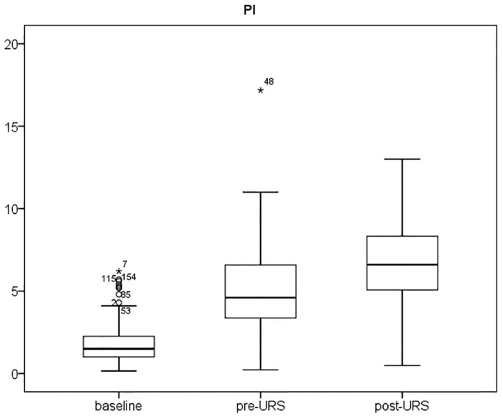
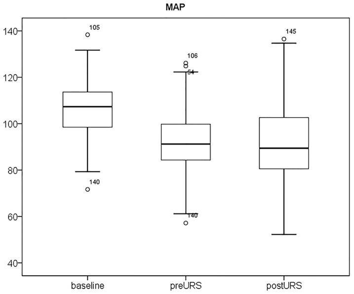
Figs. 1 and 2 show the PI and MAP at the 3 time periods. PI increased after intravenous anesthesia and increased further after stone disintegration. MAP decreased after induction of anesthesia, but there was no further change during stone evacuation.
Figure 1. Interquartile range (IQR) of Perfusion Index (PI) in patients with Grade I, II, and III hydronephrosis.
The PI increased after induction of anesthesia, and increased further after stone disintegration.
Figure 2. Interquartile range (IQR) of mean arterial pressure (MAP) in patients with Grade I, II, and III hydronephrosis.
The MAP decreased after induction of anesthesia, but there were no further changes during stone evacuation.
Discussion
Calculation of the PI by pulse oximetry provides a measure of changes in peripheral perfusion in the finger. In particular, the PI can predict postoperative shivering [15] and is an early predictor of survival after resuscitation [16]. Based on these previous studies, we conclude that changes in the PI are related to changes in peripheral microcirculation, and these are correlated with vascular status, sympathetic reactions, and function of the circulatory system.
In the present study, anesthesia and surgery of patients with renal calculi increased the measured PI in all hydronephrosis groups. Propofol relaxes vessel tone and decreases sympathetic reactions, resulting in an increase of the PI [17], and this is the likely cause of the 195–309% increase of PI in our patients after anesthesia. However, patients in all 3 of our hydronephrosis groups experienced a further 50.2–67.3% increase of PI after ureteral stone destruction, even though the level of sedation remained steady. Moreover, while PI increased after stone destruction, the MAP did not change significantly. This phenomenon was first discovered in this study, and it supports our hypothesis that release of ureteral obstruction by URS and laser lithotripsy increases microcirculation during anesthesia and that these changes can be measured by calculation of PI from a pulse oximeter. Our patients had no active changes in sympathetic tone when under anesthesia, and changes in body temperature can be ignored because of the short duration of surgery. The sudden drop of intra-renal pressure following URS laser lithotripsy is a key factor explaining the subsequent increase of the PI. Stone disintegration and the reestablishment of ureteral patency by an indwelling double-J catheter affects the circulatory system, possibly by free radicals and release of cytokines [18], [19], and this leads to microcirculatory changes that the oximeter measures concurrently. The circulatory system can be regarded as a pump system that pumps blood from the heart via the vasculature to each visceral organ. Any change in this system, including cardiac disease (pump dysfunction), vascular disease (circuit impairment), or renal obstruction(outflow stasis), may affect hemodynamic integrity and result in microcirculatory change [20].
Devarajan [21] suggested that serum creatinine should not be used as an indicator of rapid changes in kidney function because its concentration only accurately reflects kidney function in the steady state. Our comparison of patients with different grades of hydronephrosis indicated that only patients with grade I hydronephrosis had significant increases in eGFR at 14 days after URS. This indicates that the degree of hydronephrosis does not always correlate with renal function, and that eGFR may not be useful for evaluation of surgical success.
We reported the impact of changes in peripheral microcirculation induced by restoration of ureteral patency and urine flow as the percent change of PI (Table 3). The percent change of PI in the 3 hydronephrosis groups were not significantly different, suggesting no correlation between microcirculatory changes with hydronephrosis severity following stone disintegration. However, the percentage change of MAP negatively correlated with the percent change of PI (γ = −0.3, p<0.05). This may be because changes in microcirculation mainly affected the distal arterioles (which is innervated by the sympathetic system) and the smooth muscle tone in arterioles, and these preceded the observed change in MAP [22].
There can be great individual variations in PI, and several factors may interfere with measurement of PI [23], [24], so many previous studies used the pleth variabililty index (PVI) instead of PI to estimate volume status [25]–[27]. Nonetheless, our results suggest that PI can be used to assess changes in microcirculation in the perioperative period. The non-invasive nature of pulse oximetry makes it easy to use, and also makes it easy to gather information on changes in peripheral microcirculation [28], [29]. In addition, PI can indicate early changes in the sympathetic system and microperfusion in debilitated patients [30]. In this study, we successfully documented changes in microcirculation via PI following removal of ureteral obstructions. An animal study of the physiology of changes in hydronephrosis and relief of obstructive uropathy should be established to further examine the mechanisms of this effect.
In conclusion, our results showed that measurement of PI by a pulse oximeter allows monitoring of changes in peripheral microcirculation during endoscopic surgery for unilateral ureteral obstruction, but that eGFR did not change immediately after stone destruction. Use of a pulse oximeter to measure PI is simple and non-invasive, and provides important information regarding microperfusion in surgical patients. Thus, this method may improve patient safety and help clinicians to make important and prompt decisions while in the operating room.
Supporting Information
IRB protocol.
(DOCX)
IRB protocol in Chinese.
(DOCX)
CONSORT diagram.
(DOC)
STORBE checklist for cross-sectional study.
(DOC)
Acknowledgments
Dr. Ho-Shiang Huang moved from the Department of Urology, National Taiwan University Hospital to the Department of Urology, National Cheng Kung University Hospital, College of Medicine, National Cheng Kung University in August, 2013.
Funding Statement
The authors have no funding or support to report.
References
- 1. Turek Z, Sykora R, Matejovic M, Cerny V (2009) Anesthesia and the microcirculation. Seminars in Cardiothoracic & Vascular Anesthesia 13:249–258. [DOI] [PubMed] [Google Scholar]
- 2. Toyama S, Kakumoto M, Morioka M, Matsuoka K, Omatsu H, et al. (2013) Perfusion index derived from a pulse oximeter can predict the incidence of hypotension during spinal anaesthesia for Caesarean delivery. British Journal of Anaesthesia 111:235–241. [DOI] [PubMed] [Google Scholar]
- 3. Kus A, Gurkan Y, Gormus SK, Solak M, Toker K (2013) Usefulness of perfusion index to detect the effect of brachial plexus block. J Clin Monit Comput 27:325–328. [DOI] [PubMed] [Google Scholar]
- 4. van Genderen ME, Lima A, Bakker J, van Bommel J (2013) [Peripheral circulation in critically ill patients: non-invasive methods for the assessment of the peripheral perfusion]. Ned Tijdschr Geneeskd 157:A5338. [PubMed] [Google Scholar]
- 5. Ginosar Y, Weiniger CF, Meroz Y, Kurz V, Bdolah-Abram T, et al. (2009) Pulse oximeter perfusion index as an early indicator of sympathectomy after epidural anesthesia. Acta Anaesthesiol Scand 53:1018–1026. [DOI] [PubMed] [Google Scholar]
- 6. Mowafi HA, Ismail SA, Shafi MA, Al-Ghamdi AA (2009) The efficacy of perfusion index as an indicator for intravascular injection of epinephrine-containing epidural test dose in propofol-anesthetized adults. Anesth Analg 108:549–553. [DOI] [PubMed] [Google Scholar]
- 7. Chalisey A, Karim M (2013) Hypertension and hydronephrosis: rapid resolution of high blood pressure following relief of bilateral ureteric obstruction. J Gen Intern Med 28:478–481. [DOI] [PMC free article] [PubMed] [Google Scholar]
- 8. Harris RH, Yarger WE (1974) Renal function after release of unilateral ureteral obstruction in rats. Am J Physiol 227:806–815. [DOI] [PubMed] [Google Scholar]
- 9. Harris KP, Purkerson ML, Klahr S (1991) The recovery of renal function in rats after release of unilateral ureteral obstruction: the effects of moderate isotonic saline loading. Eur J Clin Invest 21:339–343. [DOI] [PubMed] [Google Scholar]
- 10. Ma MC, Huang HS, Chen CF (2002) Impaired renal sensory responses after unilateral ureteral obstruction in the rat. J Am Soc Nephrol 13:1008–1016. [DOI] [PubMed] [Google Scholar]
- 11. Otani Y, Otsubo S, Kimata N, Takano M, Abe T, et al. (2013) Effects of the Ankle-brachial Blood Pressure Index and Skin Perfusion Pressure on Mortality in Hemodialysis Patients. Intern Med 52:2417–2421. [DOI] [PubMed] [Google Scholar]
- 12. Goertz JK, Lotterman S (2010) Can the degree of hydronephrosis on ultrasound predict kidney stone size? American Journal of Emergency Medicine 28:813–816. [DOI] [PubMed] [Google Scholar]
- 13.Levey AS, Bosch JP, Lewis JB, Greene T, Rogers N, et al. (1999) A more accurate method to estimate glomerular filtration rate from serum creatinine: A new prediction equation. Annals of Internal Medicine 130:: 461–+. [DOI] [PubMed] [Google Scholar]
- 14. Levey AS, Stevens LA, Schmid CH, Zhang YP, Castro AF, et al. (2009) A New Equation to Estimate Glomerular Filtration Rate. Annals of Internal Medicine 150:604–U607. [DOI] [PMC free article] [PubMed] [Google Scholar]
- 15.Kuroki C, Godai K, Hasegawa-Moriyama M, Kuniyoshi T, Matsunaga A, et al. (2013) Perfusion index as a possible predictor for postanesthetic shivering. J Anesth. [DOI] [PubMed]
- 16. He HW, Liu DW, Long Y, Wang XT (2013) The peripheral perfusion index and transcutaneous oxygen challenge test are predictive of mortality in septic patients after resuscitation. Crit Care 17:R116. [DOI] [PMC free article] [PubMed] [Google Scholar]
- 17. Takeyama M, Matsunaga A, Kakihana Y, Masuda M, Kuniyoshi T, et al. (2011) Impact of skin incision on the pleth variability index. J Clin Monit Comput 25:215–221. [DOI] [PubMed] [Google Scholar]
- 18. Huang HS, Chen J, Chen CF, Ma MC (2006) Vitamin E attenuates crystal formation in rat kidneys: roles of renal tubular cell death and crystallization inhibitors. Kidney Int 70:699–710. [DOI] [PubMed] [Google Scholar]
- 19. Huang HS, Ma MC, Chen J (2009) Low-vitamin E diet exacerbates calcium oxalate crystal formation via enhanced oxidative stress in rat hyperoxaluric kidney. Am J Physiol Renal Physiol 296:F34–45. [DOI] [PubMed] [Google Scholar]
- 20. Auer J, Robert B, Eber B (2001) Peripheral and myocardial microcirculation. Circulation 104:E100. [PubMed] [Google Scholar]
- 21.Devarajan P (2008) Neutrophil gelatinase-associated lipocalin (NGAL): a new marker of kidney disease. Scand J Clin Lab Invest Suppl 241 89–94. [DOI] [PMC free article] [PubMed]
- 22. Lima A, van Genderen ME, Klijn E, Bakker J, van Bommel J (2012) Peripheral vasoconstriction influences thenar oxygen saturation as measured by near-infrared spectroscopy. Intensive Care Med 38:606–611. [DOI] [PMC free article] [PubMed] [Google Scholar]
- 23. Monnet X, Guerin L, Jozwiak M, Bataille A, Julien F, et al. (2013) Pleth variability index is a weak predictor of fluid responsiveness in patients receiving norepinephrine. Br J Anaesth 110:207–213. [DOI] [PubMed] [Google Scholar]
- 24. Mousa WF (2013) Effect of hypercapnia on pleth variability index during stable propofol: Remifentanil anesthesia. Saudi J Anaesth 7:234–237. [DOI] [PMC free article] [PubMed] [Google Scholar]
- 25. Broch O, Bein B, Gruenewald M, Hocker J, Schottler J, et al. (2011) Accuracy of the pleth variability index to predict fluid responsiveness depends on the perfusion index. Acta Anaesthesiol Scand 55:686–693. [DOI] [PubMed] [Google Scholar]
- 26. Hood JA, Wilson RJ (2011) Pleth variability index to predict fluid responsiveness in colorectal surgery. Anesth Analg 113:1058–1063. [DOI] [PubMed] [Google Scholar]
- 27. Tsuchiya M, Yamada T, Asada A (2010) Pleth variability index predicts hypotension during anesthesia induction. Acta Anaesthesiol Scand 54:596–602. [DOI] [PubMed] [Google Scholar]
- 28. Atef HM, Fattah SA, Gaffer ME, Al Rahman AA (2013) Perfusion index versus non-invasive hemodynamic parameters during insertion of i-gel, classic laryngeal mask airway and endotracheal tube. Indian J Anaesth 57:156–162. [DOI] [PMC free article] [PubMed] [Google Scholar]
- 29. Yamazaki H, Nishiyama J, Suzuki T (2012) Use of perfusion index from pulse oximetry to determine efficacy of stellate ganglion block. Local Reg Anesth 5:9–14. [DOI] [PMC free article] [PubMed] [Google Scholar]
- 30. van Genderen ME, van Bommel J, Lima A (2012) Monitoring peripheral perfusion in critically ill patients at the bedside. Curr Opin Crit Care 18:273–279. [DOI] [PubMed] [Google Scholar]
Associated Data
This section collects any data citations, data availability statements, or supplementary materials included in this article.
Supplementary Materials
IRB protocol.
(DOCX)
IRB protocol in Chinese.
(DOCX)
CONSORT diagram.
(DOC)
STORBE checklist for cross-sectional study.
(DOC)


