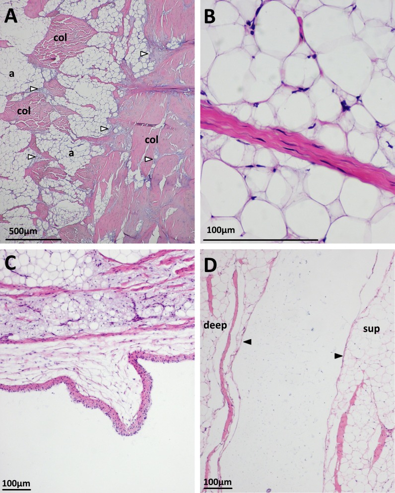
Figure 5. Some histological features of the patellar tendon in emus.
(A) Patellar tendon of an 18 month old emu. Collagen bundles (col) run along the superficial surface (right of image), interspersed with basophilic vesicular and chondroid tissue (unfilled arrows). The body of the tendon is mostly composed of adipocytes (a) and collagen bundles (col) in mixed orientations. Vesicular/chondroid tissue, when present in the body of the tendon, is associated with collagen fibre bundles. H&E. (B) Slender tenocytes within the crimped collagen fibre bundles. H&E. (C) Patellar tendon of a five week old emu, displaying a synovial villus on the deep surface, close to the sagittal midline of the tendon. H&E. (D) Patellar tendon of an 18 month old emu. The edges of the superficial (sup) and deep (deep) portions of the distal tendon have a lining layer of cells (filled arrows). H&E.

