Abstract
The pathogenesis of nonalcoholic steatohepatitis (NASH) is a two-stage process in which steatosis is the “first hit” and an unknown “second hit.” We hypothesized that “a binge” could be a “second hit” to develop NASH from obesity-induced simple steatosis. Thirty-week-old male Otsuka Long-Evans Tokushima fatty (OLETF) rats were administered 10 mL of 10% ethanol orally for 5, 3, 2, and 1 d/wk for 3 consecutive weeks. As control, male Otsuka Long-Evans Tokushima (OLET) rats were administered the same amount of alcohol. Various biochemical parameters of obesity, steatosis and NASH were monitored in serum and liver specimens in untreated and ethanol-treated rats. The liver sections were evaluated for histopathological alterations of NASH and stained for cytochrome P-4502E1 (CYP2E1) and 4-hydroxy-nonenal (4-HNE). Simple steatosis, hyperinsulinemia, hyperglycemia, insulin resistance, hypertriglycemia and marked increases in hepatic CYP2E1 and 4-HNE were present in 30-wk-old untreated OLETF rats. Massive steatohepatitis with hepatocyte ballooning was observed in the livers of all OLETF rats treated with ethanol. Serum and hepatic triglyceride levels as well as tumor necrosis factor (TNF)-α mRNA were markedly increased in all ethanol-treated OLETF rats. Staining for CYP2E1 and 4-NHE demonstrated marked increases in the hepatic tissue of all the groups of OLETF rats treated with ethanol compared with OLET rats. Our data demonstrated that “a binge” serves as a “second hit” for development of NASH from obesity-induced simple steatosis through aggravation of oxidative stress. The enhanced levels of CYP2E1 and increased oxidative stress in obesity play a significant role in this process.
INTRODUCTION
In 1980, Ludwig et al. (1) introduced the term “nonalcoholic steatohepatitis” (NASH), and subsequently the more embracing term “nonalcoholic fatty liver disease” (NAFLD) was established to cover the full spectrum of hepatic steatosis associated with insulin resistance and the metabolic syndrome (2). Alcoholic liver disease (ALD) and NAFLD are histologically indistinguishable. To distinguish between ALD and NAFLD, a cutoff limit for alcohol consumption was introduced. In general, an intake of <140 g ethanol per week or 20 g ethanol/day is acceptable for the diagnosis as NAFLD (3,4).
The molecular mechanisms involved in pathogenesis of NASH are not clear. Day and James (5) proposed a “two-hit” model to explain the progression of NASH from simple steatosis. The “first hit” constitutes the deposition of triglycerides in the cytoplasm of the hepatocyte. The disease does not progress to NASH unless additional molecular events occur (the “second hit”) that result in extensive steatosis, hepatitis, fibrosis and cell death, which are the histological hallmarks of NASH (6). The “first hit,” macrovesicular steatosis, results from increased uptake and synthesis of fatty acids in liver (7). However, steatosis alone does not appear to be progressive and occurs in many individuals who may never develop signs of progressive liver injury or NASH (4,8). The “second hit” is generally attributed to oxidative stress that triggers lipid peroxidation of hepatocyte membrane (9). This process results in production of proinflammatory cytokines and triggers activation of hepatic stellate cells, which initiate liver fibrosis (10,11). Lipid peroxidation and generation of reactive oxygen species (ROS) can also directly and adversely affect hepatocytes, resulting in necrosis and cell death (12). Increased levels of insulin and fatty acid content have an impact on ROS-mediated cell injury by catalyzing lipid peroxidation either through cytochrome P4502E1 (CYP2E1) or CYP4A (13,14) and by inhibiting mitochondrial oxidation of lipids (15).
In NASH, diverse etiologies can give rise to the same histological features as of alcoholic steatohepatitis (ASH). In both human and experimental animals, prolonged intake of ethanol induces hepatic CYP2E1 (16,17). The increased expression of CYP2E1 results in oxidative stress and production of ROS. The toxic metabolites of ethanol along with ROS may contribute to the development of steatohepatitis (17–19). In both human and rat liver, CYP2E1 is expressed predominantly in acinar zone 3 (18). Induction of CYP2E1 by ethanol is associated with increased expression of the enzyme in zone 3 and also spreads into zones 2 and 1 (18). It was observed that upregulation of CYP2E1 in simple steatosis is associated with diabetes mellitus and obesity (13,20). The increased activity of CYP2E1 in simple steatosis leads to tissue oxidative stress and production of ROS (6,21). When an obese individual consumes 20 g ethanol/day or 140 g ethanol/week, ROS along with the toxic metabolites of ethanol could contribute toward development of NASH. Ingestion of ethanol could further upregulate the expression and activity of CYP2E1 to metabolize ethanol in obese individuals.
The Otsuka Long-Evans Tokushima fatty (OLETF) rat is a useful animal model to study the biochemical, physiological and pathological alterations associated with insulin resistance, obesity and associated hepatic steatosis (22,23). OLETF rats spontaneously develop insulin resistance and steatosis from 25 to 30 wks of age (22). The biochemical and histopathological alterations observed in adult OLETF rats during obesity and steatosis are similar to the hepatic fatty degeneration present in obese individuals (24). OLETF rats have a deletion in the gene encoding cholecystokinin (CCK)-1 receptor, making the OLETF rat a CCK-1 receptor knockout model (25). CCK, the brain-gut peptide, inhibits food intake by reducing the size and duration of a meal, and its inhibitory actions are mediated through CCK-1 receptors. The aim of the present investigation was to study whether “a binge” or occasional alcohol intake could serve as the “second hit” to develop NASH from obesity-induced simple steatosis and also the molecular events involved in this process. To examine this, we used OLETF rats, which serve as a suitable animal model to study the molecular mechanisms associated with the development of NASH from simple steatosis.
MATERIALS AND METHODS
Animals
All animal experiments were carried out with the Guide for the Care and Use of Laboratory Animals published by the National Academy Press (26). The protocol was also approved by the Animal Care and Research Committee of Kanazawa Medical University on the Ethics of Animal Experiments. OLETF rats, a well-known animal model for type 2 diabetes mellitus and obesity, were procured from Otsuka Pharmaceuticals (Tokushima, Japan). OLETF rats spontaneously develop obesity, hyperglycemia, hyperinsulinemia and insulin resistance during their lifespan. OLETF rats depict insulin resistance at about the age of 10–15 wks and type 2 diabetes mellitus at the age of 25–30 wks (22). Otsuka Long-Evans Tokushima (OLET) rats, which do not develop diabetes mellitus spontaneously, were used as controls. Five-week-old male OLETF and OLET rats were housed in stainless-steel wire mesh cages at 23 ± 2°C with a relative humidity of 50 ± 10% in 12-h light–dark cycles and 10–15 air changes per hour. All animals had access to sterile drinking water ad libitum throughout the experiment. Both OLETF and OLET rats were sacrificed at the age of 10, 20 and 30 wks to evaluate any histopathological changes, auto-induction of CYP2E1 through aging and related oxidative stress.
Western Blotting for CYP2E1
The effect of aging on autoinduction of CYP2E1 in OLETF and OLET rats were evaluated by Western blotting and immunohistochemical staining for CYP2E1. About 100 mg liver tissue was homogenized in 1 mL ice-cold 50 mmol/L Tris-HCl buffer (pH 8) containing 150 mmol/L NaCl, 1 mmol/L EDTA and 1% Triton X-100. The protease inhibitors 0.5 mmol/L phenylmethylsulfonyl fluoride (PMSF), 5 μg/mL aprotinin and 1 μg/mL pepstatin were added to the buffer just before use. The homogenate was centrifuged at 10,000g for 10 min at 4°C, and the supernatant was collected. The protein concentration in the supernatant was determined by using the Coomassie plus protein assay (Pierce Biotechnology, Rockford, IL, USA), and the samples were stored at −20°C until assayed. The proteins were denatured and resolved on 4–20% sodium do-decyl sulfate–polyacrylamide gradient gel (Bio-Rad, Hercules, CA, USA) and electroblotted to an activated polyvinylidene fluoride (PVDF) membrane (Millipore, Bedford, MA, USA). Nonspecific binding sites were blocked with 5% nonfat dry milk, and the membrane was incubated overnight on a rocker at 4°C with CYP2E1 polyclonal antibody raised in rabbit against rat full-length native protein as immunogen (#ab28146; Abcam, Cambridge, MA, USA). After incubation, the membrane was washed three times and treated with horseradish peroxidase–conjugated rabbit second antibody (Biomeda, Foster City, CA, USA) at room temperature for 2 h. The membrane was washed again, treated with enhanced chemiluminescence reagent (GE Healthcare, Piscataway, NJ, USA), exposed to Kodak autoradiography film (BioMax XAR, New Haven, CT, USA) and developed. The membranes were reprobed by using Western ReProbe buffer (G-Biosciences, St. Louis, MO, USA) with a monoclonal antibody to GAPDH (Novus Biologicals, Littleton, CO, USA) that served as a control protein. The Western blotting images were quantified by using Gel-Pro analyzer software (Media Cybernetics, Silver Spring, MD, USA).
Administration of Alcohol
Thirty-week-old male OLETF rats (620 ± 15 g) were administered 10 mL of 10% ethanol or water by using an intragavage tube for 5, 3, 2 and 1 d/wk for 3 consecutive weeks on a fixed time schedule. As the treated control, 30-wk-old male OLET rats (460 ± 10 g) were administered the same amount of ethanol or water concurrently. All the animals were sacrificed on d 21 from the beginning of exposure under ether anesthesia and blood, and livers were collected immediately. The liver tissue was instantly weighed, and a small portion of the tissue from the center of the lobe (about 100 mg) was cut into small pieces and fixed in RNAlater solution (Life Technologies, Tokyo, Japan) and stored at −20°C for polymerase chain reaction (PCR) studies. The median lobe of the liver tissue was cut into 3-mm pieces and instantly fixed in 10% phosphate-buffered formalin for histopathological and immunohistochemical studies. The remaining liver tissue was flash-frozen in liquid nitrogen and stored at −80°C for protein analysis.
Measurement of ALT, AST, γ-GTP, Total Cholesterol, Triglycerides, Glucose, Adiponectin and Insulin in Serum
Blood was allowed to clot for 3–5 h at 37°C, and the serum was separated in the conventional method. Serum alanine transaminase (ALT) and aspartate transaminase (AST) and glucose were measured using an auto-analyzer. Serum γ-glutamyl transpeptidase (γ-GTP) was determined by using l-γ-glutamyl-3-carboxy-4-nitroanilide according the method of Theodorsen and Stromme (27). Total cholesterol present in the serum was measured following the enzymatic method of Kayamori et al. (28) using β-NAD. Serum triglycerides were determined by using a commercial kit (Sigma-Aldrich, St. Louis, MO, USA) according to the method of Bucolo and David (29). Adiponectin levels in the serum was determined using an ELISA kit (cat. no. K1002-1; B-Bridge International, Otsuka Pharmaceuticals, Otsuka, Japan) as per the manufacturer’s protocol. Serum insulin was measured by using a chemiluminescence insulin assay kit (Diagnostic Automation, Calabasas, CA, USA).
Measurement of Hepatic Triglycerides
Hepatic triglyceride content in the frozen liver tissue was determined as described before (30). Triglycerides present in the rat liver tissue were quantified by using a triglyceride reagent (#T2449; Sigma-Aldrich), which contains lipase for hydrolysis of triglycerides to glycerol. About 200 mg frozen liver tissue in a microfuge tube was treated with 350 μL ethanolic KOH (two parts ethanol:one part 30% KOH) and incubated overnight at 55°C. The tubes were vortexed well until the tissue was digested completely. The volume was brought to 1 mL with ethanol:H2O (1:1), mixed well and centrifuged at 1,500 × g for 5 min. Exactly 200 μL supernatant was transferred to a new microfuge tube, followed by 200 μL 1 mol/L MgCl2. It was vortexed well, incubated for 10 min on ice and centrifuged at 1,500 × g for 5 min. The resultant supernatant was transferred to a new microfuge tube. A total of 1 mL triglyceride reagent (Sigma-Aldrich) was diluted with 5 mL free glycerol reagent (#F6428; Sigma-Aldrich). Exactly, 1 mL of diluted triglyceride reagent was added to a 5-mL plastic tube followed by 30 μL of the sample prepared above. Different concentrations of glycerol standard (#G7793; Sigma-Aldrich) were made up to 30 μL with water, and 30 μL water was used as the blank. All tubes were vortexed and allowed to stand at room temperature for 15 min. The resultant color was read at 540 nm in a spectrophotometer. The hepatic triglyceride content was calculated as per determination of serum triglyceride concentration by using Sigma-Aldrich glycerol standard and represented as milligrams per gram fresh liver tissue.
Histopathological Evaluation of the Liver Tissue
The formalin-fixed liver tissues were processed in an automatic tissue processor optimized for liver tissue, embedded in paraffin blocks and cut into sections of 5-μm thickness. The sections were stained with hematoxylin and eosin (H&E) as per the standard protocol. The stained sections were examined under an Olympus BX50 microscope attached with a DP 71 digital camera (Olympus Corporation, Tokyo, Japan) and photographed.
Liver histology in the different animal groups was assessed for NASH activity by a hepatopathologist for steatosis, lobular inflammation and hepatocellular ballooning by using the NASH Clinical Research Network scoring system (31,32). Scores for steatosis (score 0–3), lobular inflammation (score 0–3) and ballooning (score 0–3) were summed to produce the NAFLD activity score (NAS), thus ranging from 0 to 9. A liver tissue with a NAS ≥5 was considered as NASH.
Immunohistochemical Staining for CYP2E1 and 4-HNE in Rat Liver
Immunohistochemical staining for CYP2E1 and 4-hydroxy-nonenal (4-HNE) was carried out on paraffin liver sections to examine the upregulation of CYP2E1 and increased production of reactive oxygen species (ROS) during ethanol treatment on OLETF and OLET rats. The liver sections were deparaffinized by using xylene and alcohol and hydrated to water. Immunohistochemistry was performed using a broad-spectrum his-tostain kit (Invitrogen, Carlsbad, CA, USA). After blocking, the liver sections were treated with CYP2E1 (a gift from Jerome M. Lasker, CYP450-GP, Vista, CA, USA) and 4-HNE (Nikken Seil, Shizuoka, Japan) primary antibodies and incubated in a moisturized slide chamber (Evergreen Scientific, Los Angeles, CA, USA) at 4°C overnight. The sections were then washed three times in cold phosphate-buffered saline and incubated with broad-spectrum biotinylated secondary antibody for 2 h at room temperature. The slides were washed again and treated with streptavidin-peroxidase conjugate and incubated for another 1 h at room temperature. The final stain was developed by using 3% 3-amino-9-ethylcarbazole (AEC) in N,N-dimethylformamide. The stained sections were washed and counterstained with Mayer’s hematoxylin for 2 min and mounted by using aqueous-based mounting medium. The slides were examined under a microscope (Olympus BX50, Tokyo, Japan) attached with a digital camera (Olympus DP71) and photographed. The staining intensity in 10 randomly selected microscopic fields were quantified by using WinRoof image analyzing software (Mitani, Fukui, Japan). Data were presented as percentage square microns, where the sample with maximum staining intensity (square microns) was considered as 100%.
Quantitative Real-Time PCR
Quantitative real-time reverse transcription (RT)-PCR was carried out to evaluate the rate of expression of tumor necrosis factor (TNF)-α and peroxisome proliferator–activated receptor-γ (PPAR-γ) in the rat hepatic tissue during chronic alcohol administration in OLETF and OLET rats. Total cellular RNA was isolated from rat liver by using an RNeasy lipid tissue Mini Kit (Qiagen, Valencia, CA, USA) in accordance with the manufacturer’s instructions. The purity of the isolated RNA was evaluated by using ultraviolet spectrometry, and the A260:A280 ratio was always >1.8. About 1–2 μg pure isolated RNA was reversely transcribed into complementary DNA (cDNA) by using Sprint RT eight-well strips (Clontech, Mountain View, CA, USA) in a total volume of 20 μL in RNAse free H2O at 42°C for 60 min. The primer set for TNF-α (TaqMan Rn00562055_m1) (NM_012675.3) and PPAR-γ (TaqMan Rn00440945_m1) (NM_001145366.1) was procured from Applied Biosystems (Carlsbad, CA, USA). The expression rate of TNF-α and PPAR-γ mRNA was quantified by using TaqMan real-time RT-PCR (7900HT Real-Time PCR system; Applied Biosystems). Each reaction was multiplexed with β-actin (Taq-Man Rn00667869_m1) (#NM_031144.2) as a housekeeping gene, and all data were normalized based on the expression levels of β-actin. All samples were run in triplicate. The quantitative PCR was performed as follows: the samples were denatured at 95°C for 20 s (1 cycle), amplification of 1 s at 95°C for denature and 20 s at 60°C for annealing (40 cycles), a final melting curve at 50°C for 1 min (1 cycle) and cooling to 25°C (1 cycle).
Statistical Analysis
Arithmetic mean and standard deviation (SD) were calculated for all the data and presented as mean ± SD. All data were analyzed and compared by using analysis of variance or Student t test. A value of P < 0.05 was considered statistically significant.
RESULTS
Biochemical Abnormalities in OLETF Rats Compared with OLET Rats Aged 30 Wks
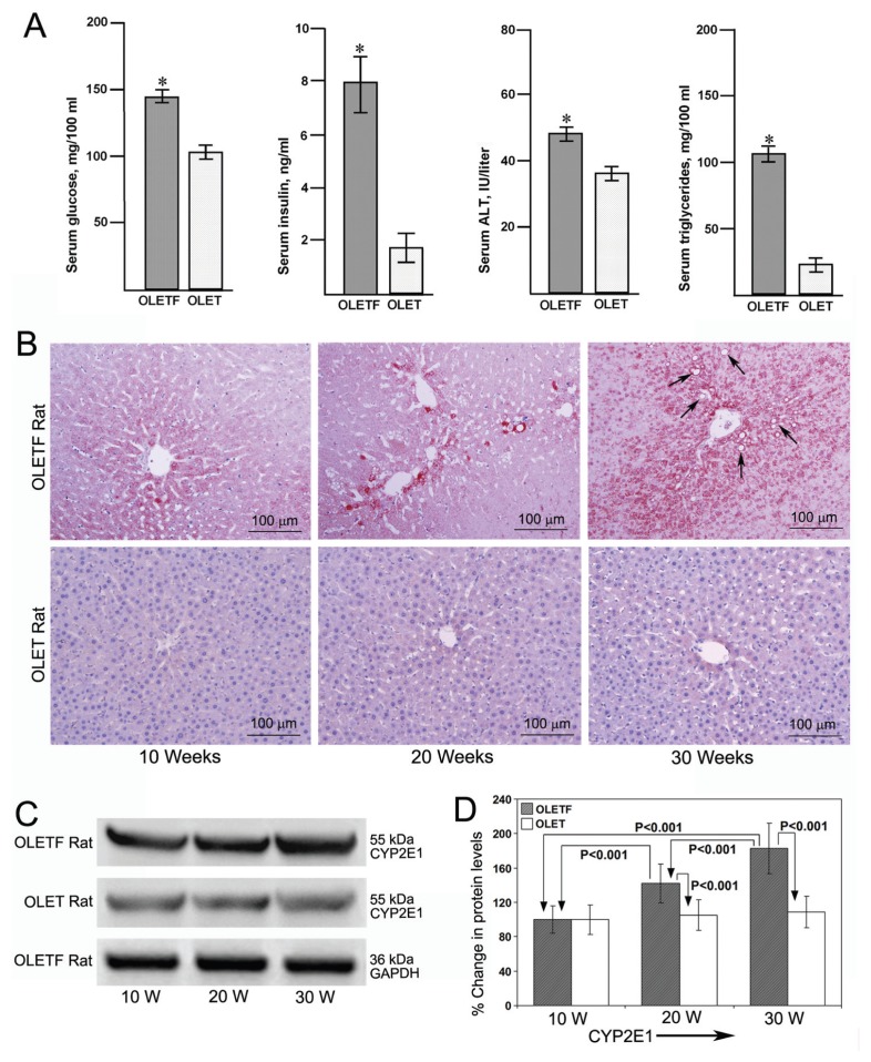
The biochemical abnormalities in OLETF rats compared with OLET rats at 30 wks of age are depicted in Figure 1A. The OLETF rats consumed an excess of 10 g chow per day compared with OLET rats, and the mean body weight of OLETF rats increased to 620 ± 15 g, which was significantly higher (P < 0.001) than that of OLET rats (460 ± 10 g). We observed a significant increase (P < 0.001) in the levels of glucose, insulin, ALT and triglycerides in the serum of 30-wk-old OLETF rats compared with OLET rats, indicating insulin resistance, diabetes, steatosis (Figure 1B, arrow) and increased hepatic uptake of free fatty acids. There was no difference in serum levels of AST, γ-GTP and total cholesterol between OLETF and OLET rats at 30 wks of age (data not shown).
Figure 1.
Serological and histological findings and CYP2E1 protein levels in OLETF and OLET rats aged 30 wks. (A) Serum levels of glucose, insulin, alanine transaminase (ALT) and triglycerides (TG) in OLETF and OLET rats aged 30 wks. *P < 0.001, OLETF rats versus OLET rats (n = 10). (B) Immunohistochemical staining for CYP2E1 in liver tissues from OLETF and OLET rats aged 10, 20 and 30 wks (×100). Marked increase in the staining pattern of CYP2E1 in comparable with aging in OLETF rats but not in OLET rats. (C) Western blotting for CYP2E1 in the liver tissues from OLETF and OLET rats aged 10, 20 and 30 wks. Data are representative of eight rats per each group. (D) Quantitative analysis of Western blot images. Data are mean ± SD of eight images per group. *P < 0.001, OLETF rats aged 20 and 30 wks versus 10 and 30 wks versus 20 wks and OLETF rats versus OLET rats at 20 and 30 wks.
Expression of CYP2E1 in the Livers of OLETF and OLET Rats Aged 10, 20 and 30 Wks
Immunohistochemical staining for CYP2E1 in the liver tissue of 10-, 20- and 30-wk-old OLETF and OLET rats is depicted in Figure 1B. There was a remarkable increase in the staining intensity of CYP2E1 on par with aging and increase of body weight in OLETF rats compared with OLET rats. At 30 wks, the untreated OLETF rat livers showed marked staining for CYP2E1 in the pericentral area along with fatty degeneration. Staining for CYP2E1 was moderately increased in the pericentral area in OLET rats during aging (Figure 1B). Western blotting for CYP2E1 protein levels in the liver tissue of 10-, 20- and 30-wk-old OLETF and OLET rats are presented in Figure 1C. There was a significant increase in the protein levels of CYP2E1 in OLETF rats during aging but not in OLET rats. Reprobing the Western blot images for GAPDH demonstrated equal loading of proteins. The results of the quantitative evaluation of Western blot images are represented in Figure 1D. In OLETF rats, CYP2E1 protein levels were significantly higher (P < 0.001) at 20 and 30 wks compared with 10 wks, but not in OLET rats. CYP2E1 in OLETF rats at 30 wks were also significantly higher (P < 0.001) compared with 20 wks. CYP2E1 protein levels were significantly higher (P < 0.001) in OLETF rats compared with OLET rats at 10, 20 and 30 wks (Figure 1D). Because the mean CYP2E1 protein level at 10 wks in both OLETF and OLET rats was indicated as 100%, we could not show the level of significance between OLETF and OLET rats at 10 wks.
Evaluation of the Liver Tissue of OLETF and OLET Rats Untreated and Treated with Ethanol
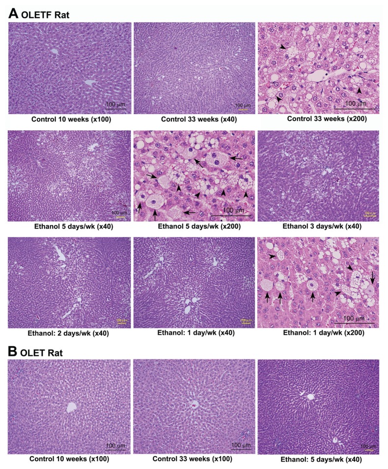
The histopathological images of untreated and ethanol-treated OLETF and OLET rat livers are presented in Figure 2. There was no histopathological alteration in the liver tissue of 10-wk-old control OLETF rats (Figure 2A). In the livers of 33-wk-old untreated OLETF rats, fatty degeneration and microvesicular steatosis (arrowhead) were present, especially in the pericentral area (Figure 2A). Massive steatohepatitis with focal hepatic necrosis was present in the livers of all OLETF rats treated with ethanol for 5, 3, 2 and 1 d/wk. There was numerous hepatocyte ballooning (arrow) and microvesicular steatosis (arrowhead), especially in the pericentral areas. The histopathological changes were more intense in rats receiving ethanol for 5 d/wk. The 33-wk-old OLET rats untreated and treated with ethanol for 5, 3, 2 and 1 d/wk did not show any pathological alterations compared with 10-wk-old control OLET rats (Figure 2B). Because there was no pathological alteration, the images are not shown for 3, 2 and 1 d/wk ethanol-treated OLET rats.
Figure 2.
Histology of untreated and ethanol-treated OLETF and OLET rats. (A) H&E staining of liver tissues from 10- and 33-wk-old untreated OLETF rats and 30-wk-old OLETF rats treated with 10 mL of 10% ethanol for 5, 3, 2 and 1 d/wk for 3 wks. There was no histological alteration in 10-wk-old OLETF rats. Fatty degeneration and microvesicular steatosis (arrowhead) were present in 33-wk-old untreated OLETF rats. Massive steatohepatitis with numerous hepatocyte ballooning (arrow) and microvesicular steatosis (arrowhead) were present in all OLETF rats treated with ethanol, irrespective of number of days treated. (B) H&E staining of liver tissues from 10- and 33-wk-old untreated OLET rats and 30-wk-old OLET rats treated with ethanol 5 d/wk for 3 wks. No histopathological changes or fatty degeneration were seen in either untreated rats or rats treated with ethanol. Scale bars, 100 μm.
The liver histological alterations assessed for NASH activity in the different group of animals are presented in Table 1. The scoring for steatosis, lobular inflammation and hepatocyte ballooning were added up to produce the NAS, ranging from 0 to 9. NAS was >5 in all OLETF rats treated with ethanol indicating NASH. NAS was <1 in all OLET rats treated with ethanol demonstrating absence of NASH activity (Table 1).
Table 1.
NASH activity scoring in control and ethanol-treated OLETF and OLET rats.
| OLETF rats | OLET rats | |||||||
|---|---|---|---|---|---|---|---|---|
|
|
|
|||||||
| Parameters | Control, 33 wks | Ethanol, 5 d/wk | Ethanol, 3 d/wk | Ethanol, 2 d/wk | Ethanol, 1 d/wk | Control, 33 wks | Ethanol, 5 d/wk | Ethanol, 1 d/wk |
| Steatosis | 1.1 ± 0.2 | 2.1 ± 0.3a | 1.9 ± 0.3a | 2.0 ± 0.4a | 1.9 ± 0.3 | 0.0 ± 0.0 | 0.0 ± 0.0 | 0.0 ± 0.0 |
| Lobular inflammation | 0.0 ± 0.0 | 1.4 ± 0.2a | 1.3 ± 0.2a | 1.1 ± 0.2a | 0.8 ± 0.1a | 0.0 ± 0.0 | 0.5 ± 0.1 | 0.0 ± 0.0 |
| Hepatocyte ballooning | 0.2 ± 0.0 | 2.6 ± 0.4a | 2.7 ± 0.4a | 2.5 ± 0.3a | 2.4 ± 0.3a | 0.0 ± 0.0 | 0.0 ± 0.0 | 0.0 ± 0.0 |
| NAS (0–9) | 1.3 ± 0.2 | 6.1 ± 0.9a | 5.9 ± 0.9a | 5.6 ± 0.9a | 5.1 ± 0.7a | 0.0 ± 0.0 | 0.5 ± 0.1 | 0.0 ± 0.0 |
Data are means ± SD of eight rats per group. Scores for steatosis (score 0–3), lobular inflammation (score 0–3) and hepatocyte ballooning (score 0–3) were summed to produce the NAS, thus ranging from 0 to 9.
P < 0.001 versus OLETF untreated control rats.
Serum ALT, AST, Glucose, Insulin and Adiponectin Levels in OLETF and OLET Rats Untreated and Treated with Ethanol
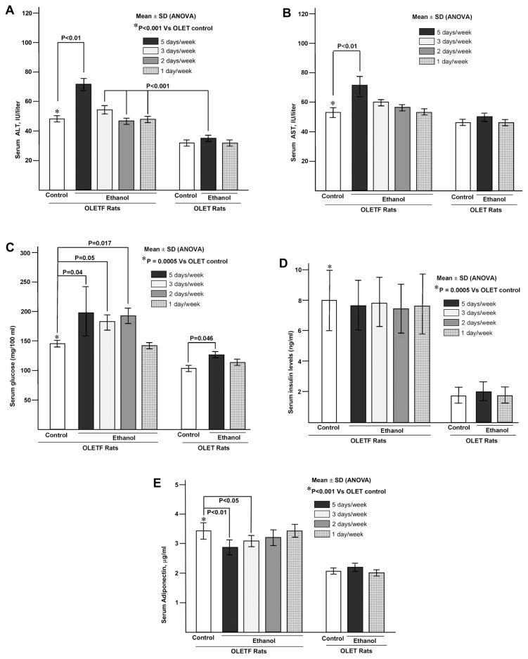
Serum ALT, AST, glucose, insulin and adiponectin levels in OLETF and OLET untreated control rats and rats treated with ethanol are presented in Figures 3A–E, respectively. Serum ALT levels in 33-wk-old untreated OLETF rats were significantly higher (P < 0.001) compared with OLET rats. Serum ALT was significantly increased (P < 0.01) in OLETF rats treated with ethanol for 5 d/wk compared with untreated control, whereas on other days, the difference was not significant. However, serum ALT levels in OLETF rats treated with ethanol for 3, 2 and 1 d/wk was significantly higher (P < 0.001) than that of OLET rats treated with ethanol 5 d/wk. Serum ALT levels were not different between OLET rats treated with ethanol and 33-wk-old untreated OLET rats. Serum AST levels were significantly elevated (P < 0.01) in OLETF rats treated with ethanol for 5 d/wk compared with untreated control, whereas on other days, the difference was not significant. Serum AST levels in 33-wk-old untreated OLETF rats were not significantly different from untreated OLET rats.
Figure 3.
Serum ALT, AST, glucose and insulin levels in OLETF and OLET untreated rats and rats treated with ethanol. All data are mean ± SD of 10 rats in each group. (A) Serum ALT levels. *P < 0.001, OLETF untreated versus OLET untreated rats; *P < 0.001, OLETF rats versus OLET rats, ethanol for 5 d/wk; P < 0.01, OLETF rats ethanol 5 d/wk versus OLETF control rats. (B) Serum AST levels. P < 0.01, OLETF rats, ethanol 5 d/wk versus OLETF control rats. (C) Serum glucose levels. *P = 0.0005, OLETF control versus OLET control rats. Glucose levels were significantly higher in OLETF rats treated with ethanol for 5, 3 and 2 d/wk versus OLETF control rats. (D) Serum insulin levels. *P = 0.0005, OLETF control versus OLET control rats. Serum insulin levels were not altered significantly in any groups of OLETF rats treated with ethanol compared with OLETF control rats. (E) Serum adiponectin levels. *P < 0.001, OLETF control versus OLET control rats. P < 0.01 and P < 0.05, OLETF 5 d/wk and OLETF 3 d/wk versus OLETF control rats. Serum adiponectin levels did not alter in any groups of ethanol-treated OLET rats compared with untreated controls.
Serum glucose levels in OLETF control rats was significantly higher (P = 0.0005) compared with OLET control rats. Serum glucose levels were significantly higher in OLETF rats treated with ethanol for 5, 3 and 2 d/wk compared with untreated control (Figure 3C), but not in rats treated with ethanol for 1 d/wk. A significant difference (P = 0.046) was present in OLET rats treated with ethanol (5 d/wk) compared with untreated OLET rats. However, there was no difference between OLET rats treated with ethanol once a week and control OLET rats. Serum insulin level in OLETF untreated rats was significantly higher (P = 0.0005) than in OLET untreated rats. Serum insulin level was not altered in OLETF rats treated with ethanol for 5, 3, 2 and 1 d/wk compared with untreated OLETF control rats (Figure 3D). There was no difference in serum insulin levels between OLET rats treated with ethanol and untreated control OLET rats (Figure 3D).
The mean serum adiponectin level in OLETF untreated control rats was significantly higher (P < 0.001) than in OLET untreated control rats. Serum adiponectin levels were significantly decreased in OLETF rats treated with ethanol for 5 d/wk (P < 0.01) and 3 d/wk (P < 0.05) compared with untreated control (Figure 3E), but not in rats treated with ethanol for 2 and 1 d/wk. However, the reduced levels of serum adiponectin on all days of ethanol-treated rats were significantly higher (P < 0.001) compared with untreated or ethanol-treated OLET rats. There was no difference in serum adiponectin levels between OLET controls and rats treated with ethanol (Figure 3E).
Serum and Hepatic Triglycerides, Serum γ-GTP and Total Cholesterol in OLETF and OLET Rats Untreated and Treated with Ethanol
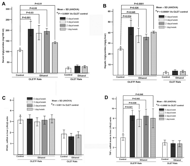
Serum and hepatic triglyceride levels in OLETF and OLET untreated and treated rats are presented in Figures 4A and B, respectively. Serum triglyceride level in untreated OLETF control rats was significantly higher (P = 0.0001) than in OLET control rats. In OLETF rats treated with ethanol for 5, 3, 2 and 1 d/wk, serum triglyceride levels were significantly higher compared with untreated control (Figure 4A). There was no difference in serum triglyceride levels between untreated OLET rats and OLET rats treated with ethanol on both 5 and 1 d/wk. Hepatic triglycerides content in OLETF control rats was significantly higher (P = 0.00001) compared with OLET control rats. Hepatic triglyceride contents were also significantly higher in OLETF rats treated with ethanol for 5, 3, 2 and 1 d/wk compared with OLETF untreated control (Figure 4B). There was no difference in hepatic triglyceride levels between OLET rats treated with ethanol (both 5 and 1 d/wk) and untreated OLET rats (Figure 4B).
Figure 4.
Serum TG, hepatic TG, expression of PPAR-γ mRNA and TNF-α mRNA in the liver tissue of OLETF and OLET untreated rats and rats treated with ethanol. All data are mean ± SD of 10 rats in each group. (A) Serum TG. *P = 0.0001, OLETF untreated versus OLET untreated rats. (B) Hepatic TG levels. *P = 0.00001, OLETF untreated versus OLET untreated rats. (C) Expression of PPAR-γ mRNA in the liver. *P < 0.001, OLETF untreated versus OLET untreated rats. In both OLETF and OLET rats, PPAR-γ mRNA was not altered after treatment with ethanol compared with their untreated controls. (D) Expression of TNF-α mRNA in the liver. *P = 0.0005, OLETF untreated versus OLET untreated rats. TNF-α mRNA was significantly increased in all groups of OLETF rats treated with ethanol compared with OLETF untreated rats.
There was no significant difference in serum γ-GTP and total cholesterol levels in OLETF untreated control rats compared with OLET control rats (data not shown). There was also no difference in serum γ-GTP and total cholesterol in OLETF rats treated with ethanol for 5, 3, 2 and 1 d/wk compared with OLETF untreated control rats (data not shown). In OLET rats also, serum γ-GTP and total cholesterol were not altered between untreated controls and rats treated with ethanol 5 d/wk (data not shown).
Expression of PPAR-γ and TNF-α mRNA in the Liver Tissue of OLETF and OLET Rats Untreated and after Treatment with Ethanol
The nuclear receptor PPAR-γ regulates fatty acid and glucose metabolism and activates various genes involved in lipogenesis at the transcriptional level. PPAR-γ and TNF-α mRNA expression in the liver tissue of OLETF and OLET untreated and treated rats are presented in Figures 4C and D, respectively. Hepatic PPAR-γ mRNA expression was significantly higher (P < 0.001) in OLETF control rats than in OLET controls. However, there was no difference in hepatic PPAR-γ mRNA levels in both OLETF and OLET rats after treatment with ethanol compared with their respective controls. TNF-α mRNA expression was significantly higher (P = 0.005) in OLETF control rats than in OLET control rats. TNF-α mRNA expression was also significantly higher in OLETF rats treated with ethanol for 5, 3, 2 and 1 d/wk compared with OLETF untreated rats (Figure 4D). There was no difference in TNF-α mRNA expression between OLET rats treated with ethanol and untreated OLET controls (Figure 4D).
Expression of CYP2E1 OLETF and OLET Rats Untreated and Treated with Ethanol
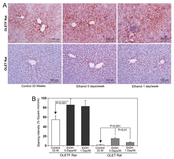
Expression of CYP2E1 in OLETF and OLET untreated and treated rats are presented in Figure 5A. In untreated OLTEF rats, there was marked staining of CYP2E1 in the pericentral areas along with steatosis. A strong staining for CYP2E1 was present, even in centrilobular areas. There was slight staining for CPY2E1 in the pericentral areas of 33-wk-old OLET untreated rats, which is normal. There was a striking increase in CYP2E1 staining along with steatohepatitis in all the groups of OLETF rats treated with ethanol. In the OLET rat groups, staining for CYP2E1 increased in the livers of rats treated with ethanol for 5 d/wk only. Quantitative analysis of the staining intensity of CYP2E1 is presented in Figure 5B. In OLETF rats, staining intensity of CYP2E1 was significantly higher (P < 0.001) in all groups of rats treated with ethanol compared with untreated controls. There was no significant difference in the staining intensity of CYP2E1 between 5, 3, 2 and 1 d/wk of ethanol administration in OLETF rats (Figure 5B). There was an increase (P < 0.001) in the staining intensity of CYP2E1 in the livers of OLET rats treated with ethanol for 5 d/wk. The images are presented for the animals treated with ethanol for 5 and 1 d/wk only, because the staining intensity is almost the same in all the groups of ethanol-treated OLETF rats.
Figure 5.
Immunohistochemical staining of CYP2E1 in the liver tissues from OLETF and OLET rats without and with ethanol (EtOH) treatment. The images are representative of eight rats per group. (A) Marked increase of CYP2E1 staining in the pericentral areas of both untreated and ethanol-treated OLETF rats. (B) Quantitative data of CYP2E1 staining intensity. Data are mean ± SD of eight rats per group. P < 0.001, untreated versus all groups of ethanol-treated in both OLETF and OLET rats. Original magnification 100×.
Staining of 4-HNE in OLETF and OLET Rat Livers After Treatment with Ethanol
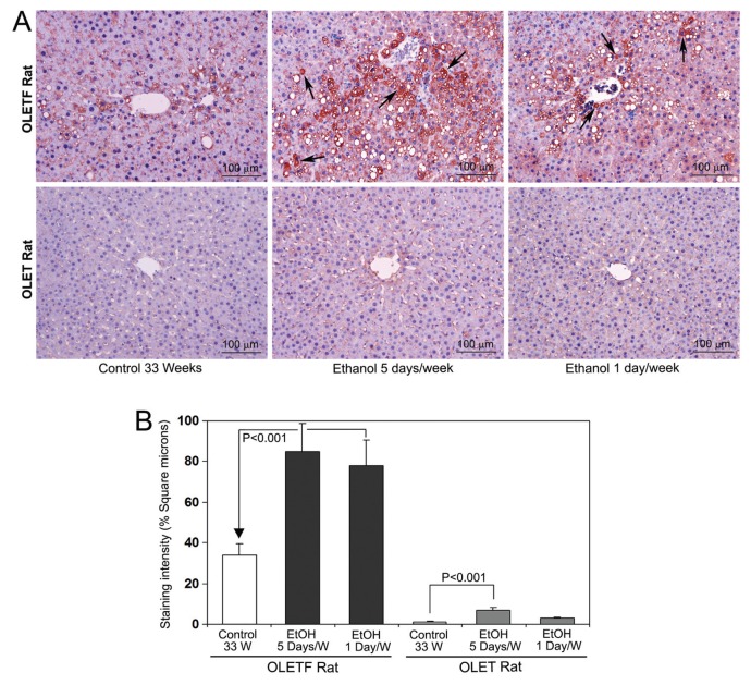
Staining of 4-HNE in the liver tissue of OLETF and OLET untreated and treated rats is demonstrated in Figure 6A. Marked staining for 4-HNE was present in the pericentral areas along with steatosis in untreated OLETF rats. Staining for 4-HNE was remarkably increased in all the groups of OLETF rats treated with ethanol. The OLETF rat livers also showed 4-HNE adduct aggresomes within hepatocytes, which was more prominent in rat livers treated with ethanol for 5 d/wk (arrow) (33). There was moderate staining for 4-HNE in the livers of OLET rats treated with ethanol for 5 d/wk (Figure 6A). Quantitative analysis of the staining intensity of 4-HNE is presented in Figure 6B. The staining intensity was significantly high (P < 0.001) in all the groups of OLETF rats treated with ethanol compared with untreated control. There was an increase (P < 0.01) in the staining intensity of 4-HNE in the livers of OLET rats treated with ethanol for 5 d/wk, which is comparable with the increase of CYP2E1.
Figure 6.
Immunohistochemical staining of 4-HNE in the liver tissues from OLETF and OLET rats without and with ethanol (EtOH) treatment. The images are representative of eight rats per group. (A) Marked increase in the staining intensity of 4-HNE in both untreated and ethanol-treated OLETF rats compared with OLET rats. There were enormous amounts of 4-HNE adduct aggresomes within hepatocytes (arrow). (B) Quantitative data of HNE staining intensity. Data are mean ± SD of eight rats per group. P < 0.001, untreated versus all groups of ethanol-treated OLETF rats. Original magnification 100×.
DISCUSSION
In the present study, we hypothesized that “a binge” or occasional alcohol intake could be a “second hit” for pathogenesis of NASH from simple steatosis in obese individuals. To test our hypothesis, we gave 10 mL of 10% ethanol to obese and wild-type rats. When compared with the animal body weight, the amount of alcohol we administered was 1.6 g/kg to OLETF rats and 2.1 g/kg to OLET rats. Even though the amount of alcohol administered to OLET rats was 30% higher compared with OLETF rats, OLET rats did not develop NASH or any biochemical abnormalities. In addition, in all OLET rats treated with ethanol, NAS was <1, indicating the absence of NASH activity. Thus, here we demonstrate that “a binge” or occasional alcohol intake could be a “second hit” to develop NASH from obesity-induced simple steatosis.
It was reported that obesity and overweight is directly associated with hepatic steatosis in patients (34). Obesity-induced simple steatosis could develop into NASH and then to liver cirrhosis and hepatocellular carcinoma (35). In the current study, we observed hyperglycemia, insulin resistance and well-developed steatosis in 30-wk-old OLETF rats. Moreover, markedly high levels of insulin, ALT and triglycerides were present in the serum of OLETF rats compared with OLET rats. These data proved that the OLETF rat is an appropriate model for study of the mechanism of the pathogenesis of NASH in patients with obesity, steatosis, insulin resistance and hyperlipidemia.
The amount of ethanol administered in the current study is much less compared with the Lieber-DeCarli liquid diet (36) to induce steatosis in rats (80–100 mL liquid diet containing 5% ethanol) (20–25 g ethanol/kg body weight/day). In the present study, 10 mL of 10% ethanol, 5 d/wk for 3 wks, did not produce any significant alteration in the serum levels of ALT, AST, γ-GTP, total cholesterol, triglycerides and insulin and hepatic triglyceride content as well as liver histopathology in OLET rats. On the other hand, OLETF rats treated with ethanol just for 1 d/wk for 3 wks produced massive steatohepatitis in the liver along with related histopathological and biochemical alterations. We observed similar results in the animals administered ethanol for 5, 3 and 2 d/wk for 3 wks. These results strongly suggest that “a binge” or occasional alcohol intake can serve as a “second hit” to develop NASH from simple steatosis in obese individuals.
Obesity is mostly associated with insulin resistance, which leads to increased blood glucose, type 2 diabetes mellitus and impairment of hepatic metabolism. The decreased glucose absorption promotes influx of free fatty acids into the liver, increased de novo lipogenesis and a decrease in hepatic clearance of triglycerides due to the impaired β-oxidation system (37,38). This scenario causes a marked elevation of hepatic triglycerides along with hepatic inflammation, influx of mononuclear cells, hepatocyte ballooning and steatosis. The process also leads to an increase of serum triglycerides and ALT. In the present study, we have observed well-developed steatosis in untreated adult OLEFT rats accompanied with up-regulation of hepatic CYP2E1 and also 4-HNE. It was reported that chronic administration of ethanol to rats resulted in decreased levels of serum adiponectin by suppressing PPAR-γ expression (39). In addition, chronic ethanol feeding to rats suppressed the secretion of adiponectin from isolated epididymal adipocytes, and the CYP2E1-dependent ROS production disrupts adiponectin secretion (40). In the current study, we observed a decrease of serum adiponectin levels in rats treated with ethanol for 5 and 3 d/wk. The highly increased levels of CYP2E1 levels and ROS production observed in the present study, especially in rats treated with ethanol for 5 and 3 d/wk, would be responsible for the decreased serum levels of adiponectin.
The transcription factor PPAR-γ plays a significant role in lipogenesis in the liver through transcriptional activation of various genes involved in adipogenesis (41). PPAR-γ mRNA expression in the liver was significantly higher in untreated OLEFT rats compared with OLET rats. However, ethanol administration did not alter the levels of PPAR-γ mRNA in both OLETF and OLET rats, probably because the amount of ethanol administered in the current study was relatively less. TNF-α is involved in acute systemic inflammation, obesity and pathogenesis of insulin resistance and type 2 diabetes mellitus (42). We have observed a marked increase in the expression of TNF-α mRNA in the liver tissue of OLETF rats treated with ethanol. Untreated OLETF rats also exhibited a significant increase of TNF-α mRNA levels compared with untreated OLET rats. It was observed that hepatic macrophages from ob/ob mice, an animal model that spontaneously develops NASH, express significantly greater levels of TNF-α mRNA (43). Moreover, TNF-α promoter polymorphism is higher in NASH patients with insulin resistance than in patients negative for TNF-α polymorphism (44). Most importantly, it was observed that TNF-α–deficient obese mice had lower levels of circulating free fatty acids and were protected from the obesity-induced insulin resistance (45).
In the current study, we demonstrated that CYP2E1 is markedly increased on par with aging and increase of body weight in untreated OLETF rats but not in OLET rats. It was observed that CYP2E1 is upregulated in NASH associated with type 2 diabetes mellitus and obesity (11,18). It was also shown that binge ethanol exposure upregulates CYP2E1 and increases liver injury in obese rats (46). However, the authors administered ethanol 4 g/kg body weight at every 12 h for 3 d consecutively to obese rats, which is fivefold higher than the dosage used in the current study. In addition, they reported that CYP2E1 is downregulated in untreated obese rats, and also ethanol induction of CYP2E1 in obese rats was much less compared with lean rats. In the present study, staining intensity for CYP2E1 in 33-wk-old untreated OLETF rats was strikingly higher compared with untreated OLET rats. Furthermore, there was a marked increase in the staining intensity of CYP2E1 in the hepatic tissue of OLETF rats treated with ethanol compared with untreated controls. Our results and the data from previous studies (43) suggest that when an obese man consumes 20 or 140 g ethanol/week, the increased level of CYP2E1 metabolizes ethanol into acetaldehyde and induces the production of ROS, which in turn contributes to the development of NASH. This result suggests that the molecular mechanisms involved in the pathogenesis of NASH may be similar to that of ASH, involving induction of CYP2E1 and subsequent events. The unchecked ASH and NASH can develop into fibrosis and then progress to cirrhosis and ultimately to hepatocellular carcinoma.
Studies on wild-type and CYP2E1 null mice demonstrated that both intestinal and hepatic CYP2E1 induced by ethanol is critical in binge alcohol-mediated oxidative stress, gut leakage, endotoxemia, impaired fat metabolism and inflammation contributing to hepatic apoptosis and steatohepatitis (47). It was observed that in a well-characterized population with biopsy-proven NAFLD, modest alcohol consumption was associated with decreased prevalence of steatohepatitis (48,49). Furthermore, it was reported that light-to-moderate alcohol consumption protects against insulin resistance in severely obese patients with steatosis and steatohepatitis (50). The above studies suggest that moderate alcohol consumption <20 g/d is not harmful and may be beneficial against several complications.
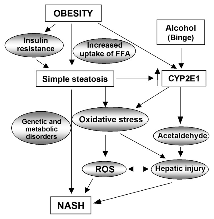
A schematic representation of the mechanism of pathogenesis of NASH from obesity-induced simple steatosis is presented in Figure 7. Obesity results in insulin resistance and induces type 2 diabetes mellitus. It also leads to increased uptake and deposition of free fatty acids in liver, which results in hepatic fatty degeneration and simple steatosis. Hepatic steatosis results in upregulation of CYP2E1 that leads to oxidative stress and production of ROS. When an obese individual with simple steatosis consumes excess alcohol on an occasion (a binge), the increased CYP2E1 metabolizes ethanol into acetaldehyde, which in turn produces oxidative stress and ROS. The excessive amount of ROS along with the toxic acetaldehyde induces pathogenesis of NASH in obese individuals. Furthermore, the acetaldehyde- and ROS- induced hepatocyte injury contributes to this process. Therefore, obesity-induced steatosis serves as the “first hit” and a binge serves as the “second hit” toward the pathogenesis of NASH in obese individuals.
Figure 7.
Schematic representation of the mechanism of pathogenesis of NASH from obesity-induced simple steatosis. Obesity results in insulin resistance and increased hepatic uptake of free fatty acids (FFA), which induces simple steatosis. Steatosis upregulates CYP2E1 and also generates oxidative stress. When an obese individual consumes excessive alcohol on an occasion (binge drinking), the increased CYP2E1 metabolizes ethanol into acetaldehyde through the microsomal ethanol oxidizing system, which in turn aggravates oxidative stress and produces ROS. The excess ROS along with toxic acetaldehyde causes hepatic injury and induces pathogenesis of NASH. Therefore, obesity plays as the “first hit” and a binge serves as the “second hit” toward pathogenesis of NASH in obese individuals.
CONCLUSION
The results of the present study demonstrated that “a binge” or occasional alcohol intake serves as a “second hit” for development of NASH in obese individuals with simple steatosis. During “a binge,” the upregulated CYP2E1 in steatosis metabolizes ethanol and produces reactive metabolic intermediates and ROS, which subsequently induce pathogenesis of NASH through aggravation of oxidative stress. Our findings indicate that occasional alcohol intake is a risk factor for pathogenesis of NASH in obese individuals with simple steatosis and should be avoided.
ACKNOWLEDGMENTS
The authors thank Mr. Mitsuru Araya for assistance in animal experiments. This work was supported in part by a grant for specially promoted research from Kanazawa Medical University (SR2012-04) and by a grant from the Ministry of Health, Labor, and Welfare of Japan.
Footnotes
Online address: http://www.molmed.org
DISCLOSURE
The authors declare that they have no competing interests as defined by Molecular Medicine, or other interests that might be perceived to influence the results and discussion reported in this paper.
REFERENCES
- 1.Ludwig J, Viggiano TR, McGill DB, Oh BJ. Nonalcoholic steatohepatitis: Mayo Clinic experiences with a hitherto unnamed disease. Mayo Clin Proc. 1980;55:434–8. [PubMed] [Google Scholar]
- 2.Sanyal AJ. AGA technical review in nonalcoholic fatty liver disease. Gastroenterology. 2002;123:1705–25. doi: 10.1053/gast.2002.36572. [DOI] [PubMed] [Google Scholar]
- 3.Younossi ZM. Nonalcoholic fatty liver disease. Curr Gastroenterol Rep. 1999;1:57–62. doi: 10.1007/s11894-999-0088-1. [DOI] [PubMed] [Google Scholar]
- 4.Teli MR, James OF, Burt AD, Bennett MK, Day CP. The natural history of nonalcoholic fatty liver: a follow-up study. Hepatology. 1995;22:1714–9. [PubMed] [Google Scholar]
- 5.Day CP, James OFW. Steatohepatitis: a tale of two “hits”? Gastroenterology. 1998;114:842–5. doi: 10.1016/s0016-5085(98)70599-2. [DOI] [PubMed] [Google Scholar]
- 6.Browning JD, Horton JD. Molecular mediators of hepatic steatosis and liver injury. J Clin Invest. 2004;114:147–52. doi: 10.1172/JCI22422. [DOI] [PMC free article] [PubMed] [Google Scholar]
- 7.Burt AD, Mutton A, Day CP. Diagnosis and interpretation of steatosis and steatohepatitis. Semin Diagn Pathol. 1998;15:246–58. [PubMed] [Google Scholar]
- 8.Matteoni CA, et al. Nonalcoholic fatty liver disease: a spectrum of clinical and pathological severity. Gastroenterology. 1999;116:1413–9. doi: 10.1016/s0016-5085(99)70506-8. [DOI] [PubMed] [Google Scholar]
- 9.Chitturi S, Farrell GC. Etiopathogenesis of nonalcoholic steatohepatitis. Semin Liver Dis. 2001;21:27–41. doi: 10.1055/s-2001-12927. [DOI] [PubMed] [Google Scholar]
- 10.Paschos P, Paletas K. Non alcoholic fatty liver disease and metabolic syndrome. Hippokratia. 2009;13:9–19. [PMC free article] [PubMed] [Google Scholar]
- 11.Mandrekar P, Ambade A, Lim A, Szabo G, Catalano D. An essential role for monocyte chemoattractant protein-1 in alcoholic liver injury: regulation of proinflammatory cytokines and hepatic steatosis in mice. Hepatology. 2011;54:2185–97. doi: 10.1002/hep.24599. [DOI] [PMC free article] [PubMed] [Google Scholar]
- 12.Jaeschke H, et al. Mechanisms of hepatotoxicity. Toxicol Sci. 2002;65:166–76. doi: 10.1093/toxsci/65.2.166. [DOI] [PubMed] [Google Scholar]
- 13.Raucy JL, et al. Induction of cytochrome P450IIE1 in the obese overfed rat. Mol Pharmacol. 1991;39:275–80. [PubMed] [Google Scholar]
- 14.Leclercq IA, et al. CYP2E1 and CYP4A as microsomal catalysts of lipid peroxides in murine nonalcoholic steatohepatitis. J Clin Invest. 2000;105:1067–75. doi: 10.1172/JCI8814. [DOI] [PMC free article] [PubMed] [Google Scholar]
- 15.Fong DG, Nehra V, Lindor KD, Buchman AL. Metabolic and nutritional considerations in nonalcoholic fatty liver. Hepatology. 2000;32:3–10. doi: 10.1053/jhep.2000.8978. [DOI] [PubMed] [Google Scholar]
- 16.Lu Y, Cederbaum AI. CYP2E1 and oxidative liver injury by alcohol. Free Radic Biol Med. 2008;44:723–38. doi: 10.1016/j.freeradbiomed.2007.11.004. [DOI] [PMC free article] [PubMed] [Google Scholar]
- 17.Takahashi T, Lasker JM, Rosman AS, Lieber CS. Induction of cytochrome P-4502E1 in the human liver by ethanol is caused by a corresponding increase in encoding messenger RNA. Hepatology. 1993;17:236–45. [PubMed] [Google Scholar]
- 18.Tsutsumi M, Lasker JM, Shimizu M, Rosman AS, Lieber CS. The intralobular distribution of ethanol-inducible P450IIE1 in rat and human liver. Hepatology. 1989;10:437–46. doi: 10.1002/hep.1840100407. [DOI] [PubMed] [Google Scholar]
- 19.Baker SS, Baker RD, Liu W, Nowak NJ, Zhu L. Role of alcohol metabolism in non-alcoholic steatohepatitis. PLoS One. 2010;5:e9570. doi: 10.1371/journal.pone.0009570. [DOI] [PMC free article] [PubMed] [Google Scholar]
- 20.Favreau LV, Malchoff DM, Mole JE, Schenkman JB. Responses to insulin by two forms of rat hepatic microsomal cytochrome P-450 that undergo major (RLM6) and minor (RLM5b) elevations in diabetes. J Biol Chem. 1987;262:14319–26. [PubMed] [Google Scholar]
- 21.Gonzalez FJ. Role of cytochromes P450 in chemical toxicity and oxidative stress: studies with CYP2E1. Mutat Res. 2005;569:101–10. doi: 10.1016/j.mrfmmm.2004.04.021. [DOI] [PubMed] [Google Scholar]
- 22.Sato T, Asahi Y, Toide K, Nakayama N. Insulin resistance in skeletal muscle of the male Otsuka Long-Evans Tokushima Fatty rat, a new model of NIDDM. Diabetologia. 1995;38:1033–41. doi: 10.1007/BF00402172. [DOI] [PubMed] [Google Scholar]
- 23.Rector RS, et al. Cessation of daily exercise dramatically alters precursors of hepatic steatosis in Otsuka Long-Evans Tokushima Fatty (OLETF) rats. J Physiol. 2008;586:4241–9. doi: 10.1113/jphysiol.2008.156745. [DOI] [PMC free article] [PubMed] [Google Scholar]
- 24.Ota T, et al. Insulin resistance accelerates a dietary rat model of nonalcoholic steatohepatitis. Gastroenterology. 2007;132:282–93. doi: 10.1053/j.gastro.2006.10.014. [DOI] [PubMed] [Google Scholar]
- 25.Moran TH, Bi S. Hyperphagia and obesity of OLETF rats lacking CCK1 receptors: developmental aspects. Dev Psychobiol. 2006;48:360–7. doi: 10.1002/dev.20149. [DOI] [PubMed] [Google Scholar]
- 26.Institute of Laboratory Animal Resources; Commission on Life Sciences; National Research Council. Guide for the Care and Use of Laboratory Animals. Washington (DC): National Academy Press; 1996. [Google Scholar]
- 27.Theodorsen L, Stromme JH. Gamma-glutamyl-3-carboxy-14-nitroanilide: the substrate of choice for routine determinations of gamma-glutamyl-transferase activity in serum? Clin Chim Acta. 1976;72:205–10. doi: 10.1016/0009-8981(76)90074-7. [DOI] [PubMed] [Google Scholar]
- 28.Kayamori Y, Hatsuyama H, Tsujioka T, Nasu M, Katayama Y. Endpoint colorimetric method for assaying total cholesterol in serum with cholesterol dehydrogenase. Clin Chem. 1999;45:2158–63. [PubMed] [Google Scholar]
- 29.Bucolo G, David H. Quantitative determination of serum triglycerides by the use of enzymes. Clin Chem. 1973;19:476–82. [PubMed] [Google Scholar]
- 30.Fishman S, et al. Resistance to leptin action is the major determinant of hepatic triglyceride accumulation in vivo. FASEB J. 2007;21:53–60. doi: 10.1096/fj.06-6557com. [DOI] [PubMed] [Google Scholar]
- 31.Kleiner DE, et al. Design and validation of a histological scoring system for nonalcoholic fatty liver disease. Hepatology. 2005;41:1313–21. doi: 10.1002/hep.20701. [DOI] [PubMed] [Google Scholar]
- 32.Roth CL, et al. Vitamin D deficiency in obese rats exacerbates nonalcoholic fatty liver disease and increases hepatic resistin and Toll-like receptor activation. Hepatology. 2012;55:1103–11. doi: 10.1002/hep.24737. [DOI] [PubMed] [Google Scholar]
- 33.Amidi F, et al. M-30 and 4HNE are sequestered in different aggresomes in the same hepatocytes. Exp Mol Pathol. 2007;83:296–300. doi: 10.1016/j.yexmp.2007.09.001. [DOI] [PMC free article] [PubMed] [Google Scholar]
- 34.Hu KQ, et al. Overweight and obesity, hepatic steatosis, and progression of chronic hepatitis C: a retrospective study on a large cohort of patients in the United States. J Hepatol. 2004;40:147–54. doi: 10.1016/s0168-8278(03)00479-3. [DOI] [PubMed] [Google Scholar]
- 35.Farrell GC, Larter CZ. Nonalcoholic fatty liver disease: from steatosis to cirrhosis. Hepatology. 2006;43(2 Suppl 1):S99–112. doi: 10.1002/hep.20973. [DOI] [PubMed] [Google Scholar]
- 36.Lieber CS, DeCarli LM. The feeding of ethanol in liquid diets. Alcohol Clin Exp Res. 1986;10:550–3. doi: 10.1111/j.1530-0277.1986.tb05140.x. [DOI] [PubMed] [Google Scholar]
- 37.Tamura S, Shimomura I. Contribution of adipose tissue and de novo lipogenesis to nonalcoholic fatty liver disease. J Clin Invest. 2005;115:1139–42. doi: 10.1172/JCI24930. [DOI] [PMC free article] [PubMed] [Google Scholar]
- 38.Charlton M, Sreekumar R, Rasmussen D, Lindor K, Nair KS. Apolipoprotein synthesis in nonalcoholic steatohepatitis. Hepatology. 2002;35:898–904. doi: 10.1053/jhep.2002.32527. [DOI] [PubMed] [Google Scholar]
- 39.Tian C, et al. Long term intake of 0.1% ethanol decreases serum adiponectin by suppressing PPARγ expression via p38 MAPK pathway. Food Chem Toxicol. 2014;65:329–34. doi: 10.1016/j.fct.2014.01.007. [DOI] [PubMed] [Google Scholar]
- 40.Tang H, et al. Ethanol-induced oxidative stress via the CYP2E1 pathway disrupts adiponectin secretion from adipocytes. Alcohol Clin Exp Res. 2012;36:214–22. doi: 10.1111/j.1530-0277.2011.01607.x. [DOI] [PMC free article] [PubMed] [Google Scholar]
- 41.Deng T, et al. A peroxisome proliferator-activated receptor gamma (PPARgamma)/PPARgamma coactivator 1beta autoregulatory loop in adipocyte mitochondrial function. J Biol Chem. 2011;286:30723–31. doi: 10.1074/jbc.M111.251926. [DOI] [PMC free article] [PubMed] [Google Scholar]
- 42.Moller DE. Potential role of TNF-alpha in the pathogenesis of insulin resistance and type 2 diabetes. Trends Endocrinol Metab. 2000;11:212–7. doi: 10.1016/s1043-2760(00)00272-1. [DOI] [PubMed] [Google Scholar]
- 43.Lin HZ, Yang SQ, Kujhada F, Ronnet G, Diehl AM. Metformin reverses fatty liver disease in obese, leptin deficient mice. Nat Med. 2000;6:998–1003. doi: 10.1038/79697. [DOI] [PubMed] [Google Scholar]
- 44.Valenti L, et al. Tumor necrosis factor alpha promoter polymorphisms and insulin resistance in nonalcoholic fatty liver disease. Gastroenterology. 2002;122:274–80. doi: 10.1053/gast.2002.31065. [DOI] [PubMed] [Google Scholar]
- 45.Uysal KT, Wiesbrock SM, Marino MW, Hotamisligil GS. Protection from obesity-induced insulin resistance in mice lacking TNF-alpha function. Nature. 1997;389:610–4. doi: 10.1038/39335. [DOI] [PubMed] [Google Scholar]
- 46.Carmiel-Haggai M, Cederbaum AI, Nieto N. Binge ethanol exposure increases liver injury in obese rats. Gastroenterology. 2003;125:1818–33. doi: 10.1053/j.gastro.2003.09.019. [DOI] [PubMed] [Google Scholar]
- 47.Abdelmegeed MA, et al. CYP2E1 potentiates binge alcohol-induced gut leakiness, steatohepatitis, and apoptosis. Free Radic Biol Med. 2013;65:1238–45. doi: 10.1016/j.freeradbiomed.2013.09.009. [DOI] [PMC free article] [PubMed] [Google Scholar]
- 48.Dunn W, et al. Modest alcohol consumption is associated with decreased prevalence of steatohepatitis in patients with non-alcoholic fatty liver disease (NAFLD) J Hepatol. 2012;57:384–91. doi: 10.1016/j.jhep.2012.03.024. [DOI] [PMC free article] [PubMed] [Google Scholar]
- 49.Dunn W, Xu R, Schwimmer JB. Modest wine drinking and decreased prevalence of suspected nonalcoholic fatty liver disease. Hepatology. 2008;47:1947–54. doi: 10.1002/hep.22292. [DOI] [PMC free article] [PubMed] [Google Scholar]
- 50.Cotrim HP, et al. Effects of light-to-moderate alcohol consumption on steatosis and steatohepatitis in severely obese patients. Eur J Gastroenterol Hepatol. 2009;21:969–972. doi: 10.1097/MEG.0b013e328328f3ec. [DOI] [PubMed] [Google Scholar]


