

Abstract
The development of chelating agents for copper radionuclides in positron emission tomography radiopharmaceuticals has been a highly active and important area of study in recent years. The rapid evolution of chelators has resulted in highly specific copper chelators that can be readily conjugated to biomolecules and efficiently radiolabeled to form stable complexes in vivo. Chelators are not only designed for conjugation to monovalent biomolecules but also for incorporation into multivalent targeting ligands such as theranostic nanoparticles. These advancements have strengthened the role of copper radionuclides in the fields of nuclear medicine and molecular imaging. This review emphasizes developments of new copper chelators that have most greatly advanced the field of copper-based radiopharmaceuticals over the past 5 years.
Keywords: PET, copper 64, chelator, molecular imaging, radiopharmaceutical
Introduction
Positron emission tomography (PET) is one of the molecular imaging modalities that probes physiological changes noninvasively, and its hybridization with anatomical imaging modalities, such as computed tomography (CT) and magnetic resonance imaging have made great contributions to the diagnosis of disease over the past few decades.1 The success of PET imaging relies on a synergistic effort of biomarker identification, radiopharmaceutical discovery, and development and instrument innovation. Along with [18F]FDG, which images glucose metabolism, there are a plethora of probes for imaging other biological processes and biomarkers, although only a handful are approved for human use.2
Because of the short half-lives of the traditional positron-emitting radionuclides, such as 18F and 11C, radionuclides with longer half-lives are needed for monitoring in vivo processes with longer biological half-lives using longer circulating antibodies and nanoparticles. Radiometals such as 64Cu (T1/2 = 12.7 h), 86Y (T1/2 = 14.74 h), and 89Zr (T1/2 = 78.4 h), fall into this category.
The development of bifunctional chelators plays a pivotal role in metal-based radiopharmaceuticals.3,4 Copper has unique chemical characteristics that make the use of its radiometals particularly interesting.5 Copper has many important biological roles in vivo, such as electron transfer, catalysis, and structural shaping.6 Many copper-containing compounds are biologically active and have anti-inflammatory and antiproliferative properties.7,8 Copper radionuclides, such as 60Cu, 61Cu, 62Cu, 64Cu, and 67Cu (Table 1) offer versatile choices for applications in imaging,9 therapy,10,11 or theranostics. Copper-64, with a half-life of 12.7 h, and its unique decay profile (β+: 18%; β−: 38%; electron capture: 44%),12 is well suited for radiolabeling nanoparticles, antibodies, antibody fragments, peptides, or small molecules for PET imaging, radio-therapy, and drug discovery and development.13 Copper-64 chloride is produced using a biomedical cyclotron in high specific activity and is commercially available.14,15
Table 1.
| Isotope | t1/2 (h) | method of production | decay mode | Eβ+/− (keV) |
|---|---|---|---|---|
| 60Cu | 0.4 | cyclotron, 60Ni(p,n)60Cu | β+ (93%); EC (7%) | 3920, 3000; 2000 |
| 61Cu | 3.3 | cyclotron, 61Ni(p,n)61Cu | β+ (62%); EC (38%) | 1220, 1150; 940, 560 |
| 62Cu | 0.16 | cyclotron, 62Zn/62Cu generator | β+ (98%); EC (2%) | 2910 |
| 64Cu | 12.7 | cyclotron, 64Ni(p,n)64Cu | β+ (18%); EC (44%) β−(38%) |
653 579 |
| 67Cu | 62.01 | reactor, 67Zn(n, p) | β− (100%) | 577, 484, 395 |
In the development of Cu-64 radiopharmaceuticals, chelator design is essential, because it is required for complex Cu(II), and for many imaging probes, to attach it to biomolecules and generate kinetically and thermodynamically stable systems. The development of copper chelators is especially challenging as there are many copper chelating proteins in vivo (e.g., ceruloplasmin, superoxide dismutase, metallothionein, copper transporters, and chaperones) that potentially can displace the copper ion from the chelator.16 Of the different oxidation states of copper, Cu(II) is most common for Cu-64 radiopharmaceuticals. With a 3d9 electronic configuration, Cu(II) has borderline hardness, favoring binding to nitrogen donors. The coordination number ranges from four to six, forming complexes that have square planar, square pyramidal, trigonal bipyramidal, or octahedral geometry. Jahn–Teller distortion occurs and this is observable by the X-ray crystallography in the molecular structures of Cu(II) complexes.
This mini review will focus on recent developments of copper chelators for use in PET radiopharmaceuticals (primarily in the past 5 years).17–19 It includes innovations in chelators with novel structures and modification of known chelators with functional groups amenable for bioconjugation (Figures 1–6). Methods for in vitro evaluation of copper chelate stability, nonchelating nanocarriers of copper-64, and factors involved in the specific activity of copper radionuclides are also discussed.
Figure 1.
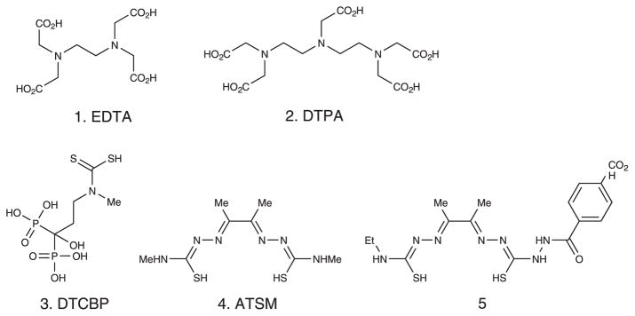
Examples of acyclic chelators.
Figure 6.

Examples of strain-promoted clickable chelators.
Acyclic chelators
Classical acyclic copper chelators include ethylenediaminetetraacetic acid (1. EDTA), diethylene triamine pentaacetic acid (2. DTPA), and their derivatives. Dithiocarbamates have been used for chelating copper, although these are typically used for chelating technetium and rhenium in nuclear medicine.20 Torres Martin de Rosales et al. reported a dual-modality PET–MRI agent based on 64Cu-dithiocarbamatebisphosphonate (3. DTCBP conjugated to iron oxide nanoparticles.21 This was successfully used to image draining lymph nodes in a mouse model. Although the copper complex with DTCBP is not highly kinetically stable, with a half-life of less than 5 min in 5 M HCl at 90°C compared with 154 h for Cu-CB-TE2A, the in vivo imaging showed retention of the probe in popliteal and iliac lymph nodes.22 Bis(thiosemicarbazones) such as diacetyl-bis(N4-methylthiosemicarbazone) (4. H2ATSM) were developed as copper chelator mainly for hypoxia imaging and have been previously reviewed extensively.23,24 The application of bis(thiosemicarbazones) as bifunctional chelators is rare. However, Hueting et al. reported their evaluation of them, such as 5, using a bombesin derivative as the model peptide.25
Nonbridged macrocyclic chelators
Because of the macrocyclic effect, macrocyclic chelators generally form kinetically more stable Cu(II) complexes than acyclic chelators. The classical macrocycles used to chelate Cu (II) for PET imaging are cyclen (6) and cyclam (7) modified with N-acetic acid arms, with the most commonly used being DOTA (8) and 1,4,8,11-tetraazacyclododecane-1,4,8,11-tetraacetic acid (TETA) (9).26–28 However, in vivo instability of these complexes has been demonstrated.29 Svobodová, et al. efficiently synthesized a dimethyl-diphosphonate cyclam derivative 10 that has square-pyramidal or trigonal-bipyramidal geometry with copper depending on the extent of protonation of the chelator, leaving one phosphoric acid arm that is not chelated with Cu(II).30 This complex is kinetically labile, losing the copper quickly under acidic conditions. Barnard, et al. made a diamide macrocyclic chelator (11) and showed that the proton on the nitrogen of the amide was deprotonated, forming a distorted square planar complex with Cu(II). A biodistribution study with the 64Cu-labeled compound showed rapid blood clearance but with much higher liver uptake and retention than CB-TE2A, indicating its instability in vivo.31 Pandya et al. reported a synthesis of TE2A (12), and the acid decomplexation and reduction potential measurements indicated that Cu-TE2A is more stable than Cu-TETA, on the basis of rat biodistribution data showing significantly lower retention of 64Cu-TE2A than 64Cu-TETA in kidney and liver at 24 h post injection, suggesting a lower extent of demetallation in vivo.32 In a follow-up paper, a new synthesis of the salt-free TE2A was reported that was conjugated to c(RGDyK), but no biodistribution data were reported.33 Zhang et al. reported a multivalency platform based on the DOTA chelator using c(RGDyK) with no space between the c and (RGDyK) as a model peptide.34 Interestingly, the dimer outperformed the corresponding monomer and trimer as a PET-imaging agent. Ferreira et al. compared p-SCN-Bn-DOTA (13) with two newly developed chelators, p-SCN-Bn-PCTA (14) and p-SCN-Bn-Oxo (15) conjugated with trastuzumab.35 Both 14-trastuzumab and 15-trastuzumab were superior to the DOTA analogue, with respect to labeling efficiency, serum stability, and tumor uptake. Subsequently, Cooper et al. reported a systematic comparison of eight chelators including 13, 14, and 15, after conjugation with antibody rituximab.36 They were able to label the DOTA conjugate at low temperatures in 20 min, and the labeled conjugate was stable in serum for up to 48 h. They speculated that the discrepancy with other reports on DOTA-based antibody conjugates came from the source of Cu-64 they used, emphasizing the importance of the radionuclide purity, especially for less copper-selective chelators such as DOTA.
Cross-bridged macrocyclic chelators
Because the in vivo stability of Cu(II) complexes is vital for PET imaging, the incorporation of 1,8-ethylene cross-bridged macrocyclic chelators into 64Cu radiopharmaceuticals was an important advance. Among several cyclen and cyclam-based cross-bridged macrocyclic chelators, the lead chelator was CB-TE2A (16), which showed improved in vivo stability over DOTA and TETA.37–39 However, because of the requirement of harsh radiolabeling conditions (95°C for 60 min), the application of this chelator is limited to heat insensitive molecules, precluding its application in antibodies or other proteins.40 It is noteworthy that Yoo and colleagues developed an efficient synthesis of propylene cross-bridged TE2A (19).41
Compound 19 showed high in vitro and in vivo stability and can be labeled under milder conditions than the corresponding ethylene cross-bridged 16. A large series of propylene cross-bridged tetraaza macrocyclic compounds were patented by the same authors.42
To keep the two pendant arms of CB-TE2A available for chelating copper and neutralizing the positive charge when one of the carboxylates is utilized for conjugation to a biomolecule, Lewis et al. introduced a 4-nitrobenzyl group at the 6-position of the cyclam backbone, which was converted to an amine-reactive isothiocyanate (20) for easy conjugation to biomolecules.43 The radiolabeling and stability studies of the radio-Cu(II) chelate have not been reported to our knowledge. Boswell et al. functionalized cross-bridged cyclam with orthogonally protected 2-bromopentanedioic acid to get an asymmetrically substituted cross-bridged macrocyclic chelator (21).44 However, the labeling conditions were similar to those used for CB-TE2A, that is, requiring high temperatures. Liu et al. went further to generate the bis-pentanedioic acid modified cross-bridged cyclam (22).45 Using c(RGDyK) as a model peptide ligand, they tested the concept of multivalency on the basis of this platform. The results showed that the bivalent probe was superior in uptake and retention in tumors than the corresponding monomer.
Additional efforts to find a chelator that could be labeled at lower temperatures resulted in the phosphonate-armed chelators, such as CB-TE2P (17) and CB-TE1A1P (18), which can be labeled at room temperature within 1 h in high specific activity.46,47 The pharmacokinetics of these Cu(II) phosphonate CB complexes were compared with copper complexes of CB-TE2A, diamsar, and NOTA, and exhibited rapid blood clearance with minimum retention in liver, kidney, and bone marrow.46 This is in contrast with the noncross-bridged analogues, such as 10 published by Svobodová, et al.30 One of the phosphonate cross-bridged chelators, CB-TE1A1P, was conjugated to the somatostatin analog, Y3-TATE,48 and the α4β1 integrin-targeting agent, LLP2A.49 64Cu-CB-TE1A1P-Y3-TATE could be labeled in ~90% purity within 1 h at 40°C, whereas 64Cu-CB-TE1A1P-LLP2A was 100% labeled by 1 h at room temperature, likely because of a longer linker between the chelator and the biomolecule. The biodistributions of both compounds were improved over CB-TE2A analogs, having greater tumor uptake and improved contrast.48,49
Sarcophagine-based chelators
Sarcophagine chelators were pioneered by Sargeson and colleagues in the late 1970s.50 A comparison of CB-TE2A and diamsar (23) conjugated RGD peptides was performed, and the data suggested that a linker between the diamsar chelator and the peptide would improve the targeting and biodistribution.51 SarAr (24) includes the diamsar chelator as well as a linker, allowing for improved labeling efficiency after conjugation.52 Ma et al. functionalized both of the primary amines with succinic or glutaric anhydride (25) and conjugated the chelators with Tyr3-octreotate or with two equivalents of bombesin. The labeling conditions were explored, but no biological data are available on the bioconjugates as of the time of this writing.53–55
Click chemistry applied to chelator-biomolecule conjugation
Click chemistry, as first reported by K. Barry Sharpless in 2001,56 has been embraced by the PET radiopharmaceutical field as a new way to incorporate radionuclides via prosthetic groups or chelators.57 The most commonly used approach is the Huisgen 1,3-dipolar cycloaddition, also called copper-catalyzed alkyneazide cycloaddition (CuAAC). This reaction benefits from high chemoselectivity, fast reaction kinetics, and high stereoselectivity and results in high purity products in high yield in a wide spectrum of solvents. A series of click-to-chelate metal chelators were developed on the basis of Huisgen 1,3-dipolar cycloaddition chemistry.58 However, applications of CuAAC click chemistry for copper chelators is technically challenging because it is difficult to remove the copper from the chelator under conditions that can be tolerated by the bioconjugate, especially for sensitive biomolecules such as antibodies or peptides. Knör et al. synthesized ethynylphenyl-DOTA (26) and 4-acetylphenyl-DOTA (27) for chemoselective conjugation with azido-functionalized and aminooxy-functionalized Tyr3-octreotate, respectively.59 They removed the chelated copper from the final product through precipitation with sodium sulfide. Lebedev et al. applied a similar strategy on propargyl derivatized tert-butyl protected DOTA (28) and a CB-cyclam-based chelator (29).60 It is noteworthy that the newly formed triazole functioned as a new chelating unit that resulted in the ability to label 64Cu to the cross-bridged chelator under milder labeling conditions than CB-TE2A.
Strain-promoted copper-free click chemistry is especially attractive for conjugating copper chelators in that there is no need to remove the catalyst from the clicked product. Baumhover et al. functionalized DOTA and NOTA with a monofluorocyclooctyne (30 and 31) and conjugated them with an azide-modified peptide.61 Chen et al. modified diamsar with commercially available DBCO-PEG4-NHS ester and conjugated it (32) with an azido-modified RGD peptide,62 and the PET imaging and biodistribution results showed high specific uptake in the tumor with relatively low uptake in other tissues.
In vitro evaluation of copper chelators
Factors that influence the in vivo stability of copper chelators have been evaluated.63 Through the pharmacokinetic evaluation of three cyclen-based and three cyclam-based chelators, it was concluded that negatively charged and neutral complexes have superior clearance profiles compared with positively charged complexes. More recently, the in vivo stability of copper complexes with CB-TE2A, CB-TE2P, CB-TE1A1P, NOTA, and diamsar were evaluated.46 For more precise measurement on in vivo stability, the Anderson group has performed metabolism studies to determine the extent of transchelation of 64Cu from radiometal chelates37 and from radiometalchelate-peptide conjugates.29 However, it is not always feasible or practical to apply animal biodistribution and/or metabolism studies for the evaluation of all the possible chelators. In vitro or in silico assays that mimic the in vivo environment that influences the copper dissociation are needed to predict the in vivo stability of the chelator conjugates. Traditionally, acid-promoted copper decomplexation was used as a measure of kinetic stability of the copper-chelator complex.64 This proved to give results that were somewhat in accordance with in vivo stability of the chelates than the dissociation constants, which measure thermodynamic stability. Cyclic voltammetry has also been applied to measure the reduction potentials of copper-chelator complexes,64 taking into consideration that the loss of copper in vivo may be attributable to the reduction of Cu(II) to Cu(I) with subsequent Cu(I) dissociation from the chelator.64 Packard and colleagues reported a simple method for measuring the copper exchange rate in vitro,65 where excess cold copper was used to exchange with chelated 64Cu at different pHs and temperatures. The results from three chelators were consistent with the trend of in vivo stability.
However, a larger pool of chelators is needed to validate further to support this copper exchange assay. It is also questionable whether a highly kinetically stable Cu(II) chelate will provide meaningful data in this assay.
Nonchelating nanocarriers
Besides chelating copper, nonchelating methods to complex copper have been reported, such as the use nanoparticles to enclose copper. Petersen et al. patented this novel ‘labeling’ technique, which uses liposomes to entrap 64Cu without the use of ionophores.66 Tumors were imaged through the enhanced permeation and retention mechanism. Copper has also been incorporated into metal matrices. Using this strategy, Zhou et al. synthesized a 64Cu-doped CuS nanoparticle agent for PET imaging and photothermal ablation of tumor through the enhanced permeation and retention effect.67
Factors involved in the specific activity of copper radionuclides
A critical factor in labeling chelator-biomolecule conjugates with copper radionuclides is the specific activity (amount of radioactivity per unit mass), which has been reported to be in the range of 94 to 310 mCi/μg for 64Cu.14 The effective specific activity is the ability to label a specific Cu(II) chelator at a particular temperature, pH, and for a specific amount of time. Therefore, the highest achievable specific activity is limited by the chelator used and the purity of the produced 64Cu, which includes the presence of other transition metals such as Ni, Zn, Fe, and Co. Zeng and Anderson recently reported a novel way to determine the specific activity of copper radionuclides using liquid chromatography–mass spectrometry analysis.68
This study demonstrates the importance of the specificity of chelators for Cu(II) compared with other transition metals, as well as their ability to efficiently chelate Cu(II) at a low concentration and form complexes with good kinetic and thermodynamic stability.
Conclusion
Recent studies comparing peptide, peptidomimetic, or antibody bioconjugates with different chelators all point to the conclusion that is the choice of chelator is critical, as it influences the radiolabeling of the bioconjugate, targeting and pharmacokinetics.36,69,70 As new copper chelators are developed, we will have more choices that allow a better match with specific nanoparticles, antibodies, antibody fragments, peptides, peptidomimetics, or small molecules. DOTA is still the most widely used chelator, even though it is well known that 64Cu-DOTA complex has only modest in vivo stability, likely because of its commercial availability, Food and Drug Administration approval, and mild labeling conditions.71 Overall, cross-bridged cyclam derivatives are still the chelators with the best kinetic stability in vivo. The modifications of either the cross-bridge or the pendant arms improved the labeling condition without compromising the in vivo stability. With great efforts in copper chelator development, there has been less work done so far translating them toward clinical use. With comparable efforts in the translational direction, we would expect more impact of copper-64 in the field of nuclear medicine.
Figure 2.

Examples of nonbridged macrocyclic chelators.
Figure 3.

Examples of cross-bridged macrocyclic chelators.
Figure 4.

Examples of sarcophagine-based chelators.
Figure 5.

Examples of clickable chelators.
Acknowledgments
The authors would like to acknowledge NCI grants R01CA093375 and R01 CA064475 for funding of research on chelatorbiomolecule conjugates in cancer targeting.
Biographies
 Zhengxin Cai received his BS degree from the School of Pharmaceutical Science and Technology, Tianjin University (2005). His graduate study was focused on total synthesis of natural products in the laboratory of Professor Michael Harmata at the University of Missouri–Columbia. After receiving his PhD (2011), he has been doing postdoctoral research in the laboratory of Professor Carolyn J. Anderson at the University of Pittsburgh. His research focus now is on the design, synthesis, and biological evaluation of 64Cu- and 18F-labeled radiopharmaceuticals.
 Carolyn J. Anderson, PhD received her BS in Chemistry from UW-Superior and her PhD in Inorganic Chemistry from Florida State University. She did her postdoctoral research at Washington University in St. Louis (1990–1992), where she remained until 2011 before moving to the University of Pittsburgh to become Professor of Radiology and Director of the Molecular Imaging Laboratory. Her research interests include the development and evaluation of novel radiometal-based radiopharmaceuticals for diagnostic imaging and targeted radiotherapy of cancer.
Footnotes
This article is published in the Journal of Labelled Compounds and Radiopharmaceuticals as a special issue on ‘Current Developments in PET and SPECT Imaging’, edited by Jonathan R. Dilworth, University of Oxford and Sofia I. Pascu, University of Bath.
Conflict of Interest
The authors have no conflicts of interest.
References
- 1.James ML, Gambhir SS. Physiol Rev. 2012;92:897. doi: 10.1152/physrev.00049.2010. [DOI] [PubMed] [Google Scholar]
- 2.Gillings N. Magn Reson Mater Phys, Biol Med. 2013;26:149. [Google Scholar]
- 3.Bartholomä MD. Inorg Chim Acta. 2012;389:36. [Google Scholar]
- 4.Wadas TJ, Wong EH, Weisman GR, Anderson CJ. Chem Rev. 2010;110:2858. doi: 10.1021/cr900325h. [DOI] [PMC free article] [PubMed] [Google Scholar]
- 5.Coles MP. Annu Rep Prog Chem, Sect A: Inorg Chem. 2012;108:220. [Google Scholar]
- 6.Puig S, Thiele DJ. Curr Opin Chem Biol. 2002;6:171. doi: 10.1016/s1367-5931(02)00298-3. [DOI] [PubMed] [Google Scholar]
- 7.Tardito S, Marchio L. Curr Med Chem. 2009;16:1325. doi: 10.2174/092986709787846532. [DOI] [PubMed] [Google Scholar]
- 8.Iakovidis I, Delimaris I, Piperakis SM. Molecular Biology International. 2011;2011:1. doi: 10.4061/2011/594529. [DOI] [PMC free article] [PubMed] [Google Scholar]
- 9.Williams H, Robinson S, Julyan P, Zweit J, Hastings D. Eur J Nucl Med Mol Imag. 2005;32:1473. doi: 10.1007/s00259-005-1906-9. [DOI] [PubMed] [Google Scholar]
- 10.Connett JM, Anderson CJ, Guo LW, Schwarz SW, Zinn KR, Rogers BE, Siegel BA, Philpott GW, Welch MJ. Proc Natl Acad Sci U S A. 1996;93:6814. doi: 10.1073/pnas.93.13.6814. [DOI] [PMC free article] [PubMed] [Google Scholar]
- 11.Novak-Hofer I, Schubiger A. Eur J Nucl Med Mol Imag. 2002;29:821. doi: 10.1007/s00259-001-0724-y. [DOI] [PubMed] [Google Scholar]
- 12.Qaim SM. Q J Nucl Med Mol Imaging. 2008;52:111. [PubMed] [Google Scholar]
- 13.Smith SV. Expert Opin Drug Discov. 2007;2:659. doi: 10.1517/17460441.2.5.659. [DOI] [PubMed] [Google Scholar]
- 14.McCarthy DW, Shefer RE, Klinkowstein RE, Bass LA, Margeneau WH, Cutler CS, Anderson CJ, Welch MJ. Nucl Med and Biol. 1997;24:35. doi: 10.1016/s0969-8051(96)00157-6. [DOI] [PubMed] [Google Scholar]
- 15.Obata A, Kasamatsu S, McCarthy DW, Welch MJ, Saji H, Yonekura Y, Fujibayashi Y. Nucl Med and Biol. 2003;30:535. doi: 10.1016/s0969-8051(03)00024-6. [DOI] [PubMed] [Google Scholar]
- 16.Boal AK, Rosenzweig AC. Chem Rev. 2009;109:4760. doi: 10.1021/cr900104z. [DOI] [PMC free article] [PubMed] [Google Scholar]
- 17.Zeglis BM, Lewis JS. Dalton Trans. 2011;40:6168. doi: 10.1039/c0dt01595d. [DOI] [PMC free article] [PubMed] [Google Scholar]
- 18.Wadas TJ, Wong EH, Weisman GR, Anderson CJ. Curr Pharm Des. 2007;13:3. doi: 10.2174/138161207779313768. [DOI] [PubMed] [Google Scholar]
- 19.Anderson CJ, Ferdani R. Cancer Biother Radiopharm. 2009;24:379. doi: 10.1089/cbr.2009.0674. [DOI] [PMC free article] [PubMed] [Google Scholar]
- 20.Berry DJ, Torres Martin de Rosales R, Charoenphun P, Blower PJ. Mini Rev Med Chem. 2012;12:1174. doi: 10.2174/138955712802762112. [DOI] [PMC free article] [PubMed] [Google Scholar]
- 21.Torres Martin de Rosales R, Tavaré R, Paul RL, Jauregui-Osoro M, Protti A, Glaria A, Varma G, Szanda I, Blower PJ. Angew Chem Int Ed. 2011;50:5509. doi: 10.1002/anie.201007894. [DOI] [PMC free article] [PubMed] [Google Scholar]
- 22.The nanoparticle was injected in mouse’s footpads.
- 23.Barnard P, Bayly S, Betts H, Bonnitcha P, Christlieb M, Dilworth J, Holland J, Pascu S. Q J Nucl Med Mol Imaging. 2008;52:174. [PubMed] [Google Scholar]
- 24.Dilworth JR, Hueting R. Inorg Chim Acta. 2012;389:3. [Google Scholar]
- 25.Hueting R, Christlieb M, Dilworth JR, Garayoa EG, Gouverneur V, Jones MW, Maes V, Schibli R, Sun X, Tourwe DA. Dalton Trans. 2010;39:3620. doi: 10.1039/b925128f. [DOI] [PubMed] [Google Scholar]
- 26.Lattuada L, Barge A, Cravotto G, Giovenzana GB, Tei L. Chem Soc Rev. 2011;40:3019. doi: 10.1039/c0cs00199f. [DOI] [PubMed] [Google Scholar]
- 27.Anderson CJ, Dehdashti F, Cutler PD, Schwarz SW, Laforest R, Bass LA, Lewis JS, McCarthy DW. J Nucl Med. 2001;42:213. [PubMed] [Google Scholar]
- 28.Silversides JD, Allan CC, Archibald SJ. Dalton Trans. 2007;0:971. doi: 10.1039/b615329a. [DOI] [PMC free article] [PubMed] [Google Scholar]
- 29.Bass LA, Wang M, Welch MJ, Anderson CJ. Bioconjugate Chem. 2000;11:527. doi: 10.1021/bc990167l. [DOI] [PubMed] [Google Scholar]
- 30.Svobodová I, Havlíčková J, Plutnar J, Lubal P, Kotek J, Hermann P. Eur J Inorg Chem. 2009;2009:3577. [Google Scholar]
- 31.Barnard PJ, Holland JP, Bayly SR, Wadas TJ, Anderson CJ, Dilworth JR. Inorg Chem. 2009;48:7117. doi: 10.1021/ic900307f. [DOI] [PMC free article] [PubMed] [Google Scholar]
- 32.Pandya DN, Kim JY, Park JC, Lee H, Phapale PB, Kwak W, Choi TH, Cheon GJ, Yoon YR, Yoo J. Chem Commun. 2010;46:3517. doi: 10.1039/b925703a. [DOI] [PubMed] [Google Scholar]
- 33.Pandya D, Kim J, Kwak W, Park J, Gawande M, An G, Ryu E, Yoo J. Nucl Med Mol Imaging. 2010;44:185. doi: 10.1007/s13139-010-0031-2. [DOI] [PMC free article] [PubMed] [Google Scholar]
- 34.Zhang X, Liu H, Miao Z, Kimura R, Fan F, Cheng Z. Bioorg Med Chem Lett. 2011;21:3423. doi: 10.1016/j.bmcl.2011.03.110. [DOI] [PMC free article] [PubMed] [Google Scholar]
- 35.Ferreira CL, Yapp DT, Lamsa E, Gleave M, Bensimon C, Jurek P, Kiefer GE. Nucl Med Biol. 2008;35:875. doi: 10.1016/j.nucmedbio.2008.09.001. [DOI] [PubMed] [Google Scholar]
- 36.Cooper MS, Ma MT, Sunassee K, Shaw KP, Williams JD, Paul RL, Donnelly PS, Blower PJ. Bioconjugate Chem. 2012;23:1029. doi: 10.1021/bc300037w. [DOI] [PMC free article] [PubMed] [Google Scholar]
- 37.Boswell CA, Sun X, Niu W, Weisman GR, Wong EH, Rheingold AL, Anderson CJ. J Med Chem. 2004;47:1465. doi: 10.1021/jm030383m. [DOI] [PubMed] [Google Scholar]
- 38.Anderson TJWCJ, Wong EH, Weisman GR. Q J Nucl Med Mol Imaging. 2008;52:185. [PMC free article] [PubMed] [Google Scholar]
- 39.Odendaal AY, Fiamengo AL, Ferdani R, Wadas TJ, Hill DC, Peng Y, Heroux KJ, Golen JA, Rheingold AL, Anderson CJ, Weisman GR, Wong EH. Inorg Chem. 2011;50:3078. doi: 10.1021/ic200014w. [DOI] [PMC free article] [PubMed] [Google Scholar]
- 40.Wadas TJ, Anderson CJ. Nat Protocols. 2007;1:3062. doi: 10.1038/nprot.2006.431. [DOI] [PubMed] [Google Scholar]
- 41.Pandya DN, Dale AV, Kim JY, Lee H, Ha YS, An GI, Yoo J. Bioconjugate Chem. 2012;23:330. doi: 10.1021/bc200539t. [DOI] [PubMed] [Google Scholar]
- 42.Yoo JS, Pandya D. 2,476,683 A2. EP Patent. 2012
- 43.Lewis EA, Boyle RW, Archibald SJ. Chem Commun. 2004;40:2212. doi: 10.1039/b406906d. [DOI] [PubMed] [Google Scholar]
- 44.Boswell CA, Regino CAS, Baidoo KE, Wong KJ, Bumb A, Xu H, Milenic DE, Kelley JA, Lai CC, Brechbiel MW. Bioconjugate Chem. 2008;19:1476. doi: 10.1021/bc800039e. [DOI] [PMC free article] [PubMed] [Google Scholar]
- 45.Liu W, Hao G, Long MA, Anthony T, Hsieh JT, Sun X. Angew Chem Int Ed. 2009;48:7346. doi: 10.1002/anie.200903556. [DOI] [PMC free article] [PubMed] [Google Scholar]
- 46.Ferdani R, Stigers DJ, Fiamengo AL, Wei L, Li BTY, Golen JA, Rheingold AL, Weisman GR, Wong EH, Anderson CJ. Dalton Trans. 2012;41:1938. doi: 10.1039/c1dt11743b. [DOI] [PMC free article] [PubMed] [Google Scholar]
- 47.Stigers DJ, Ferdani R, Weisman GR, Wong EH, Anderson CJ, Golen JA, Moore C, Rheingold AL. Dalton Trans. 2010;39:1699. doi: 10.1039/b920871b. [DOI] [PubMed] [Google Scholar]
- 48.Guo Y, Ferdani R, Anderson CJ. Bioconjugate Chem. 2012;23:1470. doi: 10.1021/bc300092n. [DOI] [PMC free article] [PubMed] [Google Scholar]
- 49.Jiang M, Ferdani R, Shokeen M, Anderson CJ. Nucl Med Biol. 2013;40:245. doi: 10.1016/j.nucmedbio.2012.10.010. [DOI] [PMC free article] [PubMed] [Google Scholar]
- 50.Creaser II, Harrowfield JM, Herlt AJ, Sargeson AM, Springborg J, Geue RJ, Snow MR. J Am Chem Soc. 1977;99:3181. [Google Scholar]
- 51.Wei L, Ye Y, Wadas TJ, Lewis JS, Welch MJ, Achilefu S, Anderson CJ. Nucl Med Biol. 2009;36:277. doi: 10.1016/j.nucmedbio.2008.12.008. [DOI] [PMC free article] [PubMed] [Google Scholar]
- 52.Di Bartolo N, Sargeson AM, Smith SV. Org Biomol Chem. 2006;4:3350. doi: 10.1039/b605615f. [DOI] [PubMed] [Google Scholar]
- 53.Smith SV. Q J Nucl Med Mol Imaging. 2008;52:193. [PubMed] [Google Scholar]
- 54.Ma MT, Karas JA, White JM, Scanlon D, Donnelly PS. Chem Commun. 2009;45:3237. doi: 10.1039/b903426a. [DOI] [PubMed] [Google Scholar]
- 55.Ma MT, Cooper MS, Paul RL, Shaw KP, Karas JA, Scanlon D, White JM, Blower PJ, Donnelly PS. Inorg Chem. 2011;50:6701. doi: 10.1021/ic200681s. [DOI] [PubMed] [Google Scholar]
- 56.Kolb HC, Finn MG, Sharpless KB. Angew Chem Int Ed. 2001;40:2004. doi: 10.1002/1521-3773(20010601)40:11<2004::AID-ANIE2004>3.0.CO;2-5. [DOI] [PubMed] [Google Scholar]
- 57.Marik J, Sutcliffe JL. Tetrahedron Lett. 2006;47:6681. [Google Scholar]
- 58.Mindt TL, Struthers H, Brans L, Anguelov T, Schweinsberg C, Maes V, Tourwé D, Schibli R. J Am Chem Soc. 2006;128:15096. doi: 10.1021/ja066779f. [DOI] [PubMed] [Google Scholar]
- 59.Knör S, Modlinger A, Poethko T, Schottelius M, Wester HJ, Kessler H. Chem Eur J. 2007;13:6082. doi: 10.1002/chem.200700231. [DOI] [PubMed] [Google Scholar]
- 60.Lebedev AY, Holland JP, Lewis JS. Chem Commun. 2010;46:1706. doi: 10.1039/b924784j. [DOI] [PMC free article] [PubMed] [Google Scholar]
- 61.Baumhover NJ, Martin ME, Parameswarappa SG, Kloepping KC, O’Dorisio MS, Pigge FC, Schultz MK. Bioorg Med Chem Lett. 2011;21:5757. doi: 10.1016/j.bmcl.2011.08.017. [DOI] [PMC free article] [PubMed] [Google Scholar]
- 62.Chen K, Wang X, Lin WY, Shen CKF, Yap LP, Hughes LD, Conti PS. ACS Med Chem Lett. 2012;3:1019. doi: 10.1021/ml300236m. [DOI] [PMC free article] [PubMed] [Google Scholar]
- 63.Jones-Wilson TM, Deal KA, Anderson CJ, McCarthy DW, Kovacs Z, Motekaitis RJ, Sherry AD, Martell AE, Welch MJ. Nucl Med Biol. 1998;25:523. doi: 10.1016/s0969-8051(98)00017-1. [DOI] [PubMed] [Google Scholar]
- 64.Woodin KS, Heroux KJ, Boswell CA, Wong EH, Weisman GR, Niu W, Tomellini SA, Anderson CJ, Zakharov LN, Rheingold AL. Eur J Inorg Chem. 2005;2005:4829. [Google Scholar]
- 65.Maheshwari V, Dearling JLJ, Treves ST, Packard AB. Inorg Chim Acta. 2012;393:318. [Google Scholar]
- 66.Petersen AL, Henriksen JR, Rasmussen PH, Andresen TL. 2012/079582 A1. WO. 2012
- 67.Zhou M, Zhang R, Huang M, Lu W, Song S, Melancon MP, Tian M, Liang D, Li C. J Am Chem Soc. 2010;132:15351. doi: 10.1021/ja106855m. [DOI] [PMC free article] [PubMed] [Google Scholar]
- 68.Zeng D, Anderson CJ. Chem Commun. 2013;49:2697. doi: 10.1039/c3cc39071c. [DOI] [PubMed] [Google Scholar]
- 69.Fani M, Del Pozzo L, Abiraj K, Mansi R, Tamma ML, Cescato R, Waser B, Weber WA, Reubi JC, Maecke HR. J Nucl Med. 2011;52:1110. doi: 10.2967/jnumed.111.087999. [DOI] [PubMed] [Google Scholar]
- 70.Dearling JLJ, Voss SD, Dunning P, Snay E, Fahey F, Smith SV, Huston JS, Meares CF, Treves ST, Packard AB. Nucl Med Biol. 2011;38:29. doi: 10.1016/j.nucmedbio.2010.07.003. [DOI] [PMC free article] [PubMed] [Google Scholar]
- 71.Cheng Z, Xiong Z, Subbarayan M, Chen X, Gambhir SS. Bioconjugate Chem. 2007;18:765. doi: 10.1021/bc060306g. [DOI] [PMC free article] [PubMed] [Google Scholar]
- 72.Lederer CM, Shirley VS, editors. Table of Isotopes. 7. John Wiley & Sons; New York: 1978. [Google Scholar]