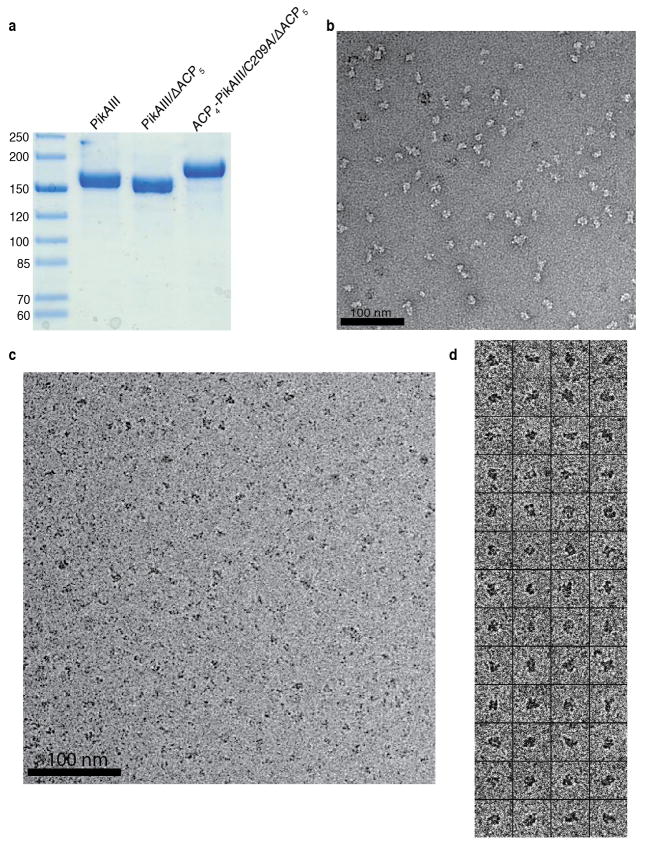
Extended Data Figure 1.
PikAIII sample preparation and raw EM images. a, SDS-PAGE gel of each purified form of PikAIII examined by cryo-EM. The numbers on the left indicate molecular weight in kDa. b, Raw EM image of holo-PikAIII particles embedded in negative stain. c, Raw cryo-EM image of holo-PikAIII particles. d, Boxed-out particle projections of holo-PikAIII.

