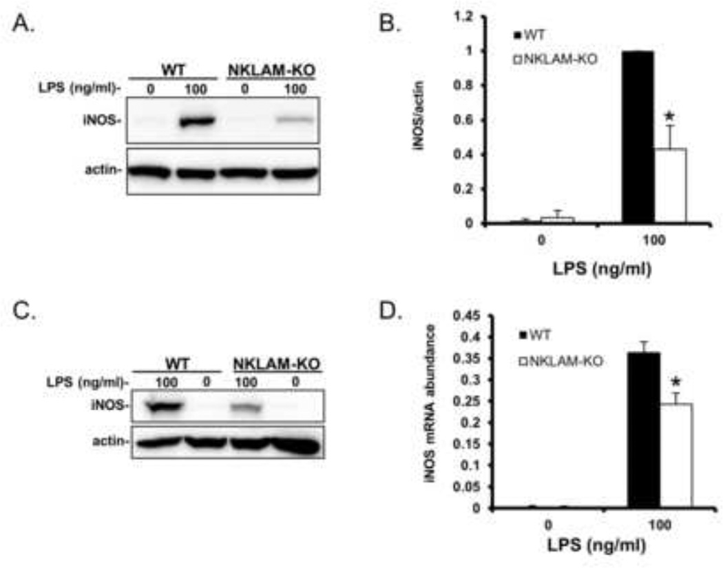
Figure 2. iNOS expression in WT and NKLAM-KO BMDM stimulated with LPS.
A) WT and NKLAM-KO BMDM were treated with 100 ng/ml LPS for18 h and whole cell lysates were made and immunoblotted for iNOS. B) Densitometric analysis for Fig. 2A; * p ≤ 0.05, n = 3. C) Splenic macrophages isolated from WT and NKLAM-KO mice were untreated or treated with 100 ng/ml LPS for 18 h and whole cell lysates were immunoblotted for iNOS protein. For all immunoblots, beta actin was used as a loading control. D) WT and NKLAM-KO BMDM were stimulated with 100 ng/ml LPS for 2 h. iNOS mRNA expression was determined by quantitative real-time PCR and the data are expressed as 2−ΔCt; * p ≤ 0.03, n = 4.

