Abstract
Background:
In the present era, because of the life-style, the disorders such as hyperacidity and gastric ulcers are found very frequently. Satwa (starch) obtained from the rhizomes of two plants namely Curcuma angustifolia Roxb. and Maranta arundinacea Linn. are used in folklore practice for the treatment of above complaints under the name Tugaksheeree.
Aim:
To compare the anti-ulcerogenic activity of the above two drugs in pyloric ligation induced gastric ulcer in albino rats.
Materials and Methods:
A total of 18 Wistar strain albino rats of both sexes grouped into three groups. Group C served as pyloric ligated control group, Group I received starch of C. angustifolia suspension and Group II received starch of M. arundinacea for seven days. On 8th day pylorus was ligated. After ligation the animals were deprived of food and water and sacrificed at the end of 14 h. The collected gastric contents were used for biochemical estimation and ulcer index was calculated from excised stomach.
Results:
Both the test drugs showed statistically significant decrease in the volume, increase in the pH, reduced the free acidity of gastric juice and decreased the peptic activity. The starch of C. angustifolia reduced a total acidity non-significantly while M. arundinacea reduced it significantly. Among the two drugs the M. arundinacea has effectively reduced the peptic activity, which is statistically significant. M. arundinacea shown statistically significant increase of total carbohydrates.
Conclusion:
Both the test drugs proved anti-ulcer activity and prevents the chance of gastric ulcer. Among these two M. arundinacea is more effective.
Keywords: Curcuma angustifolia, gastric ulcer, Maranta arundinacea, pyloric ligation, starch, Tugaksheeree
Introduction
Tugaksheeree is the drug described since Samhita period. It is an important drug and has been the ingredient of many medicinal formulations such as Chyavanaprasha,[1] Pippalyadyavaleha[2] etc., Satwa (starch) obtained from the rhizomes of two plants Curcuma angustifolia Roxb. (Family-Zingiberaceae) and Maranta arundinacea Linn. (Family-Marantaceae) which are used in the name of Tugaksheeree.[3,4,5] They are used in folklore practice in the conditions like hyperacidity and also as a nutritional food supplement. Until now, no scientific data is available about the efficacy of these drugs. Hence, the present study has been undertaken with the following aims and objectives:
To assess the effect of both the test drugs on pyloric ligation induced gastric ulcer
To assess relative efficacy of two plants as anti-ulcerogenic drugs.
Materials and Methods
Procurement of test drugs
The starch of C. angustifolia was collected from Purva Mandala District of Madhya Pradesh and the starch of M. arundinacea was collected from Dakshina Kannada District of Karnataka and authenticated in Pharmacognosy Laboratory of Institute for Post Graduate Teaching and Research in Ayurveda, Jamnagar.
Animals:
Wistar strain albino rats of both sexes weighing 180-260 g obtained from the animal house attached to the Pharmacology Laboratory, Institute for Post Graduate Teaching and Research in Ayurveda, Jamnagar was used for the experimental study. They were housed at 22 ± 2°C with constant humidity 50-60%, on a 12 h natural day and night cycles. They were fed with diet Amrut brand rat pellet feed supplied Pranav Agro Industries and tap water ad libitum. All the experiments were carried out after obtaining permission from Institutional Animal Ethics Committee (IAEC/07-08/01/07).
Dose fixation and schedule
The human dose of both the test drugs is 12 g/day. Considering adult human dose of both C. angustifolia and M. arundinacea starch, the dose for experimental study was calculated by converting the human dose to animal dose based on the body surface area ratio using the table of Paget and Barnes.[6]
Route of drug administration
The test drugs were administered by the oral route with the help of No. 6 gastric catheter sleeved onto a syringe.
Animal grouping
The selected 18 animals were grouped into three groups randomly irrespective of sex and each group comprises six animals.
Group C received tap water and served as pyloric ligated control group
Group I received starch of C. angustifolia as suspension in water in a dose of 1100 mg/kg body weight for seven consecutive days
Group II received starch of M. arundinacea as suspension in water in a dose of 1100 mg/kg body weight for seven consecutive days.
Procedure
The drugs were administered orally once daily for seven consecutive days to respective groups and vehicle to the control group. On the 8th day pylorus was ligated.[7] The rats were housed in individual metabolic cages to prevent caprophagy and overnight fasted with access to water ad libitum. Rats were anesthetized with ether anesthesia and the portion of the abdomen was opened in layer by a small midline incision just below and lateral to the xiphoid process. Pyloric portion of the stomach was slightly lifted out avoiding traction to the pylorus or damage to its blood supply. The pylorus was ligated with cotton thread and stomach was replaced carefully. The incision was closed with interrupted sutures in layers. The animals were deprived of both food and water during the post-operative period and were sacrificed by an over dose of ether at the end of 14 h after pyloric ligation. Abdominal cavity was reopened carefully and the stomach was excised after ligating the terminal portion of the esophagus to prevent the loss of gastric contents during excision [Figure 1]. Gastric contents were drained in to tubes and centrifuged at 3000 rpm for 15 min. Volume of gastric juice was noted and used for biochemical estimation.
Figure 1.
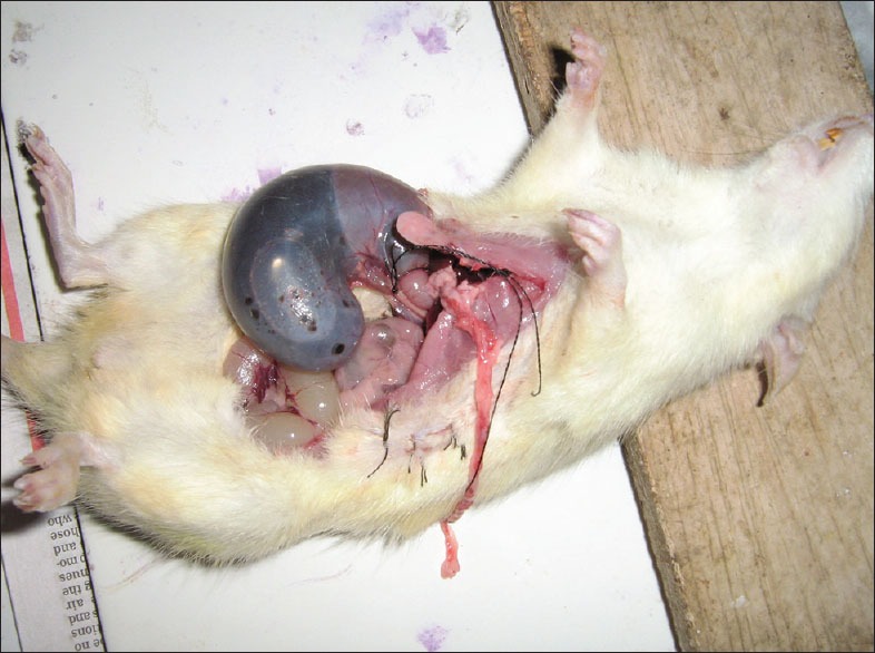
Stomach with gastric juice and ulcer after pyloric ligation
Assessment of stomach ulcer
The stomach was excised, cleaned and opened along its greater curvature and the inner surface gently washed with cold saline solution and examined for ulceration with a magnifying lens. Severity of ulcer and the total number of ulcer in each rat was recorded for calculating ulcer index. Ulcer index was calculated as follows:[8]
0-No visible ulcer
1-Approximate ulcerated area up to 0-1 mm diameter
2-Approximate ulcerated area up to 1-2 mm diameter
3-Approximate ulcerated area up to 3 mm diameter
4-Approximate ulcerated area more than 3 mm diameter
5-Perforation of the gastric wall.
Histopathological study
For histopathological study,[9] full thickness biopsy specimen were fixed in 10% formalin solution prior to dehydrating, wax embedding, sectioning and staining with hematoxylin and eosin, for histological evaluation of gastric damage by light microscopy.
Biochemical investigations
The following estimations were performed in gastric juice obtained from the sacrificed animals.
Other parameters
Change in body weight
Absolute and relative volume of gastric juice
pH of gastric juice
Ulcer index.
Statistical analysis
All the values were expressed as mean ± standard error of the mean. Data was analyzed by unpaired t- test. A level of P < 0.05 was considered to be statistically significant and the value of P < 0.01 or P < 0.001 was considered to be statistically highly significant. Level of significance was noted and interpreted accordingly.
Observations and Results
Effect of C. angustifolia and M. arundinacea starch on body weight in pyloric ligated rats
Data related to effect of test drugs on body weight in pyloric ligated rats has been given in Table 1. In comparison to their initial body weight all the groups showed a moderate increase in body weight. When the data of treated groups were compared with the pyloric control group an apparent and statistically non-significant increase in body weight was observed in both the test drug administered groups.
Table 1.
Effect of test drugs on body weight in pyloric ligated rats

Effect on volume and pH of gastric juice
Both the test drugs decreased the volume of gastric juice to statistically significant extent in comparison to pyloric ligated control rats when the values were presented as relative values. When presented as absolute values only the decrease observed with M. arundinacea was found to be statistically significant. An apparent and statistically significant increase in pH of gastric juice was observed in both the test drug administered groups in comparison to pyloric ligated control rats [Table 2].
Table 2.
Effect of test drugs on volume and pH of gastric juice in pyloric ligated rats

Effect on total and free acidity
An apparent decrease in total acidity was observed in both the test drug administered groups in comparison to pyloric ligated control rats, however only the decrease observed in M. arundinacea treated group was found to be statistically significant. An apparent and statistically highly significant decrease in free acidity was observed in both the test drug administered groups in comparison with the pyloric ligated control rats [Table 3].
Table 3.
Effect of test drugs on total and free acidity in pyloric ligated rats

Effect on peptic activity
An apparent decrease in peptic activity was observed in both the test drug administered groups in comparison to pyloric ligated control rats, however only the decrease observed in M. arundinacea treated group was found to be statistically significant [Table 4].
Table 4.
Effect of test drugs on peptic activity in pyloric ligated rats

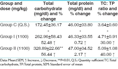
Effect on TC and TP and TC: TP ratio
An apparent increase in TC was observed in both the test drug administered groups in comparison to pyloric ligated rats; however only the decrease observed in M. arundinacea treated group was found to be statistically significant. Administration of test drugs did not affect the TP content in gastric juice of pyloric ligated rats in comparison to pyloric ligated control rats. An apparent increase in TC: TP ratio was observed in both the test drug administered groups in comparison to pyloric ligated control rats, however the observed increase in both the treated group was found to be statistically non-significant [Table 5].
Table 5.
Effect of test drugs on total carbohydrate, total protein and TC: TP ratio in pyloric ligated rats
Effect on ulcer index
An apparent decrease in ulcer index was observed in both the test drug administered groups in comparison to pyloric ligated control rats; however, the observed decrease was statistically non-significant [Table 6].
Table 6.
Effect of test drugs on ulcer index in pyloric ligated rats

Histopathological study of stomach
Microscopic examination of stomach sections obtained from different groups showed the following observations: in the stomach from pyloric ligated control group, massive destruction of the epithelial layer with hemorrhagic patches, sub-mucosal edema was observed [Figure 2]. In C. angustifolia treated group [Figure 3], the severity of the pathological changes observed was much less in comparison with the control group. In M. arundinacea treated group, the stomach cytoarchitecture depicted only minor changes [Figure 4].
Figure 2.
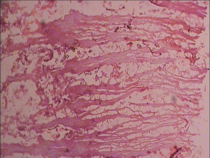
Photomicrograph of representative sections of stomach of albino rats from normal control group (1 × 100 magnification). Note: Massive destruction of the epithelial layer with hemorrhagic patches, submucosal edema
Figure 3.
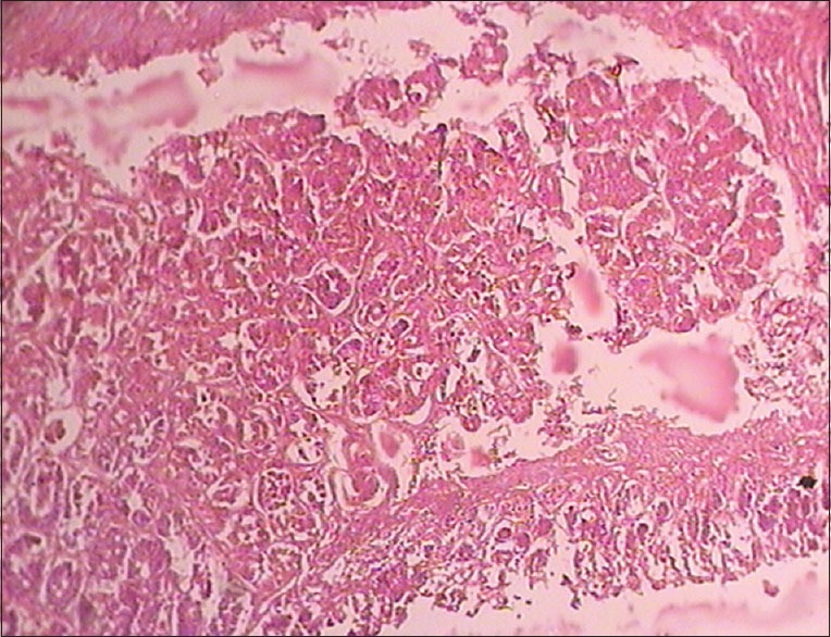
Photomicrograph of representative sections of stomach of albino rats from Curcuma angustifolia Roxb. treated group (1 × 100 magnification). Note: Comparatively less epithelial layer damage
Figure 4.
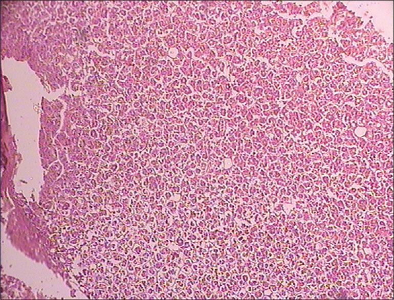
Photomicrograph of representative sections of stomach of albino rats from Maranta arundinacea Linn. treated group (1 × 100 magnification). Note: Almost normal cyto-architecture
Discussion
Ulcer is characterized by disruption of mucosal integrity leading to local defect or excavation due to active inflammation. Gastric ulcer being the most prevalent gastrointestinal disorder is considered as a major therapeutic target. The pathophysiology of ulcer is considered to be due to an imbalance between aggressive factors (acid, pepsin, Helicobacter pylori and non-steroidal anti-inflammatory drugs) and local mucosal defensive factors (mucous bicarbonate, blood flow and prostaglandins). Integrity of gastro duodenal mucosa is maintained through a homeostatic balance between these aggressive and defensive factors.[13]
In pylorus ligation model, it has been postulated that the digestive effect of accumulated gastric juice and interference with the gastric blood circulation are responsible for the induction of gastric ulcer.[14] Enhanced secretion of acid-pepsin leading to auto digestion of gastric mucosa and break down of mucosal barrier is also responsible for gastric ulcers.[15] Stimulation of pepsin secretion, with or without secretion of the acid, is the major factor in the development of gastric ulcers.[16]
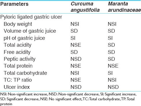
The activity profile of the test drugs C. angustifolia and M. arundinaceae against the pyloric ligated gastric ulcer is given in the form of the consolidated statement [Table 7].
Table 7.
The activity profile of the test drugs against the pyloric ligated gastric ulcer
Different aspects of the data generated during the study were analyzed to arrive at an inference with regards to the anti-gastric potential of the test drugs and if possible to suggest the probable mechanism of action.
Effect on body weight
Gain in the body weight indicates normal progressive health status of the animals and if decrease occurs it is considered to indicate interference with body functions leading to body weight loss or lower magnitude body weight gain. In the present study, loss in body weight was observed in all the groups including pyloric ligated control group, but the magnitude of loss in pyloric ligated control group is high whereas in the test formulations administered is less. The reason for this reversal of body weight loss may be due to the nutritional effect of the starches of both plants.
Effect on volume and pH of gastric juice
The volume of secretion of gastric juice is an important factor in the formation of ulcer due to the exposure of the unprotected lumen of the stomach to the accumulating acid. If the test drug is to be effective against the gastric ulcer it should have acidity decreasing effect. Acidity can be decreased by either anti-secretory effect that is to reduce the gastric juice secretion or the drug should neutralize the gastric acidity. Inhibition of acid may enhance healing of ulcers due to anti-secretory effect.
Starch of the both the test drugs showed statistically significant decrease in the volume of gastric juice. The volume of the gastric juice as mentioned above is mainly under the control of vagus. Both the drugs produced good anti-secretory effect. It would be interesting know the exact mechanism of this inhibition. Whether it is directly through the inhibition of muscarinic receptor or through ganglion blocking effect or through the direct effect on the secretory cells needs to be determined. It is also possible that the observed effect in both the drugs may be due to enhancing the function of enterogastrone or inhibition of gastrin or histamine.
Gastric juice is highly acidic with pH of 0.9-2.0. The acidity of gastric juice is due to the hydrochloric acid. Starch of both the test drugs produced a significant increase in gastric juice pH. It may be one of the contributing factors for prevention of gastric mucosal damage by over exposure to hydrochloric acid on pyloric ligation.
Effect on total and free acidity
Total acidity is the sum total of free HCl, organic acids, combined acid and acid salts. The starch of C. angustifolia reduced total acidity non-significantly while treatment with starch of M. arundinaceae reduced it significantly. This reduction in total acidity also contributes to anti-ulcer activity of test drugs.
The free acidity of the resting contents lies between 1.5 and 2.0 mEq of HCl. After the food is taken the acidity is reduced by dilution. The free acidity then steadily rises and becomes maximum 40-50 mEq of HCl in the 2nd h. Then it gradually declines. In gastric ulcer the value increases up to 3 times. Starch of the both the test drugs significantly reduced the free acidity of gastric juice in pyloric ligated rats thereby preventing the chances of gastric ulcers.
Effect on peptic activity
Both the test drug samples decreased the peptic activity indicating that they are capable of reducing the peptic activity and thereby decreasing the possibility of hyper action and secretion in the gastrium. Among the two drugs, the M.arundinaceae has better inhibitory effect on the peptic activity, which is statistically significant.
Effect on TC, TP and TC: TP ratio
The increase in TC content is due to stimulation of mucous secretion, which plays a vital role in protection of gastric mucosa which leads to a less degree of ulceration in treated group. Non-significant increase of TCs was observed in the C. angustifolia group. However in the M. arundinaceae treated group, a statistically significant increase was found indicating that the drug is capable of increasing the mucosal secretion.
Decrease in protein content signifies decreased leakage from gastric mucosa indicating increased strengthening of gastric mucosal barrier. Increase in glycoprotein content of the mucosa and cell shedding in the gastric juice were also reported as further evidence for strengthening of mucosal resistance. In the present study, no significant could be observed on the TP content of the gastric juice.
The TC: TP ratio serves as a direct index of gastric mucosal defense i.e. reflection of mucin activity. The essential basis which determines the status of mucosal defense against the offensive gastric and pepsin secretion is the quality and quantity of mucin secretion. The TC: TP ratio was not affected in C. angustifolia treated group while treatment with starch of M. arundinaceae non-significantly increased it. This indicates moderate strengthening of gastric mucosal barrier.
Effect on ulcer index
Starch from both the test drugs apparently decreased the ulcer index in pyloric ligated rats. Although the observed decrease in ulcer index is statistically non-significant they may be having good anti-ulcer activity as both the drugs significantly inhibited gastric secretion and reduced peptic activity.
Probable mechanism of action
Mucus produced by the gastric glands forms a thin protective layer over the entire mucosal surface of the stomach virtually preventing the contact of acid-pepsin with the mucosal layer. Bicarbonate secreted by the surface epithelial cells, which remains beneath and within the mucosal layer, further contributes to antacid property of the mucosal barrier. Increased blood flow to the gastric mucosa enhances the removal of acid from the mucosa. Faster regeneration and removal of mucosal cell also helps in the proper maintenance of protective mucosal barrier.
From the above resume, it becomes clear that any factor, which promotes aggressive factors and weakens defense mechanism, leads to ulcer formation. In the present study, the test drugs especially M. arundinaceae produced significant suppression of the aggressive factors in the form of suppression of secretion of gastric juice, decreasing the acid content of the gastric juice and suppressing the peptic activity. In addition to this, it also strengthens the defense mechanism as evident in the form of increased carbohydrate content and moderate elevation in the TC: TP ratio. Thus, suppression of the aggressive factors seems to be the main mechanism of action with moderate enhancement of mucosal defense mechanism enhancing effect in the test drugs especially M. arundinaceae; how this is brought about would be interesting to elucidate. The possibilities are blocking of the muscarinic acetylcholine receptors, blocking of histamine receptor or inhibition of the proton pump. It is also possible that the test drug may be promoting the release or activity of the inhibitory factors such as somatostatin and cholecystokinin. In addition, they may be acting on other factors like increased turnover of gastric epithelial cell, increased blood supply to the gastric tissue. Among the other possible mechanisms are enhancement of mucosal hydrophilicity, increase in the rate of epithelial cell renewal, increase in the formation and secretion of mucosal sulphahydryls and increased resistance of gland cells in deep mucosa to acid and peptic activity. It is also possible that the test drugs may be interfering with the entry of calcium ions, which are involved in the regulation of acid secretion in the stomach. Thus this provides an important area for future research efforts.
Conclusion
Both the test drugs contribute anti-ulcer activity by reducing total acidity; in this context, starch of M. arundinacece is more effective. Starches from both the samples have the capacity to prevent the chances of gastric ulcers by reducing the free acidity of gastric juice in pyloric ligated rats. Both the test drugs may have the property of decreasing the hyper action and secretion in the gastrium as revealed by decreased peptic activity. Both the drugs may be capable to increase the blood supply to the gastric mucosa or enhancement of gastric epithelial cells turn over by increasing the TCs content; in this property, M. arundinacece is more potent. Among the two, the starch of M. arundinacece has better anti-ulcerogenic activity than that of C. angustifolia revealed by decreasing in ulcer index.
Footnotes
Source of Support: Nil
Conflict of Interest: None declared.
References
- 1.Agnivesha, Charaka Samhita, Chikitsa Sthana. Rasayana Adhyaya, Abhayamalaki Rasayanpad 1-1/68. In: Yadavji Trikamji Acharya., editor. reprint edition. Varanasi: Chaukhambha Surabharati Prakashan; 2011. p. 379. [Google Scholar]
- 2.Vangasena, Vangasena Samhita, Amlapittarogadhikara . 84-88 commentary by Shaligramji Vaidya. In: Shankarlalji Vaidya., editor. 1st ed. Mumbai: Khemraj Shrikrishnadass; 2003. p. 655. [Google Scholar]
- 3.Sharma PV. Reprint ed. Varanasi: Chaukhambha Bharati Academy; 2003. Dravyaguna Vijnana. Part II; pp. 612–742. [Google Scholar]
- 4.Bapalal G. II. Varanasi: Chaukhambha Bharati Academy; 2005. Vaidya, Nighatu Adrasha; pp. 558–80. [Google Scholar]
- 5.Lucas DS. II. Varanasi: Chaukhambha Vishwabharathi; 2008. Dravyaguna Vijnana; pp. 672–73. [Google Scholar]
- 6.Paget GE, Berns JM. Evaluation of Drug Activities: Pharmacometrics. In: Lawranle DR, Bacharch AL, editors. Vol. 1. New York: Academic Press; 1969. [Google Scholar]
- 7.Shay H, Komarov SA, Fels SE, Meraze D, Gruenstein M, Siplet H. A simple method for the uniform production of gastric ulceration in rat. Gastroenterology. 1945;5:43–61. [Google Scholar]
- 8.Kulkarni SK. Handbook of Experimental Pharmacology. 3rd ed. Delhi: Vallabh Prakashan; 1999. To study the antisecretory and ulcer protective effect of cimetidine in pylorus-ligated rats; pp. 148–50. [Google Scholar]
- 9.Raghuramulu N, Nair KM, Kalyanasundaram S, editors. Hyderabad, India: National Institute of Nutrition (NIN); 1983. A Manual of Laboratory Techniques; pp. 246–53. [Google Scholar]
- 10.Debnath PK, Gode KD, Das DG, Sanyal AK. Effects of propranolol on gastric secretion in albino rats. Br J Pharmacol. 1974;51:213–6. doi: 10.1111/j.1476-5381.1974.tb09649.x. [DOI] [PMC free article] [PubMed] [Google Scholar]
- 11.Nair RB. Ph. D Thesis. Trivandrum: University of Kerala; 1976. In investigations on the venom of South Indian scorpion Heterometrus scaber; p. 39. [Google Scholar]
- 12.Lowry OH, Rosebrough NJ, Farr AL, Randall RJ. Protein measurement with the Folin phenol reagent. J Biol Chem. 1951;193:265–75. [PubMed] [Google Scholar]
- 13.Dhamani P, Kumar KV, Srivastava S, Palit G. Ulcer healing effect of anti-ulcer agents: A comparative study. Internet J Acad Physician Assist. 2003;3(2) Available on www.ispub.com/IJAPA/3/2/7604 . [Google Scholar]
- 14.Brodie DA, Hanson HM. A study of the factors involved in the production of gastric ulcers by the restraint technique. Gastroenterology. 1960;38:353–60. [PubMed] [Google Scholar]
- 15.Goel RK, Bhattacharya SK. Gastroduodenal mucosal defence and mucosal protective agents. Indian J Exp Biol. 1991;29:701–14. [PubMed] [Google Scholar]
- 16.Anoop A, Jegadeesan M. Biochemical studies on the anti-ulcerogenic potential of Hemidesmus indicus R. Br. var. indicus. J Ethnopharmacol. 2003;84:149–56. doi: 10.1016/s0378-8741(02)00291-x. [DOI] [PubMed] [Google Scholar]


