Abstract
Purpose
To identify the genetic basis of achromatopsia (ACHM) in four patients from four unrelated Polish families.
Methods
In this study, we investigated probands with a clinical diagnosis of ACHM. Ophthalmologic examinations, including visual acuity testing, color vision testing, and full-field electroretinography (ERG), were performed in all patients (with the exception of patient p4, who had no ERG). Direct DNA sequencing encompassing the entire coding region of the CNGB3 gene, eight exons of the GNAT2 gene, and exons 5–7 of the CNGA3 gene was performed. Segregation analysis for the presence and independent inheritance of two mutant alleles was performed in the three families available for study.
Results
All patients showed typical achromatopsia signs and symptoms. Sequencing helped detect causative changes in the CNGB3 gene in all probands. Eight different mutations were detected in the CNGB3 gene, including five novel mutations: two splice site mutations (c.1579–1G>A and c.494–2A>T), one nonsense substitution (c.1194T>G), and two frame-shift mutations (c.393_394delGCinsTCCTGGTGA and c.1366delC). We also found three mutations: one splice site (c.1578+1G>A) and two frame-shift deletions that had been previously described (c.819_826del and c.1148delC). All respective parents were shown to be heterozygous carriers for the mutation detected in their children.
Conclusions
The present study reports five novel mutations in the CNGB3 gene, and thus broadens the spectrum of probably pathogenic mutations associated with ACHM. Together with molecular data, we provide a brief clinical description of the affected individuals.
Introduction
Achromatopsia (ACHM; OMIM 216900) or rod monochromacy is a rare autosomal recessive cone disorder with a prevalence of 1 in 30,000 individuals. ACHM is characterized by reduced visual acuity, nystagmus, photophobia, a small central scotoma, and total loss of color discrimination. Although visual acuity is usually stable over time, nystagmus and hypersensitivity to bright light may decrease slightly. Most individuals have complete achromatopsia with a total lack of function of three types of cone photoreceptors. Incomplete achromatopsia is characterized by the residual ability to discriminate colors. Electroretinographic recordings (ERGs) show normal function of rod photoreceptors and no recordable or only residual cone function [1].
To date, five genes have been shown to be associated with ACHM. They code for cyclic nucleotide-gated cation channel beta-3 (CNGB3; chr.8q21-q22; OMIM 605080), cyclic nucleotide-gated cation channel alpha-3 (CNGA3; chr.2q11; OMIM 600053), guanine nucleotide-binding protein, alpha transducing activity polypeptide 2 (GNAT2; chr.1p13; OMIM 139340), phosphodiesterase 6C, cGMP-specific, cone alpha prime (PDE6C; chr.10q24; OMIM 600827), and the most recently associated gene, phosphodiesterase 6H, cGMP-specific, gamma (PDE6H; chr.12p12.3; OMIM 601190) [2-6]. Mutations in the CNGB3 gene underlie approximately 50% of ACHM cases. Most of the CNGB3 mutations are frame-shift deletions, insertions, nonsense, or splice-site mutations [3,7,8]. Twenty-five percent of all patients with ACHM have mutations in the CNGA3 gene [5,9,10]. The proteins encoded by these genes are expressed in the cone photoreceptor cells and are crucial for the cone phototransduction cascade [1]. Most mutant proteins encoded by CNGB3 are thought to be null alleles [11,12]. In vitro expression analyses have shown that most CNGA3 mutations lead to the lack of channel activity. Mutant proteins are synthesized but are retained in the endoplasmic reticulum and, therefore, impair cellular traffic [9,13]. Functional analysis with heterologous expression of mutant cyclic nucleotide-gated (CNG) channels and animal models helped to understand the underlying pathogenic mechanism in ACHM. Analysis of a Cngb3 knockout mouse model also shows reduced biosynthesis of Cnga3 and impaired cone CNG channel function. Ding et al. suggested that CNGB3 mutations may contribute, by the downregulation of CNGA3, to the pathogenic mechanism leading to human cone disease [14].
New progress in gene therapy provides great hope for individuals who have ACHM. In 2010, Komáromy et al. restored visual function by using recombinant adenoassociated virus (rAAV)-mediated gene replacement therapy in two canine models of CNGB3 ACHM [15]. Because the treatment was effective only in young dogs (<0.5 years old), the researchers then modified and examined combined gene therapy with the administration of ciliary neurotrophic factor (CNTF) and achieved successful cone rescue among all age groups [16]. At the same time, CNGB3 gene replacement using AAV was applied in several mouse models and showed long-term improvement in retinal function with significant rescue of cone ERG amplitudes [17-19]. Recently, scientists from University College London (UCL) announced they intended to create and test a newly engineered viral vector applicable for use in clinics and conduct a phase I/II clinical trial for gene therapy in patients with ACHM who carry mutations in the CNGB3 gene. The project is expected to end in 2018 (Gateway to Research).
The group of patients comprised ten independent Polish probands with a diagnosis of congenital achromatopsia. Known mutations in the CNGA3 gene were identified in six patients (Table 1). Therefore, in the present study, we report four patients with ACHM, compound heterozygotes with at least one novel CNGB3 gene alteration.
Table 1. Identified CNGA3 gene mutations.
| Patient | Mutation | Effect of mutation | Allelic state | References |
|---|---|---|---|---|
| p5 |
c.829C>T (allele 1) |
p.R277C |
heterozygous |
Wissinger et al., Am. J. Hum. Genet., 2001 |
| c.1641C>A (allele 2) |
p.F547L |
heterozygous |
Kohl et al., Nat Genet., 1998 |
|
| p6 |
c.829C>T (allele 1) |
p.R277C |
heterozygous |
Wissinger et al., Am. J. Hum. Genet., 2001 |
| c.1641C>A (allele 2) |
p.F547L |
heterozygous |
Kohl et al., Nat Genet., 1998 |
|
| p7 |
c.1641C>A |
p.F547L |
homozygous |
Kohl et al., Nat Genet., 1998 |
| p8 |
c.829C>T (allele 1) |
p.R277C |
heterozygous |
Wissinger et al., Am. J. Hum. Genet., 2001 |
| c.847C>T (allele 2) |
p.R283W |
heterozygous |
Kohl et al., Nat Genet., 1998 |
|
| p9 |
c.1641C>A |
p.F547L |
homozygous |
Kohl et al., Nat Genet., 1998 |
| p10 | c.830G>A | p.R277H | homozygous | Wissinger et al., Am. J. Hum. Genet., 2001 |
Methods
Clinical studies
Four patients from four unrelated Polish families from different regions of Poland and manifesting clinical features of ACHM participated in this study. Ophthalmologic examinations, including visual acuity testing, color vision testing (D-15 Panel and Ishihara Plate Test), and full-field ERG were performed in all patients (with the exception of patient p4, who had no ERG). This study conforms to the Helsinki declaration and was approved by the Poznan University of Medical Sciences Institutional Review Board. Written informed consent was obtained from all subjects or their legal guardians.
Molecular genetic analysis
Venous blood samples were taken from the patients and their parents (with the exception of family #4; the parents were not available for genetic analysis). Genomic DNA was extracted from leukocytes using the standard salting-out procedure [20]. Blood was collected into tubes containing EDTA and stored in a refrigerator until DNA isolation was performed [20]. Genomic DNA was extracted from leukocytes. In patient p4, initial microarray analysis toward Leber congenital amaurosis (LCA) was performed (Asper Biotech, Tartu, Estonia).
The genetic analysis of the patients with ACHM included PCR amplification of genomic DNA and sequencing of all coding exons and flanking intronic sequences of the CNGB3 gene (18 exons), the GNAT2 (eight exons) gene, as well as exons 5–7 of the CNGA3 gene. The primers used for PCR and sequencing and PCR conditions are included as a Appendix 1. PCR fragments were purified using ExoSAP-IT (GE Healthcare, Freiburg, Germany) and then subjected to direct DNA sequencing applying BigDye Terminator chemistry (Applied Biosystems, Darmstadt, Germany). All sequences were run on an ABI 3100 capillary sequencer (Applied Biosystems), trace files were analyzed with Sequencing Analysis 5.2 (Applied Biosystems, Life Technologies Corporation, Carlsbad, CA) and sequence variants called with SeqPilot software (JSI Medical Systems, Kippenheim, Germany). The primers used for PCR and sequencing are included as a supplementary table.
The sequences were verified by comparing them to the human genomic sequence of CNGB3 (GenBank NG_016980.1) and screened for mutations. Identified variations were referred to the Exome Variant Server (NHLBI Exome Sequencing Project ESP) and the Human Gene Mutation Database (HGMD) for CNGB3. In silico analysis using Net2Gene software was used for the splice-site mutations to predict possible 5′ and 3′ splice sites.
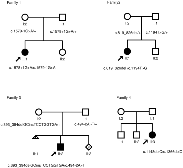
Segregation analysis for the presence and independent inheritance of two mutant alleles was performed in three families available for study. Analysis was done by sequencing the appropriate exons of the CNGB3 gene. Pedigrees of the available families with ACHM are present in Figure 1.
Figure 1.
Pedigrees and genotyping results of families with achromatopsia. The genotypes are provided for all subjects available for molecular genetic analysis. The black square and the circles represent affected male and females, respectively. White squares and circles represent unaffected family members. A triangle indicates miscarriage, and a rhombus indicates ongoing pregnancy in family 3. Arrows point to probands.
Results
Clinical features
The clinical diagnosis was based on the presence of photophobia and nystagmus, low visual acuity, total color blindness, and ERG (with the exception of patient p4, ERG was not performed). All patients showed typical ACHM signs and symptoms.
Patient p1 was a 27-year-old woman, initially referred to the eye clinic due to nystagmus and photophobia. Pendular nystagmus was noted during early infancy. P1 presented symptoms of low visual acuity 0.2 in both eyes. ERG recordings revealed normal scotopic and mildly abnormal photopic responses. The inability to distinguish colors was also observed. The fundus appearance was normal. The parents and a younger sister of the patient had no ophthalmologic problems.
Patient p2 was a 12-year-old girl. She initially had nystagmus in early infancy. Photophobia was not present. ERG recordings were consistent with the ACHM pattern: normal scotopic and non-recordable photopic responses. No response was also observed for cone-specific 30 Hz flicker stimulation (Figure 2). The patient displayed normal eye fundus. P2 presented reduced visual acuity 0.08 in the right eye and 0.1 in the left eye. Loss of color discrimination was observed. The patient has a younger brother, but neither her brother nor their parents showed any eye problems.
Figure 2.

ERG of p2. A: Photopic white flash electroretinogram (ERG) shows non-recordable response. B: Scotopic white flash ERG shows normal response. C: Photopic white 30 Hz flicker shows flat response. 1-L indicates the left eye; 2-R indicates the right eye.
Patient p3 (6-year-old boy) was diagnosed due to pendular nystagmus and photophobia in early infancy. ERG findings showed non-recordable photopic and normal scotopic responses. P3 manifested reduced visual acuity 0.08 in both eyes. In contrast to the other patients, he demonstrated only decreased color discrimination. He distinguished acute primary colors, red, blue, and green in everyday life, probably based on the contrast differences. The patient is the only child of healthy, unrelated parents, who have no ophthalmologic problems.
In patient p4 (7-year-old girl), ERG examination was not performed. Therefore, this patient was initially suspected to suffer from Leber congenital amaurosis or cone-rod dystrophy. This diagnosis was based on the patient’s clinical history, in which symptoms such as low visual acuity 0.13 in both eyes and nystagmus were apparent. However, after genetic analysis, the patient was reclassified as having ACHM. P4 also presented the inability to distinguish colors. The fundus appearance was normal in all three patients while p4 showed an optic disc pallor and disturbed foveal structure.
Generally, the clinical course of the disease was stationary, but the degree of nystagmus decreased in older patients. Ophthalmologic findings for the patients are summarized in Table 2.
Table 2. Clinical findings in Polish achromatopsia patients.
| Patient | Age (years) | Gender | Photophobia | Visual acuity | Nystagmus | ERG scotopic | ERG photopic | Other ophthalmologic symptoms |
|---|---|---|---|---|---|---|---|---|
| p1 |
27 |
female |
+ |
RE:0.2
LE:0.2 |
+ |
mildly abnormal |
absent |
loss of color discrimination,
improvement of vision in darkness, normal AF |
| p2 |
12 |
female |
- |
RE:0.08
LE:0.1 |
+ |
normal |
absent |
loss of color discrimination,
improvement of vision in darkness, hyperopia +5,0D |
| p3 |
6 |
male |
+ |
RE:0.08
LE:0.08 |
+ |
normal |
absent |
reduced color discrimination (differentiates primary colors red-blue-green), improvement of vision in darkness, hyperopic astigmatism |
| p4 | 7 | female | + | RE:0.13 LE:0.13 | + | N/A | N/A | loss of color discrimination, improvement of vision in darkness, hyperopic astigmatism, optic disc pallor |
Molecular genetic findings
In p4, additional microarray analysis was performed to confirm Leber congenital amaurosis based on an Asper Biotech test containing 641 disease-associated variants. No mutation was detected in the 13 genes that were analyzed.
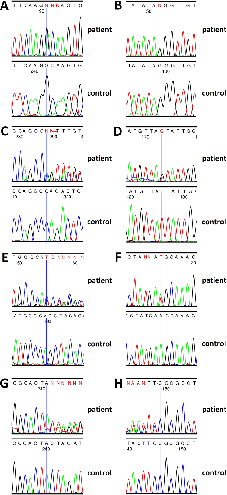
Genomic DNA samples from all four patients were subjected to molecular genetic analysis consisting of DNA sequencing of three known genes: CNGA3, CNGB3, and GNAT2. Genetic screening did not identify any mutations in CNGA3 and GNAT2. All patients were shown to carry compound heterozygous mutations in CNGB3. Among the eight different mutations that were identified in this study, three are common, known mutations while the rest are novel and have yet to be described in other patients with ACHM (Table 3). CNGB3 gene sequencing in p1 revealed a novel and a known heterozygous splice-site mutation in intron 13: c.1579–1G>A and c.1578+1G>A, respectively [3]. In silico analysis results (from the splice site prediction; Net2Gene software), together with the fact that the intronic mutation includes sequence critical for splicing, indicate that these substitutions affect the RNA splicing process. One novel nonsense substitution c.1194T>G (p.Tyr398*) in exon 11 and one previously described frame-shift mutation c.819_826del (p.Arg274Valfs*) in exon 6 was found in p2 [7]. The mutation c. 819_826del induces a frame-shift deletion of 8 bp that eliminated protein sequences, including the critical pore, S6 transmembrane, and cGMP-binding domains, resulting in a premature termination of translation. Two novel mutations, a frame-shift mutation c.393_394delGCinsTCCTGGTGA (p.Gln131Hisfs*50) in exon 4 and a splice-site mutation c.494–2A>T in intron 4, were identified in p3. Two frame-shift mutations were discovered in p4, one novel mutation c.1366delC (p.Arg456Alafs*11) in exon 12 and one known mutation c.1148delC (p.Thr383Ilefs*13) in exon 10 [7]. The p.Thr383Ilefs*13 mutation resulting from c.1148delC causes a frame-shift leading to premature termination of translation. The CNGB3 gene mutations that were identified are presented in Figure 3.
Table 3. Identified CNGB3 gene mutations.
| Patient | Mutation | Type of mutation | Effect of mutation | Allelic state | Mutation origin | References |
|---|---|---|---|---|---|---|
| p1 |
c.1578+1G>A (allele 1) |
splice site |
splicing defect |
heterozygous |
paternal |
Kohl et al., Hum Mol Genet; 2000 |
| c.1579–1G>A (allele 2) |
splice site |
splicing defect |
heterozygous |
maternal |
this study |
|
| p2 |
c.819_826del (allele 1) |
frameshift |
p.Arg274Valfs* |
heterozygous |
maternal |
Sundin et al., Nature Genet; 2000 |
| c.1194T>G (allele 2) |
nonsense |
p.Tyr398* |
heterozygous |
paternal |
this study |
|
| p3 |
c.393_394delGCinsTCCTGGTGA (allele 1) |
frameshift |
p.Gln131Hisfs*50 |
heterozygous |
maternal |
this study |
| c.494–2A>T (allele 2) |
splice site |
splicing defect |
heterozygous |
paternal |
this study |
|
| p4 | c.1148delC (allele 1) |
frameshift |
p.Thr383Ilefs*13 |
heterozygous |
no data |
Sundin et al., Nature Genet; 2000 |
| c.1366delC (allele 2) | frameshift | p.Arg456Alafs*11 | heterozygous | no data | this study |
Figure 3.
Chromatograms showing eight mutations identified in the CNGB3 gene in patients with ACHM. A: Sequence trace of part of intron 13 in an affected individual p1 carrying a heterozygous mutation c.1578+1G>A (upper panel) and a normal control individual (lower panel). B: Part of intron 13 in an affected individual p1 carrying a heterozygous mutation c.1579–1G>A (upper panel) and a normal control individual (lower panel). C: Part of exon 6 in an affected individual p2 carrying a heterozygous mutation c. 819_826del (upper panel) and a normal control individual (lower panel). D: Part of exon 11 in an affected individual p2 carrying a heterozygous mutation c.1194T>G (upper panel) and a normal control individual (lower panel). E: Part of exon 4 in an affected individual p3 carrying a heterozygous mutation c.393_394delGCinsTCCTGGTGA (upper panel) and a normal control individual (lower panel). F: Part of intron 4 in an affected individual p3 carrying a heterozygous mutation c.494–2A>T (upper panel) and a normal control individual (lower panel). G: Part of exon 10 in an affected individual p4 carrying a heterozygous mutation c.1148delC (upper panel) and a normal control individual (lower panel). H: Part of exon 12 in an affected individual p4 carrying a heterozygous mutation c.1366delC (upper panel) and a normal control individual (lower panel).
Segregation analysis, by sequencing appropriate exons and exon/intron boundaries of CNGB3, was performed in the three available families (family 1, 2, and 3). The analysis revealed that all respective parents are heterozygous carriers for the mutations detected in their children. Thus, segregation analysis was consistent with the autosomal recessive mode of inheritance (Figure 1).
Discussion
Eight different mutations were found in the CNGB3 gene in four patients with ACHM. While c.1578+1G>A, c.819_826del (p.Arg274Valfs*), and c.1148delC (p.Thr383Ilefs*13) are common mutations, c.1579–1G>A, c.1194T>G (p.Tyr398*), c.393_394delGCinsTCCTGGTGA (p.Gln131Hisfs*50), c.494–2A>T, and c.1366delC (p.Arg456Alafs*11) are mutations that, thus far, have not been reported in other patients with ACHM.
The most frequent type of mutations identified in our small group of patients were frame-shift deletions: c.819_826del, c.1148delC, c.393_394delGCinsTCCTGGTGA, and c.1366delC. This finding corroborates the observations of Kohl et al. and Sundin et al., who relied on studies of larger groups of patients [3,7,8]. Novel frame-shift deletions detected in our probands p.Gln131Hisfs*50 and p.Arg456Alafs*11, were found with second disease alleles. Mutation p.Gln131Hisfs*50 were identified with a novel splice-site alteration c.494–2A>T in intron 4 (p3). The mutation p.Gln131Hisfs*50 is located in a Ca2+/calmodulin domain (CaM). The outcome of this mutation is a frame-shift of the open reading frame that occurs after the transmembrane domain S5, which therefore eliminates the pore (P), the transmembrane domain S6, and the cGMP binding site of the protein. The fact that the splice-site mutation c.494–2A>T alters the conserved splice acceptor sequence of exon 5, together with results from in silico analysis (splice site prediction Net2Gene software), indicate that this substitution affects the RNA splicing process. Parents were also found to be healthy carriers of the mutations detected in their child (Figure 1).
The most common CNGB3 mutation, namely, p. Thr383Ilefs*13, in accordance with the reported prevalence of the mutations in patients originating from Europe, was also present in our probands. This mutation accounts for approximately 70% of all CNGB3 mutant alleles [8]. This mutation with a second disease allele p.Arg456Alafs*11 (c.1366delC) was identified in p4. The c.1366delC mutation, to date, is unique to this patient. It is present in the S6 transmembrane protein domain. Mutation c.1366delC results in a frame-shift and premature termination codon, truncating the CNGB polypeptide between transmembrane S6 and the cGMP binding site.
A nonsense substitution c.1194T>G (p.Tyr398*) in exon 11 of the CNGB3 gene was detected in p2. Mutation p.Tyr398* has not been reported to date. However, nonsense mutations localized in the same exon of the CNGB3 gene that result in a truncated protein have already been reported: 1255G>T (p.Glu419*) and 1298_1299delTG (p.Val433fs*) in patients with ACHM [8]. The mutation truncates the CNGB3 polypeptide at the pore and was found together with the previously reported mutation c.819_826del (p.Arg274Valfs*)]. Both parents carried one of these two mutations (Figure 1).
All mutations detected in our patients were found to cause premature termination of translation, giving rise to truncated CNGB3 polypeptides that probably represent null alleles. This finding is consistent with those of Kohl et al. (2005), Nishiguchi et al. (2005), and many other researchers who analyzed larger groups of patients with ACHM. The researchers stated that premature translation termination is the most common effect of the mutations described in CNGB3-related patients with ACHM [8,21].
The clinical diagnosis of achromatopsia is based on the presence of typical clinical findings as outlined in the results. However, the fundus appearance and the visual fields also contribute to the diagnosis. In contrast to other patients, p4 demonstrated optic disc pallor and disturbed foveal structure. This signs are not unusual in ACHM; some patients exhibit macular changes and atrophy. This finding is in agreement with Khan et al.’s (2007) and Thiadens et al.’s (2009) observations. The spectrum of foveal pathology and likewise fundus changes have been described in individuals with CNGB3 mutations. However, the CNGB3 mutations did not seem to correlate with specific phenotypes [5,22].
ACHM is a rare cone disorder with uncharacteristic early symptoms during childhood and, for this reason, may be confused with other retinopathies such as Leber congenital amaurosis, cone-rod dystrophy, or albinism. P4 had been previously diagnosed as suffering from Leber congenital amaurosis or cone-rod dystrophy. Diagnosis was based, inter alia, on decreased visual acuity and optic disc pallor present in early childhood. In contrast to ACHM, the most typical symptoms in these disorders is disease progression, while visual acuity is usually stable over time in ACHM. The identification of causative mutations in the CNGB3 gene aided the correct diagnosis of ACHM in p4. Similarly to many other cases in which genetic mutation is the cause of the disease, molecular analysis augmented diagnosis in clinical practice.
To conclude, our paper is the first report that provides the results of molecular analysis of the CNGB3 gene in Polish patients with ACHM. We furthermore expand the mutational spectrum associated with ACHM by describing the five novel pathogenic alterations in the CNGB3 gene. In addition, although based on a small sample, our study supports the hypothesis that CNGB3 mutations spread almost over all coding exons of the gene, what makes molecular diagnostic more difficult and requires sequencing of the entire coding region of the CNGB3 gene [8].
Acknowledgments
This study was partially supported by a grant from the Polish Ministry of Science and Higher Education (806/N-NIEMCY/2010/0)
Appendix 1. GNAT2 primers for PCR and sequencing.
To access the data, click or select the words “Appendix 1.”
References
- 1.Kohl S, Jägle H, Sharpe LT, Wissinger B. Achromatopsia. GeneReviews™. University of Washington, Seattle. 1993-2004 [Google Scholar]
- 2.Kohl S, Marx T, Giddings I, Jagle H, Jacobson SG, Apfelstedt-Sylla E, Zrenner E, Sharpe LT, Wissinger B. Total colour blindness is caused by mutations in the gene encoding the alpha-subunit of the cone photoreceptor cGMP-gated cation channel. Nat Genet. 1998;19:257–9. doi: 10.1038/935. [DOI] [PubMed] [Google Scholar]
- 3.Kohl S, Baumann B, Broghammer M, Jagle H, Sieving P, Kellner U, Spegal R, Anastasi M, Zrenner E, Sharpe LT, Wissinger B. Mutations in the CNGB3 gene encoding the beta-subunit of the cone photoreceptor cGMP-gated channel are responsible for achromatopsia (ACHM3) linked to chromosome 8q21. Hum Mol Genet. 2000;9:2107–16. doi: 10.1093/hmg/9.14.2107. [DOI] [PubMed] [Google Scholar]
- 4.Aligianis IA, Forshew T, Johnson S, Michaelides M, Johnson CA, Trembath RC, Hunt DM, Moore AT, Maher ER. Mapping of a novel locus for achromatopsia (ACHM4) to 1p and identification of a germline mutation in the alpha subunit of cone transducin (GNAT2). J Med Genet. 2002;39:656–60. doi: 10.1136/jmg.39.9.656. [DOI] [PMC free article] [PubMed] [Google Scholar]
- 5.Thiadens AAHJ, den Hollander AI, Roosing S, Nabuurs SB, Zekveld-Vroon RC, Collin RW, Baere ED, Koenekoop RK, van Schooneveld MJ, Strom TM, van Lith-Verhoeven JJC, Lotery AJ, van Moll-Ramirez N, Leroy BP, van den Born LI, Hoyng CB, Cremers FPM, Klaver CCW. Homozygosity mapping reveals PDE6C mutations in patients with early-onset cone photoreceptor disorders. Am J Hum Genet. 2009;85:240–7. doi: 10.1016/j.ajhg.2009.06.016. [DOI] [PMC free article] [PubMed] [Google Scholar]
- 6.Kohl S, Coppieters F, Meire F, Schaich S, Roosing S, Brennenstuhl C, Bolz S, van Genderen MM, Riemslag FC, European Retinal Disease Consortium Lukowski R, den Hollander AI, Cremers FP, De Baere E, Hoyng CB, Wissinger B. A nonsense Mutation in PDE6H Causes Autosomal-Recessive Incomplete Achromatopsia. Am J Hum Genet. 2012;91:527–32. doi: 10.1016/j.ajhg.2012.07.006. [DOI] [PMC free article] [PubMed] [Google Scholar]
- 7.Sundin OH, Yang JM, Li Y, Zhu D, Hurd JN, Mitchell TN, Silva ED, Maumenee IH. Genetic basis of total colourblindness among the Pingelapese islanders. Nat Genet. 2000;25:289–93. doi: 10.1038/77162. [DOI] [PubMed] [Google Scholar]
- 8.Kohl S, Varsanyi B, Antunes GA, Baumann B, Hoyng CB, Jägle H, Rosenberg T, Kellner U, Lorenz B, Salati R, Jurklies B, Farkas A, Andreasson S, Weleber RG, Jacobson SG, Rudolph G, Castellan C, Dollfus H, Legius E, Anastasi M, Bitoun P, Lev D, Sieving PA, Munier FL, Zrenner E, Sharpe LT, Cremers FP, Wissinger B. CNGB3 mutations account for 50% of all cases with autosomal recessive achromatopsia. Eur J Hum Genet. 2005;13:302–8. doi: 10.1038/sj.ejhg.5201269. [DOI] [PubMed] [Google Scholar]
- 9.Reuter P, Koeppen K, Ladewig T, Kohl S, Baumann B, Wissinger B. Achromatopsia Clinical Study Group. Mutations in CNGA3 impairs trafficking or function of cone cyclic nucleotide-gated channels, resulting in achromatopsia. Hum Mutat. 2008;29:1228–36. doi: 10.1002/humu.20790. [DOI] [PubMed] [Google Scholar]
- 10.Wissinger B, Gamer D, Jagle H, Giorda R, Marx T, Mayer S, Tippmann S, Broghammer M, Jurklies B, Rosenberg T, Jacobson SG, Sener EC, Tatlipinar S, Hoyng CB, Castellan C, Bitoun P, Andreasson S, Rudolph G, Kellner U, Lorenz B, Wolff G, Verellen-Dumoulin C, Schwartz M, Cremers FPM, Apfelstedt-Sylla E, Zrenner E, Salati R, Sharpe LT, Kohl S. CNGA3 mutations in hereditary cone photoreceptor disorders. Am J Hum Genet. 2001;69:722–37. doi: 10.1086/323613. [DOI] [PMC free article] [PubMed] [Google Scholar]
- 11.Peng C, Rich ED, Varnum MD. Achromatopsia-associated mutation in the human cone photoreceptor cyclic nucleotide-gated channel CNGB3 subunit alters the ligand sensitivity and pore properties of heteromeric channels. J Biol Chem. 2003;278:34533–40. doi: 10.1074/jbc.M305102200. [DOI] [PubMed] [Google Scholar]
- 12.Bright SR, Brown TE, Varnum MD. Disease-associated mutations in CNGB3 produce gain of function alterations in cone cyclic nucleotide-gated channels. Mol Vis. 2005;11:1141–50. [PubMed] [Google Scholar]
- 13.Faillace MP, Bernabeu RO, Korenbrot JI. Cellular processing of cone photoreceptor cyclic GMP-gated ion channels: a role for the S4 structural motif. J Biol Chem. 2004;279:22643–53. doi: 10.1074/jbc.M400035200. [DOI] [PubMed] [Google Scholar]
- 14.Ding XQ, Harry CS, Umino Y, Matveev AV, Fliesler SJ, Barlow RB. Impaired cone function and cone degeneration resulting from CNGB3 deficiency: down-regulation of CNGA3 biosynthesis as a potential mechanism. Hum Mol Genet. 2009;18:4770–80. doi: 10.1093/hmg/ddp440. [DOI] [PMC free article] [PubMed] [Google Scholar]
- 15.Komáromy AM, Alexander JJ, Rowlan JS, Garcia MM, Chiodo VA, Kaya A, Tanaka JC, Acland GM, Hauswirth WW, Aguirre GD. Gene therapy rescues cone function in congenital achromatopsia. Hum Mol Genet. 2010;19:2581–93. doi: 10.1093/hmg/ddq136. [DOI] [PMC free article] [PubMed] [Google Scholar]
- 16.Komáromy AM, Rowlan JS, Corr AT, Reinstein SL, Boye SL, Cooper AE, Gonzalez A, Levy B, Wen R, Hauswirth WW, Beltran WA, Aguirre GD. Transient photoreceptor deconstruction by CNTF enhances rAAV-mediated cone functional rescue in late stage CNGB3-achromatopsia. Mol Ther. 2013;21:1131–41. doi: 10.1038/mt.2013.50. [DOI] [PMC free article] [PubMed] [Google Scholar]
- 17.Carvalho LS, Xu J, Pearson RA, Smith AJ, Bainbridge JW, Morris LM, Fliesler SJ, Ding XQ, Ali RR. Long-term and age-dependent restoration of visual function in a mouse model of CNGB3-associated achromatopsia following gene therapy. Hum Mol Genet. 2011;20:3161–75. doi: 10.1093/hmg/ddr218. [DOI] [PMC free article] [PubMed] [Google Scholar]
- 18.Michalakis S, Mühlfriedel R, Tanimoto N, Krishnamoorthy V, Koch S, Fischer MD, Becirovic E, Bai L, Huber G, Beck SC, Fahl E, Büning H, Schmidt J, Zong X, Gollisch T, Biel M, Seeliger MW. Gene therapy restores missing cone-mediated vision in the CNGA3−/− mouse model of achromatopsia. Adv Exp Med Biol. 2012;723:183–9. doi: 10.1007/978-1-4614-0631-0_25. [DOI] [PubMed] [Google Scholar]
- 19.Pang JJ, Deng WT, Dai X, Lei B, Everhart D, Umino Y, Li J, Zhang K, Mao S, Boye SL, Liu L, Chiodo VA, Liu X, Shi W, Tao Y, Chang B, Hauswirth WW. AAV-mediated cone rescue in a naturally occurring mouse model of CNGA3-achromatopsia. PLoS ONE. 2012;7:e35250. doi: 10.1371/journal.pone.0035250. [DOI] [PMC free article] [PubMed] [Google Scholar]
- 20.Lahiri DK, Bye S, Nurnberger JI, Hodes ME, Crisp M. A non-organic and non-enzymatic extraction method gives higher yields of genomic DNA from whole-blood samples than do nine other methods tested. J Biochem Biophys Methods. 1992;25:193–205. doi: 10.1016/0165-022x(92)90014-2. [DOI] [PubMed] [Google Scholar]
- 21.Nishiguchi KM, Sandberg MA. orji N, Berson EL, Dryja TP. Cone cGMP-gated channel mutations and clinical findings in patients with achromatopsia, macular degeneration, and other hereditary cone diseases. Hum Mutat. 2005;25:248–58. doi: 10.1002/humu.20142. [DOI] [PubMed] [Google Scholar]
- 22.Khan NW, Wissinger B, Kohl S, Sieving PA. CNGB3 achromatopsia with progressive loss of residual cone function and impaired rod-mediated function. Invest Ophthalmol Vis Sci. 2007;48:3864–71. doi: 10.1167/iovs.06-1521. [DOI] [PubMed] [Google Scholar]


