Abstract
Outpatient drainage therapy is generally indicated for spontaneous pneumothoraces. A 63-year-old man, who had been attacked by a bull sustaining injuries on the right side of his chest, was referred to the emergency room with dyspnea. His chest X-ray showed a small pneumothorax. The next day, a chest X-ray demonstrated that his pneumothorax had worsened, although no hemothorax was identified. Outpatient drainage therapy with a thoracic vent was initiated. The air leak stopped on the third day and the thoracic vent was removed on the sixth day. Thoracic vents can be a useful modality for treating traumatic pneumothorax without hemothorax.
Keywords: Pneumothorax, Trauma, Chest wall
CASE REPORT
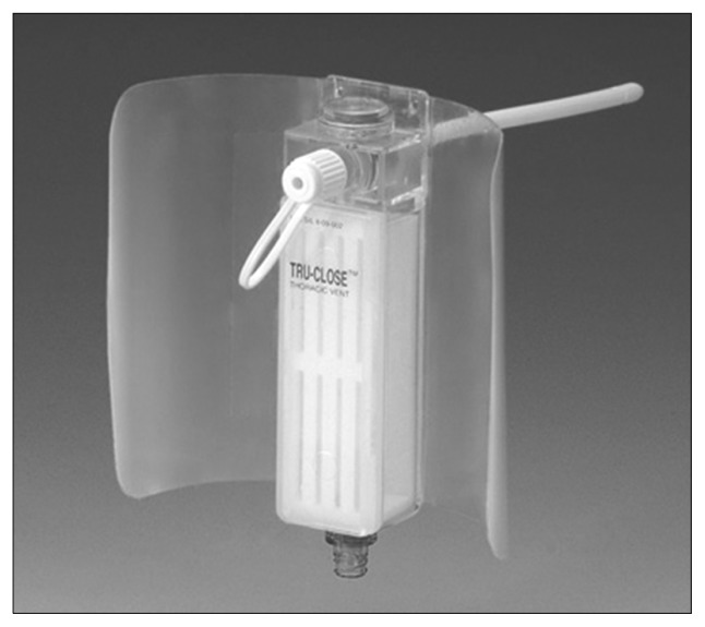
The UreSil Tru-Close Thoracic Vent (TV; UreSil Co., Ltd., Skokie, IL, USA) is used in some institutions for treating pneumothorax through outpatient drainage therapy (Fig. 1). Generally, outpatient drainage therapy with a TV is indicated for spontaneous and iatrogenic pneumothoraces. A TV is seldom a good choice for traumatic pneumothorax treatment because a concomitant hemothorax is often present. In this article, we report a case of traumatic pneumothorax that was caused by a bull attack and treated with a TV in an out-patient clinic.
Fig. 1.
Thoracic Vent. The extracorporeal dimensions of the thoracic vent are 9 cm (length)×2.5 cm (width)×2 cm (depth). The thoracic vent consists of a flexible 13 Fr urethane catheter with a removable in-line trocar connected to a one-way valve.
A 63-year-old rancher, who had been attacked by a bull an hour before arriving at the hospital, was referred to our emergency room after sustaining injuries on the right side of his chest causing dyspnea and chest pain. His chest X-ray showed right third and fourth rib fractures and a small right-sided pneumothorax (Fig. 2A). His dyspnea slightly worsened the next day and his pneumothorax was found to have progressed, although no hemothorax was found (Fig. 2B). The presence of pneumothorax without hemothorax was identified as an indication for chest drainage. Because there were no symptoms of hemothorax, we chose outpatient drainage therapy with a TV. A TV was inserted through the second intercostal space of his anterior chest wall. His right lung was fully expanded and the air leak stopped on the third day (Fig. 2C). We removed the TV on the sixth day. Only 14 mL of bloody effusion drained into the TV. The pneumothorax did not recur after the TV was removed, and no evidence of hemothorax was found.
Fig. 2.
Chest X-ray. (A) A small right-sided pneumothorax was found on day one in the hospital, (B) the right-sided pneumothorax worsened on day two in the hospital, and (C) the right lung was fully expanded on day three in the hospital. The shadow of the Thoracic Vent is shown.
DISCUSSION
Thoracic drainage is often required as a therapy for pneumothorax and is usually performed as an inpatient procedure in most institutions, although small-bore catheters have been used in some institutions [1]. In 1991, the first TV that made outpatient drainage therapy possible was reported [2]. A TV is a 10 or 13 Fr thoracic tube with a small extracorporeal box (9×2.5×2 cm) containing a one-way valve. It is useful for draining air, but not fluids because the capacity of the extracorporeal box is only 30 mL. Therefore, a TV is not indicated for pneumothorax with a significant amount of pleural effusion. Generally, a TV is indicated for spontaneous and iatrogenic pneumothoraces without pleural effusion [3].
Blunt chest trauma often results in chest organ injuries such as pneumothorax, hemothorax, rib fractures, and lung injury [4]. Bulls are one of the most dangerous farm animals, and chest trauma occurring because of bull attacks is associated with severe injuries and high mortality [5]. A traumatic pneumothorax is often accompanied with a hemothorax, meaning that outpatient drainage therapy with a TV is seldom indicated for traumatic pneumothorax. However, for patients with a pneumothorax but no hemothorax, as reported in this case, a TV is useful because it allows chest drainage for a traumatic pneumothorax to be performed without hospitalization. However, this approach should only been implemented when it is certain that no or almost no hemothorax is present. In summary, a TV can be a useful device for treating patients with traumatic pneumothorax not accompanied by hemothorax.
In conclusion, although the use of a TV is generally indicated for outpatient therapy for spontaneous and iatrogenic pneumothoraces, it can be a useful modality for treating traumatic pneumothorax without hemothorax.
Footnotes
CONFLICT OF INTEREST
No potential conflict of interest relevant to this article was reported.
REFERENCES
- 1.Noh TO, Ryu KM. Comparative study for the efficacy of small bore catheter in the patients with iatrogenic pneumothorax. Korean J Thorac Cardiovasc Surg. 2011;44:418–22. doi: 10.5090/kjtcs.2011.44.6.418. [DOI] [PMC free article] [PubMed] [Google Scholar]
- 2.Samelson SL, Goldberg EM, Ferguson MK. The thoracic vent: clinical experience with a new device for treating simple pneumothorax. Chest. 1991;100:880–2. doi: 10.1378/chest.100.3.880. [DOI] [PubMed] [Google Scholar]
- 3.Martin T, Fontana G, Olak J, Ferguson M. Use of pleural catheter for the management of simple pneumothorax. Chest. 1996;110:1169–72. doi: 10.1378/chest.110.5.1169. [DOI] [PubMed] [Google Scholar]
- 4.Shorr RM, Crittenden M, Indeck M, Hartunian SL, Rodriguez A. Blunt thoracic trauma: analysis of 515 patients. Ann Surg. 1987;206:200–5. doi: 10.1097/00000658-198708000-00013. [DOI] [PMC free article] [PubMed] [Google Scholar]
- 5.Dogan KH, Demirci S, Erkol Z, Sunam GS, Kucukkartallar T. Injuries and deaths occurring as a result of bull attack. J Agromedicine. 2008;13:191–6. doi: 10.1080/10599240802405975. [DOI] [PubMed] [Google Scholar]


