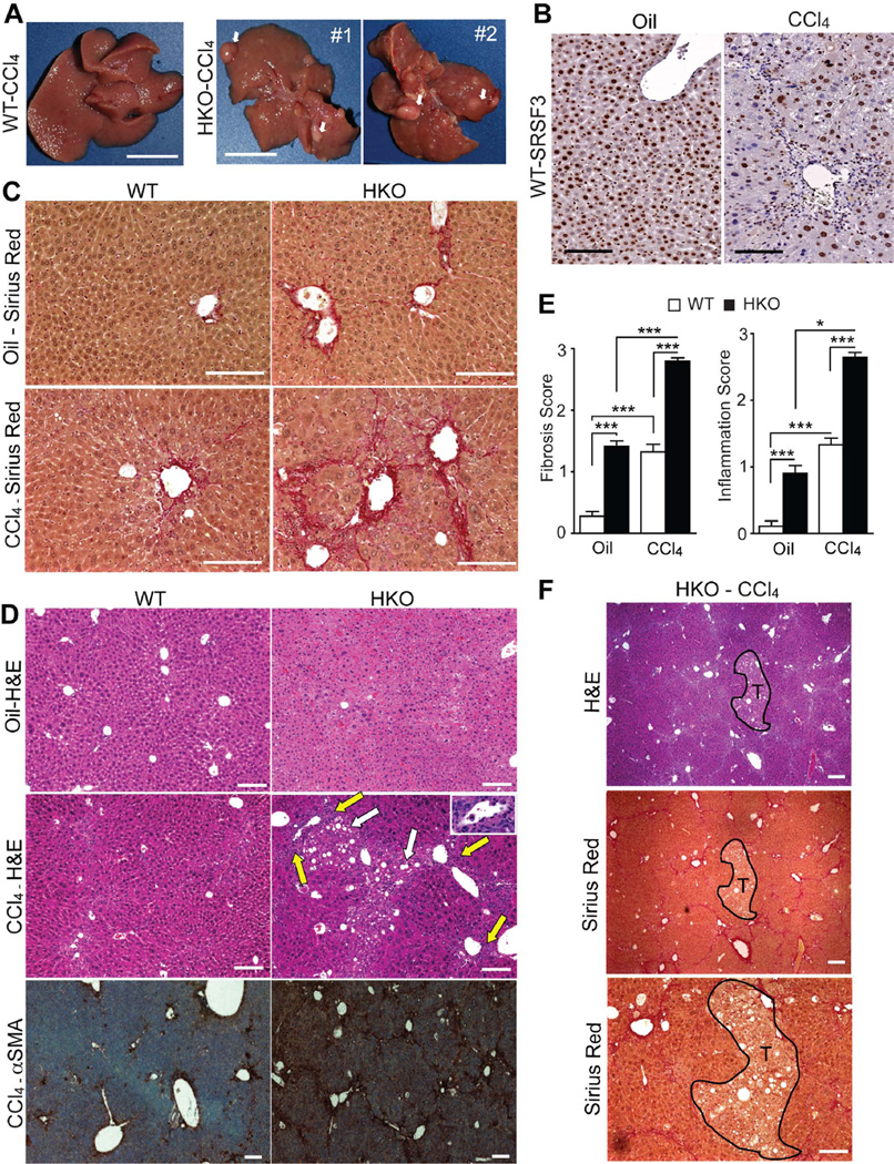
Fig. 4. SRSF3 protects against CCl4 toxicity.
(A) Gross morphology of 6 mo WT and HKO livers treated with CCl4 for 4 weeks. White arrows indicate pre-cancerous nodules in HKO liver. (B) Liver sections from WT mice treated with oil or CCl4 and stained for SRSF3. (C) Sirius Red staining for fibrosis (red) in oil and CCl4-treated mice. (D) H&E and αSMA stained liver sections from oil- or CCl4-treated mice showing steatosis (white arrows), portal inflammation (yellow arrows and inset) and myofibroblast activation (brown). (E) Mean fibrosis and inflammation scores from evaluation of multiple liver sections. (F) H&E and Sirius Red stained sections through a precancerous nodule in CCl4-treated HKO liver. Nodule is outlined in black and labeled T.

