Several studies have demonstrated that cord blood (CB) unit composition is an important predictor of outcome after CB transplantation, with higher infused doses of nucleated cells (NC) or hematopoietic stem and progenitor cells being associated with faster engraftment and better overall survival.1–3 Identification of factors predicting the hematopoietic cell content of CB units is, therefore, an important issue for CB banking strategies. Large studies have reported several maternal, fetal and obstetrical factors impacting CB cell yield.4–6 Recently, it has been documented in adult individuals that hematopoietic stem and progenitor cells circulate from the bone marrow to the peripheral blood according to a circadian rhythm,7 leading to circadian variations in their concentrations in blood (reviewed by Mendez-Ferrer8). Whether similar rhythmic release also occurs during fetal life, and whether time of day at which delivery occurs, may impact CB hematopoietic cell composition remain unknown. In this multicenter study, we analyzed factors that potentially influence the concentrations of NC and hematopoietic cells in a large series of CB samples and we observed an impact of time of day and month of delivery.
Three University centers participated in this study. CB units were collected over four consecutive years as part of a CB banking project (Ethics Committee registration number: F93/20/1728), with written informed consents obtained from the mothers. Our standard procedures for CB banking required all units to be collected in utero after vaginal delivery of single-birth term babies (>36 weeks of gestational age). After collection, a 2-mL sample of CB was taken for complete blood counts, assessment of CD34+ cell concentration by flow cytometry and Colony-Forming Units (CFU) assays. Myeloid (CFU-GM) colonies were scored by all three centers whereas erythroid (Burst Forming Unit Erythroid, BFU-E) and multilineage (CFU-Mix) colonies were only assessed in one of the centers.
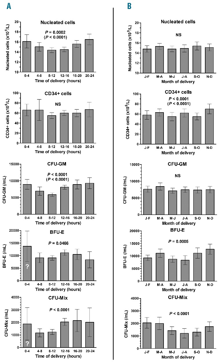
A total of 1127 CB units were analyzed. The mean (±SD) volume was 83±27 mL, obtained from babies weighing 3.420±0.420 kg after 39±1 weeks of gestation. Fifty-four percents of babies were males. The mean maternal age was 30±5 years. CB units were processed within 14.5±8.7 hours from delivery and mean cell concentrations were: 15.1±5.7 × 10⁹ NC/L, 60±58 × 106 CD34+ cells/L, 7600±6320 CFU-GM/mL, 10140±6700 BFU-E/mL and 1670±1720 CFU-Mix/mL. We observed significant variations in NC, CFU-GM, BFU-E and CFU-Mix concentrations according to time of day of delivery (Figure 1A), with the lowest values for babies born in the morning. We also examined cell concentrations according to the month of delivery (Figure 1B) and noted significant circannual variations for CD34+ cells, CFU-Mix and BFU-E. Concentrations of CFU-Mix and BFU-E were lowest during summer whereas changes in CD34+ cell concentrations were less consistent.
Figure 1.

Hematopoietic cell concentrations in cord blood (CB) according to time of day of delivery (A) and month of delivery (B). Means with 95% confidence intervals are shown. P values in parentheses are adjusted for the center. (A) Overall, 61 CB units were collected between 0:00 and 4:00, 101 between 4:00 and 8:00, 270 between 8:00 and 12:00, 378 between 12:00 and 16:00, 160 between 16:00 and 20:00 and 90 between 20:00 and 24:00. Data concerning time of delivery were missing for 67 CB units. BFU-E and CFU-Mix colonies were only measured for CB units analyzed in Center 2 (12 units collected between 0:00 and 4:00, 43 between 4:00 and 8:00, 144 between 8:00 and 12:00, 163 between 12:00 and 16:00, 31 between 16:00 and 20:00 and 16 between 20:00 and 24:00). (B) Overall, 198 CB units were collected in January-February, 224 in March–April, 247 in May–June, 143 in July–August, 146 in September–October and 169 in November–December. BFU-E and CFU-Mix colonies were only measured for CB units analyzed in Center 2 (94 units collected in January–February, 64 in March–April, 97 in May–June, 36 in July–August, 59 in September–October and 59 in November–December). J–F: January–February; M–A: March–April; M–J: May–June; J–A: July–August; S–O: September–October; N–D: November–December. NS: not significant. Question mark indicates that the measurements should be interpreted with caution because they were based on a small sample.
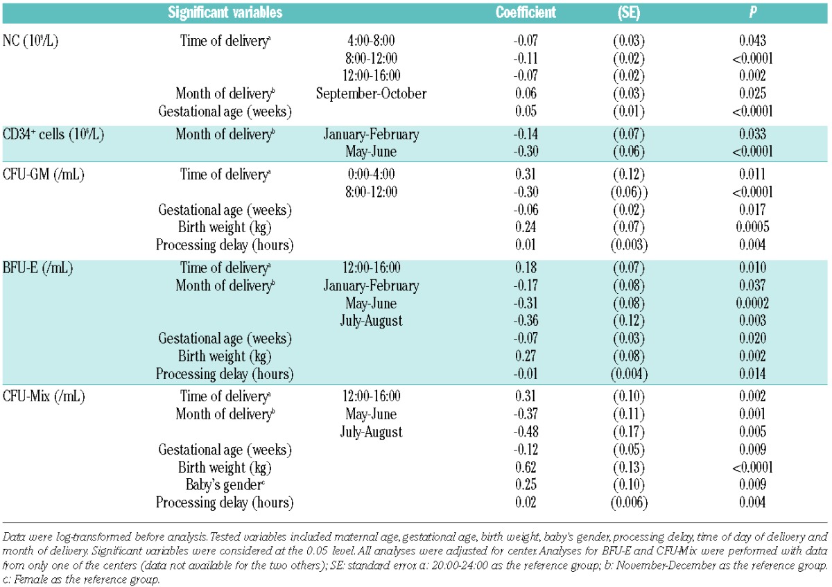
The potential influence of maternal age, gestational age, birth weight, baby’s gender, type of labor, use of epidural anesthesia, use of oxytocin, processing delay and time of delivery on NC, CD34+ cell and progenitor cell concentrations in CB was assessed in stepwise multivariate regression analysis (Table 1). NC, CFU-GM, BFU-E and CFU-Mix concentrations appeared to be determined by the time of day of delivery. Month of year was the sole factor significantly impacting CD34+ cell concentration in CB and circannual influence was also observed for BFU-E, CFU-Mix and NC concentrations. Gestational age and birth weight impacted the concentrations of NC and of both NC and hematopoietic progenitor cells, respectively, consistent with previous studies.4–6 Male gender correlated with a higher concentration of CFU-Mix. Maternal age had no influence on any parameter, in agreement with previous findings.5,6 Some groups observed that prolonged time from collection to processing was associated with a loss of hematopoietic cells,6 but within our 24-h time frame we found only a slight decrease in BFU-E concentrations. We also assessed the influence of epidural anesthesia, use of oxytocin and induced labor on CB cell concentrations in a subgroup analysis of data from two centers (data not available for one); no negative influence was observed for any. Epidural anesthesia was even associated with higher concentrations of NC (P<0.0001) and CFU-Mix (P=0.019), and use of oxytocin with higher concentrations of CFU-GM (P=0.005).
Table 1.
Maternal, fetal and time-related covariates significantly associated with cord blood parameters (derived from stepwise multivariate linear analysis).

To our knowledge, this is the first study investigating potential circadian and circannual variations in CB composition. There is increasing evidence that circadian rhythms influence stem and progenitor cell proliferation and differentiation in the bone marrow in mammals (reviewed by Mendez-Ferrer8). The underlying mechanisms remain unclear with possible roles of clock genes,9 growth factors, humoral molecules such as glucocorticoids10 and melatonin,11 and the sympathetic nervous system.12 Recently, Méndez-Ferrer et al. also reported cyclical egress of hematopoietic stem and progenitor cells from the bone marrow to the peripheral blood and demonstrated that this was, at least partly, modulated by nervous sympathetic signaling.7 Most studies in humans suggested that the peak value (called the acrophase) of hematopoietic stem/progenitor cell concentrations in the peripheral blood occurred in the afternoon or in the evening (between 3:00 p.m. and 8:00 p.m., according to the study) (reviewed by Mendez-Ferrer8). This led Lucas et al. to evaluate whether the time of day of collection could impact stem cell yield in healthy adult donors undergoing G-CSF-induced mobilization for allogeneic stem cell donation.13 Indeed, they found higher cell yields when the apheresis was performed in the afternoon rather than in the morning. By analyzing more than 1000 CB samples, our study supports the data of circadian oscillations in hematopoietic cell trafficking in humans and also suggests that these cycles are not restricted to postnatal life.
It is currently accepted that several circadian rhythms are present in the fetus (reviewed by Seron-Ferre14). Expression of clock genes has been documented in the fetal central nervous system and in other fetal organs. The synchronizing signals could be external light, which has been shown to penetrate the uterus, or maternal signals including cortisol, melatonin, glucose availability, body temperature and uterine contractures. Further studies are needed to confirm whether circadian oscillations characterize hematopoietic function during fetal life.
We also observed significant variations in CB composition according to the month of delivery. Few studies have reported circannual cycles in hematopoiesis. Some groups have shown that CFU-GM and DNA synthesis in human bone marrow were lowest in winter and that the acrophase occurred in the late summer.15
Most previous studies assessing factors impacting CB composition have considered cell contents (rather than cell concentrations) for analysis, likely because this parameter is of direct practical interest for CB unit selection for transplantation. However, the cell content of a CB unit strongly correlates with its volume4–6 and the volume of a CB unit may also depend on techniques and team aptitudes for CB collection. Hence, in our study, we chose to consider cell concentrations for our analysis, to eliminate team- and procedure-related influences. Our results have to be validated in larger studies.
In conclusion, our study may have practical implications for CB banking by raising the question of whether time of delivery may influence hematopoietic cell yield in CB units and should be taken into account for targeting CB units with the highest hematopoietic potential. It also suggests that previous observations of chronological rhythms in hematopoietic stem and progenitor cell trafficking in peripheral blood are not restricted to post-natal life.
Acknowledgments
SS is Télévie Research Assistant of the National Fund for Scientific Research (FNRS, Belgium) and has benefited from grants from the Anti-Cancer Center foundation (University of Liege, Belgium). This work was supported by grants from the FNRS (Télévie and FRSM). The authors would like to thank Walthère Dewé and Adelin Albert of the Department of Medical Statistics, University of Liège, for performing part of the statistical analysis.
Footnotes
Information on authorship, contributions, and financial & other disclosures was provided by the authors and is available with the online version of this article at www.haematologica.org.
References
- 1.Barker JN, Scaradavou A, Stevens CE. Combined effect of total nucleated cell dose and HLA match on transplantation outcome in 1061 cord blood recipients with hematologic malignancies. Blood. 2010;115(9):1843–9. [DOI] [PMC free article] [PubMed] [Google Scholar]
- 2.Page KM, Zhang L, Mendizabal A, Wease S, Carter S, Gentry T, et al. Total colony-forming units are a strong, independent predictor of neutrophil and platelet engraftment after unrelated umbilical cord blood transplantation: a single-center analysis of 435 cord blood transplants. Biol Blood Marrow Transplant. 2011;17(9):1362–74. [DOI] [PubMed] [Google Scholar]
- 3.Rodrigues CA, Sanz G, Brunstein CG, Sanz J, Wagner JE, Renaud M, et al. Analysis of risk factors for outcomes after unrelated cord blood transplantation in adults with lymphoid malignancies: a study by the Eurocord-Netcord and lymphoma working party of the European group for blood and marrow transplantation. J Clin Oncol. 2009; 27(2):256–63. [DOI] [PubMed] [Google Scholar]
- 4.Cairo MS, Wagner EL, Fraser J, Cohen G, van de Ven C, Carter SL, et al. Characterization of banked umbilical cord blood hematopoietic progenitor cells and lymphocyte subsets and correlation with ethnicity, birth weight, sex, and type of delivery: a Cord Blood Transplantation (COBLT) Study report. Transfusion. 2005;45(6):856–66. [DOI] [PubMed] [Google Scholar]
- 5.Keersmaekers CL, Mason BA, Keersmaekers J, Ponzini M, Mlynarek RA. Factors affecting umbilical cord blood stem cell suitability for transplantation in an in utero collection program. Transfusion. 2014; 54(3):545–9. [DOI] [PubMed] [Google Scholar]
- 6.Page KM, Mendizabal A, Betz-Stablein B, Wease S, Shoulars K, Gentry T, et al. Optimizing donor selection for public cord blood banking: influence of maternal, infant, and collection characteristics on cord blood unit quality. Transfusion. 2014;54(2):340–52. [DOI] [PMC free article] [PubMed] [Google Scholar]
- 7.Mendez-Ferrer S, Lucas D, Battista M, Frenette PS. Haematopoietic stem cell release is regulated by circadian oscillations. Nature. 2008; 452(7186):442–7. [DOI] [PubMed] [Google Scholar]
- 8.Mendez-Ferrer S, Chow A, Merad M, Frenette PS. Circadian rhythms influence hematopoietic stem cells. Curr Opin Hematol. 2009;16(4):235–42. [DOI] [PMC free article] [PubMed] [Google Scholar]
- 9.Tsinkalovsky O, Smaaland R, Rosenlund B, Sothern RB, Hirt A, Steine S, et al. Circadian variations in clock gene expression of human bone marrow CD34+ cells. J Biol Rhythms. 2007;22(2): 140–50. [DOI] [PubMed] [Google Scholar]
- 10.Kollet O, Vagima Y, D’Uva G, Golan K, Canaani J, Itkin T, et al. Physiologic corticosterone oscillations regulate murine hematopoietic stem/progenitor cell proliferation and CXCL12 expression by bone marrow stromal progenitors. Leukemia. 2013;27(10):2006–15. [DOI] [PubMed] [Google Scholar]
- 11.Haldar C, Haussler D, Gupta D. Effect of the pineal gland on circadian rhythmicity of colony forming units for granulocytes and macrophages (CFU-GM) from rat bone marrow cell cultures. J Pineal Res. 1992;12(2):79–83. [DOI] [PubMed] [Google Scholar]
- 12.Giudice A, Caraglia M, Marra M, Montella M, Maurea N, Abbruzzese A, et al. Circadian rhythms, adrenergic hormones and trafficking of hematopoietic stem cells. Expert Opin Ther Targets. 2010;14(5):567–75. [DOI] [PubMed] [Google Scholar]
- 13.Lucas D, Battista M, Shi PA, Isola L, Frenette PS. Mobilized hematopoietic stem cell yield depends on species-specific circadian timing. Cell Stem Cell. 2008;3(4):364–6. [DOI] [PMC free article] [PubMed] [Google Scholar]
- 14.Seron-Ferre M, Mendez N, Abarzua-Catalan L, Vilches N, Valenzuela FJ, Reynolds HE, et al. Circadian rhythms in the fetus. Mol Cell Endocrinol. 2012;349(1):68–75. [DOI] [PubMed] [Google Scholar]
- 15.Smaaland R, Sothern RB, Laerum OD, Abrahamsen JF. Rhythms in human bone marrow and blood cells. Chronobiol Int. 2002; 19(1):101–27. [DOI] [PubMed] [Google Scholar]


