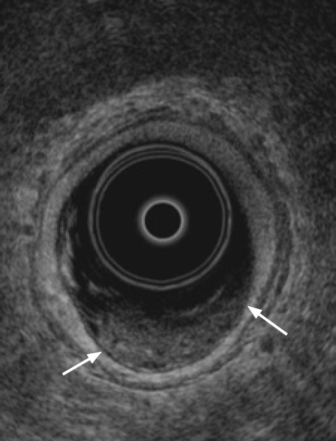
Fig. 6. T1 rectal cancer.

Axial transrectal ultrasonography shows that the hypoechoic tumor (arrows) is confined to the first inner three layers and that the hyperechoic submucosa layer is slightly thinned.

Axial transrectal ultrasonography shows that the hypoechoic tumor (arrows) is confined to the first inner three layers and that the hyperechoic submucosa layer is slightly thinned.