Abstract
Interferon (IFN)-γ has been shown to be associated with immunity to Marek’s disease virus (MDV). The overall objective of this study was to investigate the causal relationship between IFN-γ and vaccine-conferred immunity against MDV in chickens. To this end, 3 small interfering RNAs (siRNAs) targeting chicken IFN-γ, which had previously been shown to reduce IFN-γ expression in vitro, and a control siRNA were selected to generate recombinant avian adeno-associated virus (rAAAV) expressing short-hairpin small interfering RNAs (shRNAs). An MDV challenge trial was then conducted: chickens were vaccinated with herpesvirus of turkey (HVT), administered the rAAAV expressing shRNA, and then challenged with MDV. Tumors were observed in 4 out of 10 birds that were vaccinated with HVT and challenged but did not receive any rAAAV, 5 out of 9 birds that were administered the rAAAV containing IFN-γ shRNA, and 2 out of 10 birds that were administered a control enhanced green fluorescent protein siRNA. There was no significant difference in MDV genome load in the feather follicle epithelium of the birds that were cotreated with the vaccine and the rAAAV compared with the vaccinated MDV-infected birds. These results suggest that AAAV-based vectors can be used for the delivery of shRNA into chicken cells. However, administration of the rAAAV expressing shRNA targeting chicken IFN-γ did not seem to fully abrogate vaccine-induced protection.
Résumé
Il a été démontré que l’interféron (INF)-γ est associé à l’immunité contre le virus de la maladie de Marek (VMM). L’objectif général de la présente étude était d’examiner la relation causale entre l’IFN-γ et l’immunité conférée par le vaccin contre le VMM chez les poulets. Pour y parvenir, trois petits ARN interférant (siARN) ciblant l’IFN-γ, et qui avaient préalablement été montré comme étant capable de réduire l’expression in vitro de l’IFN-γ, et un siARN témoin furent choisis afin de générer du virus adéno-associé aviaire recombinant (rAAAV) exprimant de courtes boucles de siRNA (shRNA). Un essai d’infection par VMM fut alors réalisé : des poulets furent vaccinés avec de l’herpèsvirus de dinde (HVT), reçurent le rAAAV exprimant les shRNA, et par la suite challengés avec le VMM. Des tumeurs furent observées chez 4 des 10 poulets qui avaient été vacciné avec HVT et challengés mais qui n’avaient pas reçu aucun rAAAV, 5 des 9 oiseaux qui avaient reçu le rAAAV contenant l’IFN-γ avec les shRNA, et 2 des 10 oiseaux témoins qui avaient reçu un siRNA qui augmentait la protéine fluorescente verte. Il n’y avait aucune différence significative dans la charge de génome de VMM dans l’épithélium du follicule des plumes des oiseaux qui avaient été co-traités avec le vaccin et le rAAAV comparativement aux oiseaux non-vaccinés avec MMV et infectés. Ces résultats suggèrent que les vecteurs à base d’AAAV peuvent être utilisés pour la livraison de shRNA dans les cellules des oiseaux. Toutefois, l’administration de rAAAV exprimant des shRNA ciblant l’IFN-γ des oiseaux n’a pas semblé complètement abrogé la protection induite par le vaccin.
(Traduit par Docteur Serge Messier)
Introduction
Marek’s disease is a highly contagious disease of poultry caused by an oncogenic herpesvirus known as Marek’s disease virus (MDV) (1). A number of cytokines have been shown to be associated with immunity against MDV (2), interferon (IFN)-γ playing an important role (3–5). Differential expression of cytokines has been extensively investigated with the use of techniques such as microarray and reverse-transcription polymerase chain reaction (RT-PCR). However, these studies have not elucidated the functional roles played by these cytokines in immunity to Marek’s disease.
The functional role of cytokines can be studied through gain-and loss-of-function experiments both in vitro and in vivo. RNA interference (RNAi), a molecular technique by which expression of genes can be silenced with small RNA molecules [e.g., short-hairpin RNA (shRNA)], is being used as a tool for loss-of-function studies. Constructs of shRNA can be delivered by means of adeno-associated virus (AAV)-based vectors. Adeno-associated viruses were first discovered in 1965 as a contaminant of simian adenovirus (AdV) preparations (6). The small DNA-containing particles were shown to be antigenically different from AdVs. Replication of these particles occurred only when they were inoculated simultaneously with AdVs, which suggested that the particles behaved like defective viruses. Since then, AAVs have been categorized into a separate genus of the Parvoviridae family, designated Dependovirus, reflecting AAVs’ dependence on a helper virus for productive infection. Many AAV serotypes have been isolated from human and nonhuman species; however, all serotypes contain a linear single-stranded DNA (ssDNA) genome of approximately 5 kb, with 2 open reading frames, rep and cap, and inverted terminal repeats (ITRs) (7). The ITRs are the only cis-acting elements necessary for viral replication, packaging, and integration into the host genome as well as for recombinant virus generation (8).
The avian AAV (AAAV) was isolated from the Olson strain of quail bronchitis virus, an avian AdV (9). Avian AAV contains ssDNA and also requires coinfection with a helper virus for productive infection. Sequence analysis of AAAV has revealed a genome size and organization similar to that of other AAVs (10,11).
In recent years, extensive research has been conducted to characterize and use these replication-defective parvoviruses to deliver antigens or shRNA (12–14). In general, the use of AAVs is appealing because they are nonpathogenic, can infect dividing or nondividing cells, have broad tissue tropism, tend to remain episomal, and can easily be engineered as a vector. The limitation to their use is the relatively small AAAV genome, which restricts the total insert size to about 4 kb for optimal packaging.
The goals of this study were to develop an AAAV-based vector expressing shRNA targeting chicken IFN-γ and to use this system to knock down IFN-γ expression so that the biologic significance of IFN-γ in immunity against poultry pathogens, including MDV, can be evaluated.
Materials and methods
Cell culture
Human embryonic kidney cells (HEK293) were maintained in Dulbecco’s modified Eagle medium (DMEM; Invitrogen, Burlington, Ontario) supplemented with 10% fetal bovine serum (FBS; Invitrogen) and penicillin/streptomycin, 100 μg/mL, at 37°C in a 5% CO2 atmosphere. An immortal chicken fibroblast cell line, DF-1, was maintained in DMEM supplemented with 10% FBS, 1% chicken serum, and penicillin/streptomycin, 100 μg/mL, at 37°C in 5% CO2.
Experimental animals
Specific-pathogen-free eggs were obtained from the Animal Disease Research Institute, Canadian Food Inspection Agency, Ottawa, Ontario, and hatched at the Arkell Poultry Research Unit, University of Guelph, Guelph, Ontario. The hatched chicks were housed in the animal isolation facility at the Ontario Veterinary College, University of Guelph, Guelph, Ontario, during the experimental period. All animal experiments were approved by the Animal Care Committee, University of Guelph.
Virus and vaccine strains
Very virulent MDV strain RB1B (passage 9) was kindly provided by Dr. Karel A. Schat, Cornell University, Ithaca, New York, USA, and used to infect the chickens. The chickens were vaccinated subcutaneously on the day of hatch with the recommended dose of herpesvirus of turkey (HVT) (Fort Dodge Animal Health, Overland Park, Kansas, USA).
Nucleic acid isolation and quantitative real-time RT-PCR
Total DNA and RNA were extracted from all tissues, including the feather tips (for MDV genome-copy number) with the use of TRIzol reagent (Invitrogen) according to the manufacturer’s protocol. Briefly, DNA was directly extracted from the cells by adding the reagent to the cells. Tissue samples were preserved in RNAlater (Qiagen, Mississauga, Ontario) and then homogenized in 1 mL of TRIzol. After chloroform extraction, the organic phase, containing the DNA, was separated, washed with 0.1 M sodium citrate in 10% ethanol, and dissolved in 8 mM NaOH. The RNA concentration was measured by spectrophotometry at 260 nm. The total RNA was then treated with DNase by means of a DNA-free kit (Ambion, Austin, Texas, USA) according to the manufacturer’s instructions. Complementary DNA (cDNA) was prepared from 1 μg of DNase-treated total RNA by reverse transcription with the use of Moloney murine leukemia virus reverse transcriptase and Oligo(dT)12–18 primer (SuperScript First Strand Synthesis System; Invitrogen) according to manufacturer’s instructions. Real-time RT-PCR was done in a LightCycler 480 instrument (Roche Diagnostics, Laval, Quebec) in a reaction volume of 20 μL with SYBR Green 1 Master Mix (Roche Diagnostics), 0.25 μM of each primer, and 5 μL of a 1:10 dilution of cDNA. Previously published primers were used for IFN-γ and β-actin (15), and all primers were synthesized by Sigma-Aldrich Canada (Oakville, Ontario). Quantification of viral genome load and expression of cytokine genes by real-time PCR and RT-PCR was done with previously published primers and as described previously (15,16). Briefly, absolute number of MDV genome per 100 ng of feather tips was calculated based on an external standard curve. IFN-γ gene expression was calculated relative to the expression of the β-actin house-keeping gene and expressed as ratios.
Generation of recombinant AAAV (rAAAV) vectors with chicken IFN-γ-shRNA
Three Dicer-substrate RNAs (DsiRNAs) targeting chicken IFN-γ were designed and synthesized by Integrated DNA Technologies (Coralville, Iowa, USA). As previously described (16), we tested the inhibitory effect of the 3 DsiRNAs on IFN-γ expression in DF-1 and primary avian splenocytes. Subsequently we generated rAAAVs containing the following 3 sequences targeting IFN-γ (5′–3′): GGCGUGAAGAAGGUGAAAGAUAUCA, GCAAGUAGUCUAAAUCUUGUUCAAC, and CGAUGAACGAC UUGAGAAUCCAGCG. A DsiRNA targeting enhanced green fluorescent protein (EGFP-S1 DS) was tested as a control. Construction of the recombinant vectors and generation of rAAAV have been described previously (16). Briefly, for each vector plasmid, we cloned an siRNA expression cassette consisting of a nucleotide sense sequence (identical to the IFN-γ target sequence), then a loop 9 base pairs long, an antisense sequence, and a stretch of poly-thymidine (poly-T) as a polymerase III transcriptional termination signal downstream of a U6 promoter (a U6–IFN-γ or EGFP-shRNA–polyT stretch). Recombinant AAAV was then packaged with vectors for a helper-free recombinant virus production system, generously provided by Drs. Carlos Estevez and Pedro Villegas (University of Georgia, Athens, Georgia, USA). The titer was determined by real-time PCR as described by Rohr et al (17). Recombinant AAAV with an EGFP targeting sequence was also constructed to serve as a control.
In-vivo trials
There were 2 in-vivo trials in this study: the 1st was designed to assess the dose and distribution of rAAAV:IFN-γ-shRNA, and the 2nd was an MDV challenge study to determine the effect of knocking down IFN-γ.
In the 1st trial, conducted as a pilot study, 1-day-old chicks were administered rAAAV:IFN-γ-shRNA-1 (targeting sequence 1) intramuscularly in the upper third of the right pectoral muscle at a high dose (1 × 10¹⁰ genomic copies; group 1, n = 16) or at a low dose (1 × 10⁹ genomic copies; group 2, n = 16); a negative-control group (group 3, n = 8) was treated with phosphate-buffered saline (PBS). The chickens were monitored for any signs of adverse effects induced by the rAAAV. At 48 h, 120 h, 14 d, or 26 d after administration they were euthanized and the following tissues excised and stored in RNAlater: liver, spleen, lung, bursa of Fabricius, and muscle. Genomic DNA was extracted from approximately 25 to 50 mg of tissue as described. Real-time PCR was done to determine the number of genomic copies of rAAAV.
The 2nd trial involved vaccination with a commercially available HVT vaccine, followed by experimental infection with the very virulent MDV strain RB1B. According to the results of the pilot study, a high dose (1 × 10¹⁰ genomic copies) of rAAAV was administered. There were 6 groups in this trial (n = 9 to 10 per group). One group served as MDV-infected only (group MDV) and another as a negative (uninfected, unvaccinated) control. The other 4 groups were as follows: MDV-infected after administration of all 3 rAAAV:IFN-γ-shRNAs; MDV-infected after HVT vaccination; MDV-infected after HVT vaccination and administration of control shRNA; and MDV-infected after HVT vaccination and administration of all 3 rAAAV:IFN-γ-shRNAs. Briefly, 1-day-old chicks were vaccinated subcutaneously with HVT and administered rAAAV intramuscularly in the upper third of the right pectoral muscle. At 5 d of age they were infected with 250 plaque-forming units (PFU) of RB1B intra-abdominally. The chickens were then monitored daily for any clinical signs for 3 wk. Feather samples were collected from 5 birds per group at 4, 7, 10, 14, and 21 d after infection, time points selected according to an intra-abdominal MDV infection model system that corresponded to important phases of MDV pathogenesis (18). All birds were euthanized 21 d after infection, weighed, and examined for gross lesions. The weights of the bursa of Fabricius and spleen were recorded during necropsy, and the ratios of these weights to body weight were determined. The MDV genome load and transcripts in the feathers were quantified by means of real-time PCR and RT-PCR as described previously (15). The MDV genome load was described as the number of copies per 100 ng of DNA.
Data analysis
For tumor incidence data, Fisher’s exact test was used, whereas for the other experiments a 2-tailed t-test and analysis of variance were used to identify differences among groups. Differences were considered significant at P ≤ 0.05.
Results
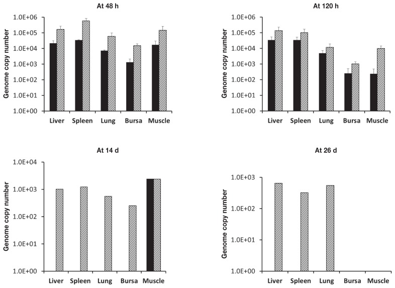
Quantitative PCR revealed that viral DNA was present in all the tissues collected after euthanasia (liver, spleen, lung, bursa of Fabricius, and muscle) within 48 h after intramuscular injection of rAAAV:IFN-γ-shRNA-1 in 1-day-old chickens (Figure 1). In the chickens that received the lower dose (1 × 10⁹ genomic copies), viral DNA was detected in muscle tissue up to 14 d after injection and in liver, spleen, and lung up to 5 d after injection. However, in the chickens that received the higher dose (1 × 10¹⁰ genomic copies), viral DNA was detectable in liver, spleen, and lung up to 26 d after injection but only up to 14 d in the bursa of Fabricius and muscle. There was a decline in the viral load over the course of the experiment with both doses. There was no unexpected death or occurrence of gross lesions in the treated birds, which suggests that administration of recombinant viruses to chickens is safe.
Figure 1.
Mean number of copies of the viral genome [and standard error (SE)] of recombinant avian adeno-associated virus (rAAAV) in various tissues of chicks (16 per group) euthanized at 48 h, 120 h, 14 d, or 26 d after administration at 1 d of age of a lower dose [1 × 10⁹ genomic copies (black bars)] or a higher dose [1 × 10¹⁰ genomic copies (hatched bars)] of an rAAAV expressing short-hairpin small interfering RNA targeting chicken interferon-γ (rAAAV:IFN-γ-shRNA).
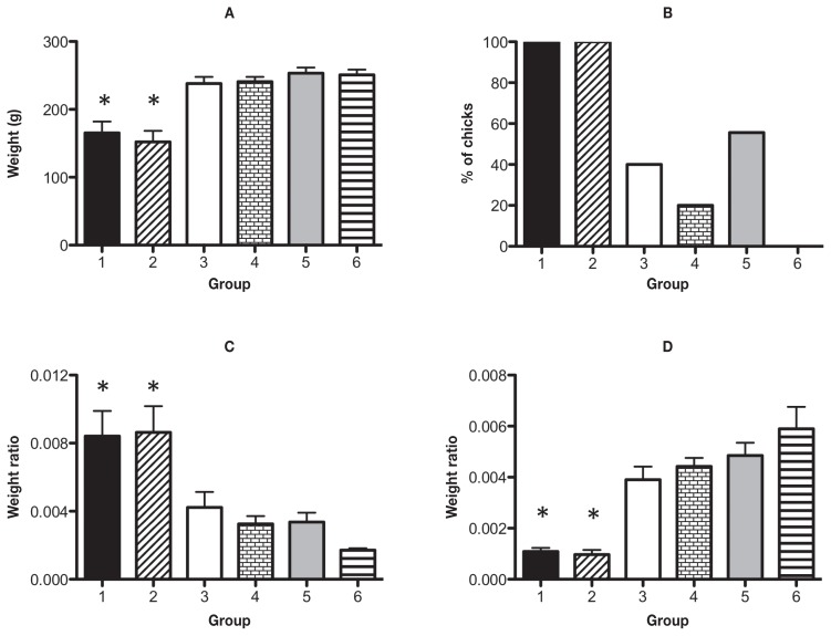
Chickens were then administered HVT and 1 × 10¹⁰ genomic copies of all 3 sequences of rAAAV:IFN-γ-shRNA or rAAAV:shRNA-EGFP on the day of hatch. Four days later they were injected intra-abdominally with 250 PFU of MDV (or an equal volume of PBS for the sham-infected group). At 21 d after the MDV challenge, the day of necropsy, the chickens receiving only MDV or MDV plus the 3 rAAAV:IFN-γ-shRNAs weighed significantly less than all the vaccinated groups as well as the control group (Figure 2A). In the unvaccinated MDV-infected group as well as the unvaccinated MDV-infected group administered all 3 rAAAV:IFN-γ-shRNAs, the tumor incidence was 100%, whereas in the infected groups that received the vaccine alone, with rAAAV:EGFP, or with rAAAV:IFN-γ-shRNA the incidence rates were 40%, 20%, and 56%, respectively (Figure 2B). None of the birds in the unvaccinated, uninfected control group had tumors or clinical signs. At necropsy the lymphoid organs were weighed, as Marek’s disease is associated with enlargement of the spleen and bursal atrophy. The spleen weight:body weight ratio showed significant enlargement of the spleen in the MDV-infected- only group and the MDV-infected group administered all 3 rAAAV:IFN-γ-shRNAs (Figure 2C). The chickens vaccinated with HVT did not show enlargement of the spleen compared with the uninfected control chickens. The lower bursa weight:body weight ratio indicated bursal atrophy in the MDV-infected-only group and the MDV-infected group administered all 3 rAAAV:IFN-γ-shRNAs (Figure 2D). In contrast, the vaccinated birds did not have significant bursal atrophy compared with the control birds. There was no significant difference in either ratio between the vaccinated-only group and the groups vaccinated and administered rAAAV.
Figure 2.
Necropsy data (mean and SE) for 6 groups of chicks (9 to 10 per group) 21 d after injection of a very virulent strain (RB1B) of Marek’s disease virus (MDV) or sham injection with phosphate-buffered saline at 5 d of age, some groups having been vaccinated with herpesvirus of turkey (HVT) on the day of hatch. A — body weight; B — frequency of gross tumors; C — ratio of spleen weight to body weight; D — ratio of weight of bursa of Fabricius to body weight. Group 1 — MDV-infected only; group 2 — MDV-infected after administration of all 3 rAAAV:IFN-γ-shRNAs (1 × 10¹⁰ genomic copies of each); group 3 — MDV-infected after HVT vaccination; group 4 — MDV-infected after HVT vaccination and administration of a control shRNA targeting enhanced green fluorescent protein (EGFP-S1 DS); group 5 — MDV-infected after HVT vaccination and administration of all 3 rAAAV:IFN-γ-shRNAs; group 6 — uninfected and unvaccinated controls. Asterisks indicate a significant difference (P ≤ 0.05) between this mean and the means for all the vaccinated groups and control group 6.
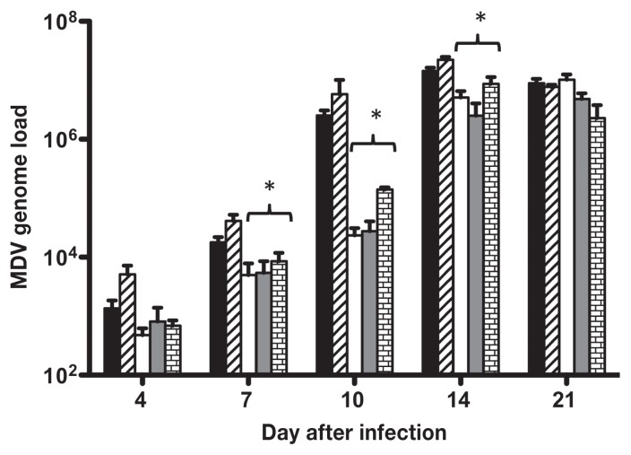
The MDV genome load was quantified in feather tips, which harbor fully infectious virus particles (19); feather tip DNA was analyzed by real-time PCR, and the results are illustrated in Figure 3. The virus was detected as early as 4 d after infection in all the infected groups. At all time points except 21 d after infection, the MDV-infected-only group and the MDV-infected group administered all 3 rAAAV:IFN-γ-shRNAs had a higher MDV genome load than the vaccinated groups. The load was no greater in the group vaccinated and treated with rAAAV than in the group vaccinated and MDV-infected only. At 7, 10, and 14 d after infection all the vaccinated birds had a significantly lower viral load than the unvaccinated birds. At 4 and 21 d after infection there was no significant difference among the groups.
Figure 3.
Mean MDV genome load (and SE) per 100 ng of DNA in feather follicle epithelium (FFE) at 4, 7, 10, 14, and 21 d after infection in groups 1 to 5. Asterisks indicate a significant difference (P ≤ 0.05) between this mean and the means for the unvaccinated groups.
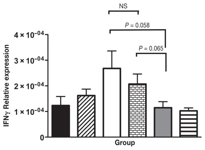
Up-regulation in the expression of IFN-γ was observed in the spleens of all the chickens that were vaccinated, MDV-infected, and/or treated with rAAAV expressing EGFP compared with the uninfected control birds (Figure 4). There was no significant difference in the expression of IFN-γ between the infected groups that received HVT only or HVT and rAAAV:EGFP, which confirmed the use of GFP as a control. The reduced expression of IFN-γ in the group that received HVT and all 3 rAAAV:IFN-γ-shRNAs approached significance when compared with the expression in the infected groups that received HVT (2.3-fold) and/or HVT plus rAAAV:EGFP (1.8-fold) (P = 0.058 and 0.065, respectively).
Figure 4.
Cytokine mRNA expression in spleen tissues of the 6 groups of chickens. IFN-γ (target) and β-actin (reference) gene expression in spleen was quantified by real time RT-PCR using SYBR. Target gene expression is presented relative to β-actin expression and normalized to a calibrator. Data is presented as mean expression (and SE) of each group. NS — not a significant difference.
Discussion
Protective immunity against MDV induced by vaccination or natural infection requires a strong cell-mediated immune response, which is associated with up-regulation of IFN-γ expression in tissues (2,20–23). In the present study, we exploited the endogenous RNAi mechanism to better understand the role of IFN-γ in vaccine-induced protection against Marek’s disease.
This is the 1st report of down-regulation of chicken IFN-γ expression with use of a recombinant AAAV in vivo. Earlier studies used rAAAVs for the expression of a reporter gene in embryonic tissues in vitro, and other studies have used AAAV vectors for the expression of microRNAs or viral proteins in vivo (12,14,24,25). Apart from the safety of AAAV-based vectors, expression cassettes up to ~ 4 kb in length can be packaged into AAV capsids without compromising infectivity (26). We selected sequences to generate rAAAVs on the basis of our earlier experiments, which examined the efficacy of various siRNAs to knock down the expression of IFN-γ (16). We chose a polymerase III promoter (U6) because it transcribes endogenous small-nuclear RNAs (snRNAs) and is most commonly used to express shRNA (27,28).
In studying the tissue tropism of rAAAV, we found the virus to be distributed in all tissues examined. Significant diversity has previously been reported in the tissue tropism of AAV serotypes 1 to 9 (29). In-vitro cellular tropism in AAAV has also been determined among various avian and nonavian cell lines, as well as in primary chicken cells and human fibroblasts. Bossis and Chiorini (10) found that rAAAV had 10 to 300 times greater transduction efficiency in avian cells compared with rAAV2, -4, and -5. In our study, viral DNA was detectable in spleen, liver, and lung up to 26 d after administration of the higher dose of AAAV. In contrast, after administration of the lower dose, viral DNA was not detectable in tissues other than muscle (the site of administration) beyond 14 d after administration. This may be due to clearance of the virus by the host immune system. The bursa of Fabricius had a relatively low amount of recombinant virus with both doses, which may suggest low tropism of AAAV towards this tissue.
We then tested the hypothesis that down-regulation of IFN-γ, via the administration of rAAAV expressing shRNA, could abrogate immunity conferred by vaccination against MDV. We also hypothesized that down-regulation of IFN-γ might exacerbate clinical and gross pathological lesions associated with Marek’s disease. Loss of body weight in MDV-infected chickens is a characteristic of the disease. Administration of rAAAV:IFN-γ-shRNA to the vaccinated MDV-challenged birds did not have any significant effect on the birds’ body weight. This suggests that the administration of shRNA after vaccination did not increase the severity of the disease or lower vaccine-induced protection. However, both unvaccinated chicken groups had a significantly lower body weight than the vaccinated groups. Birds infected with MDV display bursal atrophy and spleen enlargement (30). Administration of rAAAV:IFN-γ-shRNA to knock down the expression of IFN-γ did not have a significant effect on bursal atrophy or spleen enlargement when compared with lack of vaccination among the MDV-infected chickens. Vaccination with HVT by itself or with rAAAV:EGFP administration resulted in the protection of 60% and 80% of birds, respectively, whereas when birds were cotreated with HVT and rAAAV:IFN-γ-shRNA before MDV infection, only 44% were protected. However, the difference was not statistically significant. According to these results, IFN-γ knockdown does not seem to completely abrogate vaccine-induced immunity.
A potent activator of macrophages (31), IFN-γ also has antitumor activities (32). Previously we found that administration of rChIFN-γ, through an expression plasmid, enhanced immunity conferred by HVT, leading to a reduction in tumor incidence in MDV-infected birds (5). Other studies have also shown evidence for the role of IFN-γ in inhibiting tumor formation. For example, Plachý et al (33) injected a congenic chicken line with ChIFN-γ before infection with Rous sarcoma virus and observed a reduction in the development of tumors and their size. However, in the present study, vaccine-induced immunity conferred by HVT was not completely eliminated in the chickens that received rAAAV:IFN-γ-shRNA. It is possible that in those chickens the IFN-γ knockdown was not complete or did not occur at a time point or in tissues critical for the induction of immunity against MDV.
The feather follicle epithelium (FFE) is the only known tissue site from which fully formed infectious MDV particles can be shed into the environment (19). Studies in our laboratory have examined host responses to very virulent MDV or Marek’s disease vaccines in feathers (21,34). Earlier studies showed an increase in IFN-γ expression in the FFE of vaccinated birds that correlated with a decrease in the viral genome load in the same tissue. This led us to hypothesize that down-regulating IFN-γ may lead to an increase in viral load in the FFE. The results presented here do not show a significant difference between the 3 vaccinated groups. The lower viral load in the vaccinated birds correlated with the time of MDV latency, which begins around 6 to 7 d after infection and lasts until the late cytolytic phase, beginning around 14 d after infection (35). Increased expression of IFN-γ has been shown to correlate with increased expression of inducible nitric oxide synthase, resulting in the production of nitric oxide, which is known to inhibit viral replication (5,36). In investigating the silencing effect of rAAAV, we detected lower IFN-γ expression in the spleens of the vaccinated MDV-infected chickens administered rAAAV:IFN-γ-shRNA compared with the vaccinated MDV-infected chickens and the vaccinated MDV-infected chickens administered rAAAV:EGFP. The reduction in expression approached significance. One explanation may be that although RNAi led to a decrease in the total level of IFN-γ in the spleen, the target cells responsible for vaccine protection were not transfected with rAAAV and therefore IFN-γ expression from these cells was not affected. It is also possible that by 21 d after infection (the sampling time point) the down-regulatory effects of rAAAV:IFN-γ-shRNA on IFN-γ were diminished or completely abrogated or that the site of action of the vector was different from the tissue we sampled. Irrespective of the above finding, there was no significant difference between the vaccinated group and the group that received rAAAV:EGFP, which supported the use of the latter as a nontarget control.
These findings provide a basis for future studies aimed at using recombinant viral vectors to deliver genes or to knock down the expression of a gene of interest and to better understand the roles of certain proteins or improve vaccines.
Acknowledgments
Vectors for a helper-free recombinant virus production system were generously provided by Drs. Carlos Estevez and Pedro Villegas, University of Georgia, Athens, Georgia, USA. The study was funded by the Ontario Ministry of Agriculture, Food and Rural Affairs, the Poultry Industry Council, and the Natural Sciences and Engineering Research Council of Canada.
References
- 1.Calnek BW. Pathogenesis of Marek’s disease virus infection. Curr Top Microbiol Immunol. 2001;255:25–55. doi: 10.1007/978-3-642-56863-3_2. [DOI] [PubMed] [Google Scholar]
- 2.Haq K, Brisbin JT, Thanthrige-Don N, Heidari M, Sharif S. Transcriptome and proteome profiling of host responses to Marek’s disease virus in chickens. Vet Immunol Immunopathol. 2010;138:292–302. doi: 10.1016/j.vetimm.2010.10.007. [DOI] [PubMed] [Google Scholar]
- 3.Xing Z, Schat KA. Inhibitory effects of nitric oxide and gamma interferon on in vitro and in vivo replication of Marek’s disease virus. J Virol. 2000;74:3605–3612. doi: 10.1128/jvi.74.8.3605-3612.2000. [DOI] [PMC free article] [PubMed] [Google Scholar]
- 4.Abdul-Careem MF, Hunter BD, Parvizi P, Haghighi HR, Thanthrige-Don N, Sharif S. Cytokine gene expression patterns associated with immunization against Marek’s disease in chickens. Vaccine. 2007;25:424–432. doi: 10.1016/j.vaccine.2006.08.006. [DOI] [PubMed] [Google Scholar]
- 5.Haq K, Elawadli I, Parvizi P, Mallick AI, Behboudi S, Sharif S. Interferon-gamma influences immunity elicited by vaccines against very virulent Marek’s disease virus. Antiviral Res. 2011;90:218–226. doi: 10.1016/j.antiviral.2011.04.001. [DOI] [PubMed] [Google Scholar]
- 6.Gonçalves M. Adeno-associated virus: From defective virus to effective vector. Virol J. 2005;2:43. doi: 10.1186/1743-422X-2-43. [DOI] [PMC free article] [PubMed] [Google Scholar]
- 7.Stilwell JL, Samulski RJ. Adeno-associated virus vectors for therapeutic gene transfer. Biotechniques. 2003;34:148–50. 152, 154. doi: 10.2144/03341dd01. passim. [DOI] [PubMed] [Google Scholar]
- 8.McCarty DM, Young SM, Samulski RJ. Integration of adeno-associated virus (AAV) and recombinant AAV vectors. Annu Rev Genet. 2004;38:819–845. doi: 10.1146/annurev.genet.37.110801.143717. [DOI] [PubMed] [Google Scholar]
- 9.Yates VJ, El-Mishad AM, McCormick KJ, Trentin JJ. Isolation and characterization of an avian adenovirus-associated virus. Infect Immun. 1973;7:973–980. doi: 10.1128/iai.7.6.973-980.1973. [DOI] [PMC free article] [PubMed] [Google Scholar]
- 10.Bossis I, Chiorini JA. Cloning of an avian adeno-associated virus (AAAV) and generation of recombinant AAAV particles. J Virol. 2003;77:6799–6810. doi: 10.1128/JVI.77.12.6799-6810.2003. [DOI] [PMC free article] [PubMed] [Google Scholar]
- 11.Estevez C, Villegas P. Sequence analysis, viral rescue from infectious clones and generation of recombinant virions of the avian adeno-associated virus. Virus Res. 2004;105:195–208. doi: 10.1016/j.virusres.2004.05.010. [DOI] [PubMed] [Google Scholar]
- 12.Estevez C, Villegas P. Recombinant avian adeno-associated virus: Transgene expression in vivo and enhancement of expression in vitro. Avian Dis. 2006;50:216–221. doi: 10.1637/7174-030104R.1. [DOI] [PubMed] [Google Scholar]
- 13.Wang AP, Sun HC, Wang JY, Wang YJ, Yuan WF. Recombinant avian adeno-associated virus-mediated oviduct-specific expression of recombinant human tissue kallikrein. Poult Sci. 2008;87:777–782. doi: 10.3382/ps.2007-00372. [DOI] [PubMed] [Google Scholar]
- 14.Perozo F, Villegas P, Estevez C, Alvarado IR, Purvis LB, Williams S. Protection against infectious bursal disease virulent challenge conferred by a recombinant avian adeno-associated virus vaccine. Avian Dis. 2008;52:315–319. doi: 10.1637/8122-100207-ResNote.1. [DOI] [PubMed] [Google Scholar]
- 15.Abdul-Careem MF, Hunter BD, Nagy E, et al. Development of a real-time PCR assay using SYBR Green chemistry for monitoring Marek’s disease virus genome load in feather tips. J Virol Methods. 2006;133:34–40. doi: 10.1016/j.jviromet.2005.10.018. [DOI] [PubMed] [Google Scholar]
- 16.Haq K, Wootton SK, Barjesteh N, et al. Small interfering RNA-mediated knockdown of chicken interferon-g expression. J Interferon Cytokine Res. 2013;33:319–327. doi: 10.1089/jir.2012.0141. [DOI] [PubMed] [Google Scholar]
- 17.Rohr UP, Wulf MA, Stahn S, Steidl U, Haas R, Kronenwett R. Fast and reliable titration of recombinant adeno-associated virus type-2 using quantitative real-time PCR. J Virol Methods. 2002;106:81–88. doi: 10.1016/s0166-0934(02)00138-6. [DOI] [PubMed] [Google Scholar]
- 18.Witter RL, Schat KA. Marek’s disease. In: Saif YM, editor. Diseases of Poultry. Ames, Iowa: Iowa State University Press, Blackwell Publishing; 2003. pp. 407–467. [Google Scholar]
- 19.Calnek BW, Adldinger HK, Kahn DE. Feather follicle epithelium: A source of enveloped and infectious cell-free herpesvirus from Marek’s disease. Avian Dis. 1970;14:219–233. [PubMed] [Google Scholar]
- 20.Abdul-Careem MF, Haq K, Shanmuganathan S, et al. Induction of innate host responses in the lungs of chickens following infection with a very virulent strain of Marek’s disease virus. Virology. 2009;393:250–257. doi: 10.1016/j.virol.2009.08.001. [DOI] [PubMed] [Google Scholar]
- 21.Abdul-Careem MF, Read LR, Parvizi P, Thanthrige-Don N, Sharif S. Marek’s disease virus-induced expression of cytokine genes in feathers of genetically defined chickens. Dev Comp Immunol. 2009;33:618–623. doi: 10.1016/j.dci.2008.11.003. [DOI] [PubMed] [Google Scholar]
- 22.Haq K, Abdul-Careem MF, Shanmuganathan S, Thanthrige-Don N, Read LR, Sharif S. Vaccine-induced host responses against very virulent Marek’s disease virus infection in the lungs of chickens. Vaccine. 2010;28:5565–5572. doi: 10.1016/j.vaccine.2010.06.036. [DOI] [PubMed] [Google Scholar]
- 23.Gimeno IM, Cortes AL. Chronological study of cytokine transcription in the spleen and lung of chickens after vaccination with serotype 1 Marek’s disease vaccines. Vaccine. 2011;29:1583–1594. doi: 10.1016/j.vaccine.2010.12.079. [DOI] [PubMed] [Google Scholar]
- 24.Perozo F, Villegas P, Estevez C, Alvarado IR, Purvis LB, Saume E. Avian adeno-associated virus-based expression of Newcastle disease virus hemagglutinin-neuraminidase protein for poultry vaccination. Avian Dis. 2008;52:253–259. doi: 10.1637/8123-100207-Reg.1. [DOI] [PubMed] [Google Scholar]
- 25.Wang Y, Sun H, Shen P, Zhang X, Xia X. Effective inhibition of infectious bursal disease virus replication by recombinant avian adeno-associated virus-delivered microRNAs. J Gen Virol. 2009;90:1417–1422. doi: 10.1099/vir.0.010520-0. [DOI] [PubMed] [Google Scholar]
- 26.Dong JY, Fan PD, Frizzell RA. Quantitative analysis of the packaging capacity of recombinant adeno-associated virus. Hum Gene Ther. 1996;7:2101–2012. doi: 10.1089/hum.1996.7.17-2101. [DOI] [PubMed] [Google Scholar]
- 27.Kudo T, Sutou S. Usage of putative chicken U6 promoters for vector-based RNA interference. J Reprod Dev. 2005;51:411–417. doi: 10.1262/jrd.16094. [DOI] [PubMed] [Google Scholar]
- 28.Bannister SC, Wise TG, Cahill DM, Doran TJ. Comparison of chicken 7SK and U6 RNA polymerase III promoters for short hairpin RNA expression. BMC Biotechnol. 2007;7:79. doi: 10.1186/1472-6750-7-79. [DOI] [PMC free article] [PubMed] [Google Scholar]
- 29.Wang L, Wang H, Bell P, et al. Systematic evaluation of AAV vectors for liver directed gene transfer in murine models. Mol Ther. 2010;18:118–125. doi: 10.1038/mt.2009.246. [DOI] [PMC free article] [PubMed] [Google Scholar]
- 30.Biggs PM. The history and biology of Marek’s disease virus. Curr Top Microbiol Immunol. 2001;255:1–24. doi: 10.1007/978-3-642-56863-3_1. [DOI] [PubMed] [Google Scholar]
- 31.Weining KC, Schultz U, Münster U, Kaspers B, Staeheli P. Biological properties of recombinant chicken interferon-gamma. Eur J Immunol. 1996;26:2440–2447. doi: 10.1002/eji.1830261026. [DOI] [PubMed] [Google Scholar]
- 32.Ikeda H, Old LJ, Schreiber RD. The roles of IFN gamma in protection against tumor development and cancer immunoediting. Cytokine Growth Factor Rev. 2002;13:95–109. doi: 10.1016/s1359-6101(01)00038-7. [DOI] [PubMed] [Google Scholar]
- 33.Plachý J, Weining KC, Kremmer E, et al. Protective effects of type I and type II interferons toward Rous sarcoma virus-induced tumors in chickens. Virology. 1999;256:85–91. doi: 10.1006/viro.1999.9602. [DOI] [PubMed] [Google Scholar]
- 34.Abdul-Careem MF, Hunter BD, Sarson AJ, et al. Host responses are induced in feathers of chickens infected with Marek’s disease virus. Virology. 2008;370:323–332. doi: 10.1016/j.virol.2007.09.013. [DOI] [PubMed] [Google Scholar]
- 35.Calnek BW. Lymphomagenesis in Marek’s disease. Avian Pathol. 1998;27:S54–S64. [Google Scholar]
- 36.Cheeseman JH, Lillehoj HS, Lamont SJ. Reduced nitric oxide production and iNOS mRNA expression in IFN-gamma stimulated chicken macrophages transfected with iNOS siRNAs. Vet Immunol Immunopathol. 2008;125:375–380. doi: 10.1016/j.vetimm.2008.05.015. [DOI] [PubMed] [Google Scholar]


