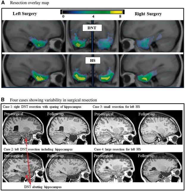
Figure 1.
Variability in surgical resection. (A) Overlay map of resections for surgical patients with DNT and hippocampal sclerosis (HS). Highest overlap of tissue removal is indicated in yellow. (B) MRI scans from four individual surgical participants preoperatively and at follow-up showing variability in resections even for the same lesion type (DNT or hippocampal sclerosis). Post-surgical resection volume and remaining ipsilesional hippocampal volumes for presented cases are as follows: Case 1: resection volume = 20.7 cm3, hippocampus = 2.4 cm3; Case 2: resection volume = 19.8 cm3, hippocampus = 0.6 cm3; Case 3: resection volume = 9.0 cm3, hippocampus = 1.1 cm3; Case 4: resection volume = 18.3 cm3, hippocampus = 0.7 cm3.

