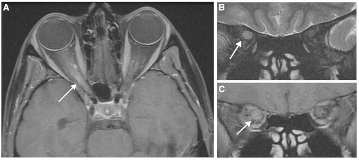
Figure 3.
Magnetic resonance optic nerve images acquired in a 30-year-old female with a 5-day history of acute right optic neuritis. (A) Axial post-contrast T1-weighted image shows swelling and enhancement of the intraorbital and intracanalicular parts of the right optic nerve. (B) Coronal T2-weighted image shows swollen hyperintense right optic nerve through posterior orbit. (C) Coronal post-contrast T1-weighted image shows gadolinium-enhancement of right optic nerve in posterior orbit. Images are courtesy of Dr Ahmed Toosy, UCL Institute of Neurology, London, UK.

