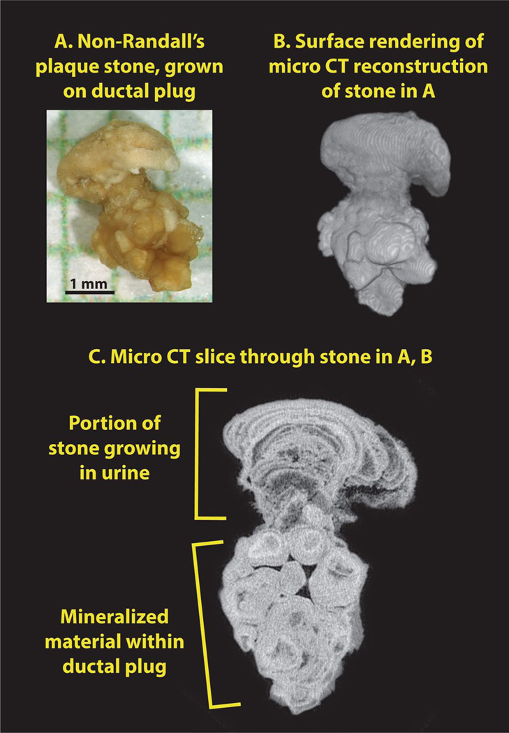
Fig. 3.
Apatite stone growing on end of BD plug. a Photograph of stone, after removal, shown on mm-grid paper. b Surface reconstruction of stone, showing head of stone on top of apatite plug. c Micro-CT image slice, showing the distinct morphologies of the apatite portion that grew in the calyceal urine and the apatite that formed within the BD lumen

