Abstract
The regulated intracellular transport of nutrient, adhesion, and growth factor receptors is crucial for maintaining cell and tissue homeostasis. Endocytosis, or endocytic membrane trafficking, involves the steps of intracellular transport that include, but are not limited to: internalization from the plasma membrane, sorting in early endosomes, transport to late endosomes/lysosomes followed by degradation, and/or recycling back to the plasma membrane via tubular recycling endosomes. In addition to regulating the localization of transmembrane receptor proteins, the endocytic pathway also controls the localization of non-receptor molecules. The non-receptor tyrosine kinase c-Src (Src), and its closely related family members Yes and Fyn, represent three proteins whose localization and signaling activities are tightly regulated by endocytic trafficking. Here, we provide a brief overview of endocytosis, Src function and its biochemical regulation. We will then concentrate on recent advances in understanding how Src intracellular localization is regulated and how its subcellular localization ultimately dictates downstream functioning. Since Src kinases are hyperactive in many cancers, it is essential to decipher the spatiotemporal regulation of this important family of tyrosine kinases.
Keywords: Cancer, cell signaling, endocytosis, Src function, Src regulation
INTRODUCTION
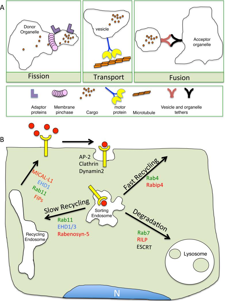
A eukaryotic cell is comprised of many distinct subcellular compartments called organelles. George Palade, the 1974 Nobel Prize winner in physiology or medicine, first observed that organelles such as the endoplasmic reticulum and the Golgi, as well as the plasma membrane, may communicate via small membrane bound vesicles and tubules(1). Nearly four decades later, James Rothman, Randy Schekman, and Thomas Sudhof were awarded the 2013 Nobel Prize for their combined work in characterizing the genetic and biochemical basis of vesicular transport. As shown in Figure 1A, communication between two intracellular organelles involves several steps. First, membrane coat proteins and adaptor proteins at the donor organelle generate a membrane bud that contains the cargo destined for the acceptor organelle. A vesicle is then derived from the membrane bud by a process called fission via the action of ‘membrane pinchases’. The newly formed vesicle travels along the microtubules tracks, and arrives at the acceptor organelle and undergoes fusion, a process mediated by protein-protein interactions between the vesicle and target organelle. The precise coordination of membrane fusion/fission and vesicular transport is particularly important for the endocytosis or endocytic trafficking of transmembrane proteins to and from the plasma membrane.
Figure 1.
Membrane dynamics and the endocytic pathway. A) Schematic diagram depicting the general process of membrane budding, membrane fission, vesicular transport along microtubules and membrane fusion. B) Schematic diagram showing the endocytic itinerary of a transmembrane receptor and its ligand (i.e., EGFR/EGF) and several key regulatory proteins involved in transport at each endocytic organelle.
Canonically, the endocytosis of a transmembrane receptor begins at the plasma membrane (Figure 1B) where ligand binding frequently induces its internalization. While there are many routes through which receptors are internalized (reviewed elsewhere(2, 3)), most itineraries converge at the early or ‘sorting’ endosome, where cargo-dependent sorting occurs. Cargo sorted into the tubular recycling endosome (TRE) compartment is typically returned to the plasma membrane, while cargo sorted to multivesicular bodies (MVB) is usually degraded via the late endosome-lysosome pathway (4). As expected, such exquisite sorting requires tight control by regulatory proteins.
One important group of endocytic regulators is the Rab family proteins consisting of more than 60 small GTPases that control the sorting, recycling or degradation of cargo proteins by regulating membrane fission/fusion, cytoskeletal transport, phosphoinositide lipid content and organelle targeting along the endocytic pathway(5). The localization of a small subset of Rab GTPases is shown in Figure 1B (green notation). In their GTP-bound, membrane-associated state, Rabs facilitate vesicular transport by recruiting effector proteins (Figure 1B; red notation). In addition to Rab proteins, the C-terminal Eps15 homology domain protein family (EHD1-4; blue notation) and their binding partners (Figure 1B; purple notation) also regulate important trafficking steps within the endocytic pathway(6). Interestingly, Rabs and EHDs are indirectly linked through mutual binding partners(7); however, the exact functional relevance of this linkage is not well understood.
While the intricate molecular regulation of membrane transport along the endocytic pathway is still under study, it is well known that endocytosis is an integral cellular process that regulates cell signaling and migration(8). Indeed, the internalization and trafficking of receptor tyrosine kinases (RTK) such as Epidermal Growth Factor Receptor (EGFR) and Platelet Derived Growth Factor Receptor (PDGFR) into MVB by the Endosomal Sorting Complexes Required for Transport (ESCRT) proteins is required for signal attenuation and receptor degradation. Disruption of RTK endocytosis can prolong signaling and lead to cancer by promoting uncontrolled cell growth and proliferation(9). It is noteworthy that localization of activated RTK to the cell surface or endosomes promotes differential signaling events, suggesting that endosomes also act as signaling hubs(10). The function of cell adhesion receptors such as integrins is also regulated by endocytosis(11).
Endocytic trafficking can also modulate cell signaling events by controlling the localization of non-receptor tyrosine kinases (NRTKs). The NRTK Src family of kinases (SFK) acts directly downstream of RTKs and thus regulates cell proliferation and migration. Src kinase activity is tightly modulated by biochemical events such as phosphorylation and protein-protein interactions (12). However, there is a growing consensus that endocytic trafficking is required for SFK function(13). Such a role for endocytic trafficking in SFK function is complicated by evidence that SFK phosphorylate and interact with several components of the endocytic pathway, in turn affecting endocytic events. In this review, we will first discuss the structure, biochemical regulation and function of SFK. We will then detail how endocytic trafficking regulates SFK localization and function. Lastly, we will conclude by summarizing how SFK also modulate endocytic events. Understanding the complex relationship between SFK and endocytosis is crucial, as Src and its endocytic regulatory components may provide novel therapeutic agents to treat Src-dependent tumors.
BODY of REVIEW
Src Family of Kinases
Src function
In the early 20th century, Peyton Rous discovered that a filterable agent, which later became known as the Rous Sarcoma Virus, caused soft tissue tumors (sarcomas) in birds(14). The transformative agent, v-Src, is a truncated form of a cellular protein kinase called c-Src (Src)(15). Src, one of the first proto-oncogenes, is the founding member of the SFK, a family of 8 closely related NRTK (Src, Fyn, Yes, Lyn, Hck, Blk, Fgr, Lck). Src is highly conserved among metazoans and a Src ortholog is even expressed in unicellular choanoflagellates (16-19). Biochemical analysis of unicellular Src has provided novel insight into the evolution of Src biochemical regulation in multicellular organisms (see below). In mammals, Src, Fyn and Yes (SYF) are ubiquitously expressed while the other family members display more restricted expression profiles. As tyrosine kinases, SFK phosphorylate proteins such as focal adhesion kinase (FAK), p190RhoGAP, and signal transducer and activator of transcription (STAT) to promote cell migration and proliferation. Given that SFK are overactive in a number of cancers, understanding Src regulatory mechanisms is a high priority for researchers(20).
SFK Structure
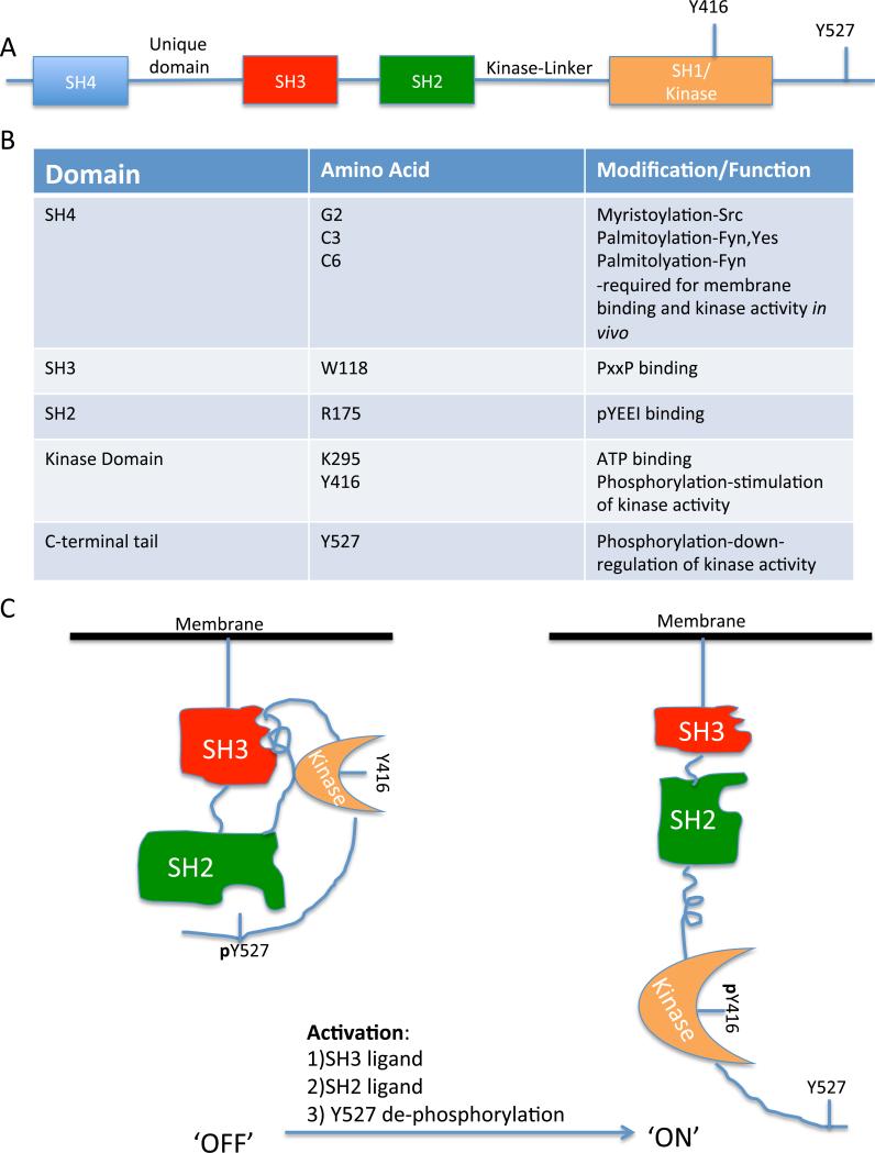
SFK share a highly conserved protein domain architecture (Fig. 2A, reviewed (21)). All SFK are myristoylated at the N-terminal Src Homology (SH) 4 domain. Some family members, such as Yes and Fyn, are also palmitoylated. N-terminal lipid modification is required for SFK membrane association and kinase activity in cells. The unique domain is the only domain that is not highly conserved between SFK members. The SH3 domain binds to PxxP proline-rich motifs (where x stands for any amino acid) while the SH2 domain binds to phospho-tyrosine (pY) residues that are flanked by acidic residues such as glutamate and followed by isoleucine (pY-E-E-I). A type-II polyproline helix lies between the SH2 and kinase domain. The kinase domain, or SH1 domain, consists of two lobes (N and C). Between the N and C lobes is a helix containing a critical tyrosine residue (Y416 in chicken or Y419 in humans(22)) that must be autophosphorylated in trans by an adjacent Src molecule to enable Src kinase activity.
Figure 2.
Src structure and regulation. A) Schematic diagram of the linear domain arrangement of c-Src. B) Table showing key amino acids within each domain, and the function or post-translational modification of these residues. C) Model depicting the inactive, autoinhibitory conformation (‘OFF’) of Y527 c-terminal phosphorylated Src and the active, open conformation (‘ON’) of Y416 phosphorylated Src. Note that modular domain color-coding is synchronized with that shown in Figure 2A.
The C-terminus of SFK contains a regulatory tyrosine (Y527 in chicken and Y530 in humans) that is phosphorylated by a regulatory kinase known as C-terminal Src kinase (CSK)(23). In keeping with the historical chicken Src numbering system used, we will abide by this numerical designation for the remainder of the review, both for human Src and other Src family members, such as Hck. Accordingly, phosphorylation at Y527 is required for Src down-regulation(24). The loss (as is the case with v-Src) or mutation of this tyrosine residue leads to constitutively active Src. A summary of critical amino acid residues in each Src modular domain and their functions can be found in Figure 2B. The domain architecture and phosphorylation of SFK is central to the regulation of their kinase activity.
It is noteworthy that CSK-mediated down-regulation of Src kinase activity appears to have evolved with metazoans(16-19). In an eloquent biochemical study, Segawa et al. compared and characterized the regulation of Src orthologs expressed in the unicellular choanoflagellate Monosiga ovata and the multicellular primitive sponge Ephydatia fluviatilis(16). CSK phosphorylation was evident in M. ovata and in E. fluviatilis; however, M. ovata Src was still active following CSK phosphorylation. In fact, ectopic expression of wild-type M. ovata Src in mammalian cells induced cellular transformation irrespective of CSK expression, indicating that exquisite Src regulation in multicellular organisms is crucial for tissue homeostasis.
Src regulation by phosphorylation and protein-protein interactions
As noted above, C-terminal phosphorylation of Src Y527 by CSK is required to turn off Src catalytic activity. This phosphorylation promotes an intramolecular interaction with its SH2 domain (Fig. 2B, ‘off’)(25). SH2-pY527 binding causes the SH3 domain to bind to the polyproline helix SH2-kinase linker region. As a result, the SH3 domain pushes against the backside of the N-lobe, which closes the cleft between the N and C lobes thus burying Y416 and preventing ATP or substrate binding. Collectively, the sequential steps of the Src intramolecular interaction are known as the ‘latch’ (SH2-pY527), ‘clamp’ (SH3-linker) and ‘switch’ (kinase domain conformation)(26). The intramolecular interactions between the Src modular domains are substantially weaker than Src intermolecular interactions, allowing for rapid induction of Src activation under the certain conditions. For example, the SH2 domain preferentially binds to pY-E-E-I; however, pY527 is followed by Q-P-G thus making it a weaker affinity substrate. Similarly, the Src SH3 domain binds with higher affinity to PxxP proline-rich motifs than it does to the kinase-linker region, which contains a type II polyproline helix rather than a PxxP motif. Thus, alleviation of the Src intramolecular interaction and full kinase activity is accomplished by: 1) dephosphorylation of pY527, 2) pY-E-E-I substrate binding to the SH2 domain, or 3) PxxP substrate binding to the SH3 domain (Fig 2B, ‘on’).
In cells, Src activation occurs downstream of activated RTKs and integrin receptors. RTK tyrosine autophosphorylation provides a high-affinity SH2 ligand for Src(27). Similarly, activation of integrin receptors induces autophosphorylation of focal adhesion kinase (FAK) at Y397 and Src SH2-pY397 binding induces Src activation(28). Src then phosphorylates several tyrosine residues on FAK that are required for focal adhesion turnover and migration(29). pY527 can be dephosphorylated by phosphatases such as receptor protein tyrosine phosphatase-alpha (RPTP-α)(30). Indeed, growth factor or integrin-induced Src activation is substantially reduced in RPTP-α knockout mice.
Hck SH3 binding to the HIV-1 protein Nef PQVP proline-rich motif best demonstrates the activation of SFK via SH3 interaction (termed SH3 displacement)(31). Hck-Nef binding (Kd= 250 nM) represents one of the strongest known SH3 binding affinities and is required for HIV replication in peripheral mononuclear cells in vitro(32, 33). Hck-Nef binding also provides insight into the interplay between the three Src regulatory mechanisms. Mutation of Y527-Q-P-G to Y-E-E-I greatly reduces Hck activation by locking its c-terminus to the SH2 domain. However, Nef binding to the Hck SH3 domain is able to induce Hck catalytic activity even in the presence of the high affinity SH2-pY527 interaction(34), suggesting that: 1) SH3 domain displacement from the linker region is sufficient to induce Hck activity and signaling, and 2) differential activation of SFK activity by SH2-pY527 or SH3-PxxP interactions may specify downstream function. While SFK conformational change represents one mode of modulating Src kinase activity, Src intracellular localization also dictates its downstream signaling events.
Endocytosis regulates SFK activation and function
Src localization
Src localization was assessed in chicken embryonic fibroblasts expressing v-Src(35). v-Src localized to both the plasma membrane and perinuclear region. Temperature-sensitive mutants (ts) of v-Src that are inactive at restrictive temperatures and active at permissive temperatures demonstrated that inactive v-Src localized in a soluble cytosolic pool while active v-Src localized to insoluble plasma membrane fractions(36). c-Src also displayed such dual localization and distribution to endosomal membrane fractions and the plasma membrane(37) however the majority of c-Src localized to endosomal membranes, which is in agreement with biochemical data suggesting that greater most of c-Src is inactive under non-stimulated conditions(38).
Localization of active Src to focal adhesions requires the N-terminal myristolyation and the SH3 domain(39). However, Src localization to focal adhesions does not require its kinase activity, although expression of a kinase-dead form of Src does alter focal adhesion morphology. Indeed, kinase-dead Src impairs focal adhesion turnover and cell migration(40). With growing evidence that Src activation and localization are tightly coupled, pertinent questions arise regarding the nature of the molecular determinants that control Src transport from the perinuclear region to the plasma membrane and focal adhesions. The majority of papers that address SFK trafficking are focused on Src; we will, however, highlight the differential trafficking of SFK members at the end of this section.
Regulation of Src activation and function by Rho GTPases, actin, and endosomes
In NIH 3T3 mouse fibroblasts, ts v-Src translocates to the plasma membrane upon temperature shift even in the absence of serum or other Src activating factors and induces cell cycle progression and transformation(41). In Swiss 3T3 fibroblasts, however, ts v-Src is retained in the perinuclear region. Swiss 3T3 cells are unique in that their actin microfilaments are rapidly depolymerized upon serum-starvation. This raised the notion that v-Src translocation depends on actin stress fibers. Indeed, serum starved Swiss 3T3 cells injected with constitutively active RhoA (which induces stress fibers) caused v-Src translocation upon temperature shift(41). In support of these findings, disruption of the actin cytoskeleton in NIH 3T3 fibroblasts with cytochalasin D also prevented v-Src translocation and inhibited v-Src induced transformation(41). In addition to RhoA, other small GTPases such as Rac and CDC42 are required to recruit Src to membrane ruffles and filopodia, respectively(42). Collectively, these studies demonstrated that Src translocates to the cell periphery along the actin cytoskeleton in a Rho GTPase-dependent fashion.
Early work using biochemical fractionation and microscopy-based localization showed that Src localizes to endosomes(37). Inhibition of the phosphatidylinositol-3-kinase (PI3K) VPS34, which disrupts early endosomal integrity, impaired Src translocation to focal adhesions, suggesting that Src translocation depends on endosomes(43). Sandilands et al. provided the first direct evidence that endosomes directly mediate Src translocation. In this landmark study, they developed a novel Src-GFP fusion protein that retained the behavior of endogenous Src(44). Previous attempts to create tagged Src constructs resulted in overactive Src, presumably by affecting its intramolecular interaction. They discovered that Src-GFP is activated on route to the plasma membrane in response to growth factor or serum-stimulated cells.
Active Src localized to vesicular structures decorated by the small GTPase RhoB. GTP-bound RhoB localizes to endosomes and lysosomes(45, 46). Interestingly, Src-GFP did not localize significantly to vesicular structures containing the closely related RhoD (see below-Fyn and Lyn traffic in RhoD vesicles). Functionally, RhoB is necessary for Src-GFP translocation and activation. In RhoB-null fibroblasts, Src is retained in the perinuclear region in its inactive state even after growth factor stimulation. RhoB regulates early endosome motility along the actin cytoskeleton by recruiting mDia1, a formin protein that promotes actin coat formation along endosomes(47). In agreement with these two studies, mDia1 is also a regulator of v-Src transport along the actin microfilaments(48). Intriguingly, RhoB motility is impaired by the loss of Src, thus it is possible that Src regulates its own transport by stimulating RhoB activation, possibly by promoting mDia1 activation.
Recycling endosomes in Src transport and activation
The perinuclear region is a crowded area of the cell. Several organelles including early endosomes, late endosomes, lysosomes, Golgi and the endocytic recycling compartment (ERC) are all localized to the perinuclear area or move into this area upon maturation (as is the case with early endosomes). Thus, the broad characterization of Src as a perinuclear cargo protein implicates several distinct or possibly overlapping organelles, which can be difficult to distinguish from one another upon perturbation of the endocytic pathway by genetic or pharmacologic manipulation. Another point of consideration is that over-expression of proteins, either Src or endocytic regulatory proteins such as Rabs, can modulate the dynamics of the endocytic pathway by promoting abnormal fission/fusion events.
While Src transport is regulated by early endosome motility and the actin cytoskeleton, Sandilands et al. also found that expression of dominant-negative Rab11, which inhibits recycling out of the perinuclear ERC, also impairs Src-GFP transport(44). These data support the notion that recycling endosomes are also crucial for Src translocation to the cell periphery.
Endocytic recycling from the ERC requires Rab11 and also EHD1 and its interacting partner Molecule Interacting with CasL-Like1 (MICAL-L1)(49-51). In HeLa cervical cancer cells, MICAL-L1 and EHD1 localize to TRE emanating outwards from the ERC. Current models suggest that MICAL-L1 functions in both TRE biosynthesis by recruiting membrane tubulators such as Syndapin-2 and EHD3, and TRE fission by recruiting membrane pinchases like EHD1(52, 53).
Recently, we demonstrated that endogenous Src decorates MICAL-L1 tubules in HeLa cells, suggesting that Src is either a cargo or regulator of MICAL-L1-decorated TRE(54). Given that inactive Src overlaps in its localization with transferrin receptor, a bona-fide cargo protein of the ERC, this suggests that Src is likely a cargo. In HeLa cells, EGF treatment causes Src to move out of the transferrin positive ERC to the cell periphery (i.e., to focal adhesions). This coincides with an increase in Src activation as measured by Src pY416, in agreement with previous findings(44). MICAL-L1- or EHD1-depletion caused Src to remain in the ERC following EGF treatment and also severely impaired Src activation(54). MICAL-L1-depletion in human fibroblasts decreased PDGF- and integrin-induced Src activation and also impaired Src-dependent processes such as PDGF-induced macropinocytosis, focal adhesion turnover, cell spreading and migration(54). Collectively, our data provide support for the idea that Src is an ERC cargo protein and that its localization, activation, and function are regulated by MICALL1 and EHD1.
Late endosomes, ESCRTs and Src trafficking: What happens to Src after activation is complete?
While our data and and studies from other laboratories clearly implicate recycling endosomes in regulating Src transport, there is also evidence that late endosomes and lysosomes regulate Src transport. In HeLa cells, over-expressed c-Src-GFP rapidly shuttles back and forth from the plasma membrane to perinuclear regions that contain the lysosomal hydrolase Cathepsin D(55). Over-expression of Src in HeLa cells induces macropinosome formation(56). Given that Src localizes to macropinosomes that eventually fuse with lysosomes, it is not surprising that over-expressed Src localizes with the lysosomal compartment. Furthermore, the ESCRT complex component Tsg101 regulates v-Src trafficking from the plasma membrane to lysosomes(57). Using conditional Tsg101 deletion mouse embryonic fibroblasts, Tu et al. showed that early loss of Tsg101 (1-3 days) induces higher levels of active v-Src compared to control cells. On the other hand, chronic Tsg101 deletion caused a decrease in active v-Src to levels below those of control cells, but also increased Src protein stability.
In human fibroblasts, transient depletion of several ESCRT components resulted in the retention of endogenous active c-Src in enlarged early endosomes containing β1 integrins(58). Functionally, the ESCRT complex is best characterized for its role in the transport of endocytic cargo to multivesicular bodies; examples of such cargo include ubiquitinated RTK and β1 integrins(59, 60). Interestingly, Src ubiquitination is required for its degradation, and both Src ubiquitination and degradation depend on its activation(61, 62). Lastly, constitutively active Src phosphorylates the ESCRT component Hrs and localizes with Hrs to enlarged early endosome structures suggesting that Src may act directly on the ESCRT complex to mediate its own downstream trafficking(63). Indeed, loss of ESCRT function results in sequestration of Src in enlarged endosomes. The signaling consequences of this phenomenon remain to be explored, although, as Tu et al. show, the chronic sequestration of active v-Src in enlarged endosomes may impair its function in mediating cell migration(57). Accordingly, we hypothesize that active Src may be ubiquitinated and transported from the plasma membrane to endosomes (or macropinosomes) and to the late endosomallysosomal compartment for degradation in an ESCRT-dependent manner.
Role of N-terminal lipid modification in differential trafficking of SFK
Despite the high level of structural and functional overlap between Src and other SFK members, important differences impart distinct regulation by trafficking. All SFK are myristolyated at the N-terminus, specifically, a glycine at position 2. However, SFK such as Fyn, Lyn and Yes are also mono- or di-palmitoylated at nearby cysteine residues. As noted above, wild-type Src-GFP preferentially localizes to RhoB containing vesicles when expressed in SYF−/− fibroblasts, although a small subset of Src-GFP also localizes to RhoD-containing endosomes(44). In contrast, Fyn-GFP expressed in SYF−/− cells localizes primarily to RhoD vesicles(64).
To test if differential lipid modifications specify RhoB vs RhoD localization, Sandilands et al. constructed a Src-GFP mutant that is palmitoylated and inversely created non-palmitoylated Fyn-GFP mutants(64). Indeed, palmitoylated Src localized to RhoD-containing early endosomes while non-palmitoylated Fyn behaved more like wild-type Src and localized to RhoB vesicles. These localization studies were confirmed using a combination of RhoB−/− MEFs and siRNA to deplete endogenous RhoD in SFY cells. In RhoB−/− cells, wild-type Src-GFP was sequestered in the perinuclear region. However, the palmitoylated Src mutant and wild-type Fyn-GFP were able to translocate from the perinuclear region to the plasma membrane(64). Conversely, while wild-type Src and non-palmitoylated Fyn translocated to the plasma membrane after RhoD depletion in SYF−/−, both the palmitoylated Src mutant and wild-type Fyn were retained in the perinuclear region(64). Yato et al. similarly found that differential lipid modifications of SFK dictate subcellular localization(65). The modes of transport for non-palmitoylated Src and palmitoylated SFK such as Fyn and Yes likely specify their distinct downstream functions. This is supported by the fact that while Src, Yes and Fyn have some overlapping functions (i.e., the loss of all three results in mouse embryonic lethality(66)), the loss of Src alone causes bone thickening (osteopetrosis) indicative of abnormal osteoclast functioning while loss of Fyn or Yes alone results in impaired thymocyte signaling and immunoglobulin receptor trafficking, respectively(67-69).
Regulation of endocytosis by SFK members
While SFK require endocytic trafficking for their activation and downstream function, they can also modulate the endocytic pathway. As noted earlier, over-expressed Src induces macropinocytosis or bulk fluid uptake in HeLa cells. v-Src also affects late endosome-lysosome fusion and biogenesis(70). The effect of over-expressed Src on the lysosomal degradative compartment likely reflects its physiologic role in resorptive processes during bone remodeling. While Src and other SFK have been implicated in regulating the endocytic transport of several cargo, we will focus on how Src specifically regulates Fibroblast Growth Factor Receptor (FGFR) and integrin internalization.
Regulation of FGFR internalization by Src
Treatment of fibroblasts with fibroblast growth factor (FGF) results in the activation and translocation of Src to the plasma membrane where it co-localizes with phosphorylated-FGFR(71). FGFR internalization and activation are impaired in SYF−/− cells and in RhoB−/− cells where inactive Src is retained in the perinuclear region. Inhibition of Src also modulates FGFR downstream signaling(71). In wild-type cells, short pulses of FGF result in the phosphorylation of Akt and ERK. Inhibition of Src (or in SYF−/− cells) impairs Akt activation. However, while Src inhibition initially attenuates ERK phosphorylation, Src inhibiton results in sustained ERK phosphorylation after long pulses of FGF (>1h) suggesting that Src also attenuates some signaling pathways(71).
How does Src regulate FGFR internalization? FGFR is internalized through clathrin-coated pits; indeed, FGF treatment increases the recruitment of clathrin to the plasma membrane, allowing for clathrin-coated pit formation and FGFR internalization(72). Clathrin is recruited to activated FGFR by epsin8. Interestingly, Src phosphorylates epsin8. Auciello et al. found that inhibition of Src in FGF-treated cells resulted in decreased epsin8 phosphorylation, decreased clathrin recruitment and thus decreased FGFR internalization(72).
FGFR and/or FGF are overexpressed in many cancers(73). Given that Src is required for FGFR internalization and signaling, and that overactive Src induces ligand-independent FGFR activation, Src may be a viable therapeutic target in FGFR-expressing cancers. Likewise, inhibition of Src by means of disrupting its trafficking (knockdown of RhoB, MICAL-L1 or EHD1) may also impede FGFR signaling events in cancer cells.
Role of Src in focal adhesion turnover and integrin internalization
Cell migration on an extracellular matrix requires highly dynamic focal adhesions(74). Focal adhesions represent the punctate attachment points of cells to an extracellular matrix. Integrin receptors, composed of alpha/beta heterodimers, bind to matrix molecules such as fibronectin, collagen and laminin via their extracellular domain. Integrin engagement with the extracellular matrix causes the intracellular recruitment of proteins to the integrin cytoplasmic tail, namely proteins such as: vinculin, talin, paxillin and FAK(75). Through these proteins, integrins are attached to the actin cytoskeletal network. During direction migration, focal adhesions must continually be assembled and disassembled at the cell front and disassembled at the cell rear and Src is central to focal adhesion disassembly(76).
Src−/− cells form focal adhesions that are highly stable(76). A similar phenotype is seen in FAK−/− cells(77), indicating that focal adhesions can form in the absence of Src and FAK but these two proteins are required for focal adhesion turnover. As noted above, recruitment of FAK to focal adhesions leads to FAK autophosphorylation at Y397. pY397 FAK serves as a binding substrate for Src, allowing for Src activation. Src then phosphorylates FAK on several tyrosine residues, most significantly, Y925(29). Prevention of FAK Y925 phosphorylation by inhibiting Src or mutation of FAK Y925F results in impaired focal adhesion disassembly and cell migration(78). The study of Ezratty et al., using a nocodazole-induced focal adhesion disassembly assay(79), was one of the first to directly implicate endocytic proteins in focal adhesion disassembly, showing that siRNA-mediated clathrin-depletion impaired focal adhesion disassembly (80). However, the significance of the link between the biochemical changes in FAK and Src and the altered integrin localization induced by focal adhesion turnover are not yet understood.
McNiven and colleagues provided an important new link when they found that Src phosphorylates the large GTPase dynamin2(81), the protein responsible for the fission of clathrin-coated pits at the plasma membrane (as well as non-clathrin invaginations). Src phosphorylation of dynamin2 Y231 results in a direct interaction between FAK and dynamin2. Src phosphorylation at this residue is critical for focal adhesion turnover, as dynamin2 Y231F mutants display increased focal adhesion stability and impaired turnover. As a result, dynamin2- depleted cells or cells expressing a dynamin2 mutant incapable of undergoing phosphorylation have increased surface levels of β1 integrins due to impaired internalization(81). It should be noted that Src phosphorylation of dynamin2 is not specific to focal adhesion turnover. Src-mediated phosphorylation of dynamin2 is also required for constitutive transferrin internalization and EGFR internalization(82, 83). Taken together, it is clear that Src modulates the internalization and trafficking of RTK such as FGFR and adhesion receptors such as integrins.
EXPERT OPINION
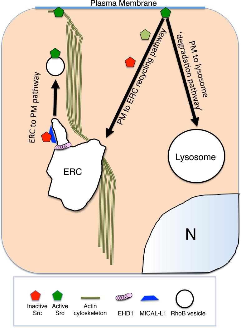
Endocytic proteins such as RhoB, Rab11, MICAL-L1 and EHD1 tightly control c-Src subcellular localization (Figure 3; ‘ERC to PM pathway’). Depletion of any of these proteins, or disruption of the actin cytoskeleton, inhibits Src movement to the cell periphery upon growth factor or adhesion receptor stimulation. Src retained inside the cell at the perinuclear ERC is unable to coordinate downstream signaling events that allow for cell migration and proliferation. While the mechanisms that control Src movement to the cell periphery have been recently highlighted, the endocytic fate of Src after activation is unknown.
Figure 3.
Schematic diagram describing Src regulation by trafficking. While the regulatory components of the Src ERC-PM pathway are partially understood, the endocytic fate of Src at the plasma membrane is less clear. Src may be recycled via the PM-ERC pathway and/or degraded via the PM to lysosome pathway depending on the cellular context (see text ‘Expert Opinion’ for details).
We predict that Src, much like other endocytic cargo, may be either degraded or recycled subsequent to its activation. In the case of v-Src, it is likely transported to the lysosome in an ESCRT-dependent manner. Several lines of evidence to support this notion: 1) v-Src is highly ubiquitinated compared to c-Src, although this ubiquitination is dependent on v-Src kinase activity, 2) ubiquitinated v-Src is degraded in a proteasome-dependent manner 3) while chronic TSG-101 depletion impairs v-Src activity, it also increases v-Src protein stability (57, 61, 62). This suggests that the fate of ubiquitinated v-Src may be analogous to that of ubiquitinated RTK, such as EGFR.
c-Src stability is significantly decreased in CSK−/− cell lines(62). This raises the idea that the fate of c-Src may be dictated by its conformational state. For example, v-Src is constitutively active and therefore constitutively ‘open’. However, phosphorylation of c-Src by CSK promotes an intramolecular interaction between its c-terminal tail with its SH2 domain; accordingly, depletion of CSK likely renders c-Src mostly in the ‘open’ conformation.
These observations support the notion that CSK expression leads to Src c-terminal phosphorylation and recycling to the ERC (Fig. 3; ‘PM to ERC’ pathway). On the other hand, we predict that v-Src (or c-Src in CSK null cells) undergoes ubiquitination-dependent degradation via the ESCRT complex (Fig. 3; ‘PM to lysosome pathway). This is further supported by the fact that in v-Src-expressing fibroblasts, expression of the E3 ligase Cbl-c inhibits cellular transformation(84). Alternatively, new lines of evidence suggest that the late-endosome/lysosome acts as a signaling platform, much like that of the early endosome and recycling endosomes. Indeed, localization of the mammalian target of Rapamycin (mTOR) protein to lysosomes is critical for its downstream functions(85, 86). Therefore, v-Src may promote differential signaling activities through its interactions with the lysosome that are important during cellular transformation.
While Src signaling and function relies heavily on the endosomal system, Src also modulates components of the endocytic pathway by controlling the internalization and trafficking of the very receptors which activate it (EGFR, FGFR, integrins). Src promotes endocytosis by phosphorylating endocytic proteins such as dynamin2 and Eps8. Moreover, Src can also be regulated by autophagy(87). In FAK−/− null mouse squamous carcinoma cells, the loss of FAK may represent cellular stress that induces autophagy, and Src localizes to autophagocytic vesicles in FAK-deleted mouse carcinoma cells. This targeting of Src requires Cbl, further supporting the notion that overactive Src promotes differential signaling activities that are important for cellular transformation and cancer cell survival. Indeed, in FAK−/− cancer cell lines, pharmacologic disruption of autophagy by 3-methyladenine or genetic disruption of autophagy by Atg5 or Atg7 depletion restored Src to focal adhesions but decreased cell viability and colony formation on soft agar. Lastly, Src also regulates the autophagic degradation of the RTK Ret in these FAK−/− cancer cell lines, which is also required for cell survival(88). Thus, targeting the autophagic pathway in some cancer cells with overactive Src may represent a novel therapeutic avenue.
In conclusion, this is an exciting time for the rapidly merging fields of cellular signaling and membrane trafficking. In this review, we have highlighted the complex relationship between the crucial Src family of cellular tyrosine kinases and the process of endocytic transport. While several facets of the Src trafficking pathway have been uncovered, it is clear that much remains to be delineated. Understanding how Src is trafficked in both normal and cancer cells has huge implications for potential treatment options in cancers in which uncontrolled Src activity promotes metastasis and decreased patient survival.
ACKNOWLEDGMENTS
The authors gratefully acknowledge NIH grant GM074876 and the Nebraska Dept. of Health for their support. The authors also thank Dr. Naava Naslavsky for her critical reading and contributions to the figures.
Abbreviations
- Csk
C-terminal Src kinase
- EGFR
Epidermal Growth Factor Receptor
- ERC
endocytic recycling compartment
- ESCRT
Endosomal Sorting Complexes Required for Transport
- FAK
focal adhesion kinase
- FGFR
Fibroblast growth factor receptor
- MVB
multivesicular bodies
- NRTK
non receptor tyrosine kinases
- RTK
receptor tyrosine kinases
- PDGFR
Platelet Derived Growth Factor Receptor
- SFK
Src family kinases
- SH
Src homology
- STAT
signal transducer and activator of transcription
- ts
temperature-sensitive
REFERENCES
- 1.Schekman R. Charting the secretory pathway in a simple eukaryote. Molecular biology of the cell. 2010;21(22):3781–3784. doi: 10.1091/mbc.E10-05-0416. [DOI] [PMC free article] [PubMed] [Google Scholar]
- 2.Howes MT, Mayor S, Parton RG. Molecules, mechanisms, and cellular roles of clathrin-independent endocytosis. Current opinion in cell biology. 2010;22(4):519–527. doi: 10.1016/j.ceb.2010.04.001. [DOI] [PubMed] [Google Scholar]
- 3.McMahon HT, Boucrot E. Molecular mechanism and physiological functions of clathrin-mediated endocytosis. Nature reviews Molecular cell biology. 2011;12(8):517–533. doi: 10.1038/nrm3151. [DOI] [PubMed] [Google Scholar]
- 4.Huotari J, Helenius A. Endosome maturation. The EMBO journal. 2011;30(17):3481–3500. doi: 10.1038/emboj.2011.286. [DOI] [PMC free article] [PubMed] [Google Scholar]
- 5.Pfeffer S, Aivazian D. Targeting Rab GTPases to distinct membrane compartments. Nature reviews Molecular cell biology. 2004;5(11):886–896. doi: 10.1038/nrm1500. [DOI] [PubMed] [Google Scholar]
- 6.Naslavsky N, Caplan S. EHD proteins: key conductors of endocytic transport. Trends in cell biology. 2011;21(2):122–131. doi: 10.1016/j.tcb.2010.10.003. [DOI] [PMC free article] [PubMed] [Google Scholar]
- 7.Zhang J, Naslavsky N, Caplan S. Rabs and EHDs: alternate modes for traffic control. Bioscience reports. 2012;32(1):17–23. doi: 10.1042/BSR20110009. [DOI] [PMC free article] [PubMed] [Google Scholar]
- 8.Sigismund S, Confalonieri S, Ciliberto A, Polo S, Scita G, Di Fiore PP. Endocytosis and signaling: cell logistics shape the eukaryotic cell plan. Physiological reviews. 2012;92(1):273–366. doi: 10.1152/physrev.00005.2011. [DOI] [PMC free article] [PubMed] [Google Scholar]
- 9.Mosesson Y, Mills GB, Yarden Y. Derailed endocytosis: an emerging feature of cancer. Nature reviews Cancer. 2008;8(11):835–850. doi: 10.1038/nrc2521. [DOI] [PubMed] [Google Scholar]
- 10.Palfy M, Remenyi A, Korcsmaros T. Endosomal crosstalk: meeting points for signaling pathways. Trends in cell biology. 2012;22(9):447–456. doi: 10.1016/j.tcb.2012.06.004. [DOI] [PMC free article] [PubMed] [Google Scholar]
- 11.Caswell PT, Vadrevu S, Norman JC. Integrins: masters and slaves of endocytic transport. Nature reviews Molecular cell biology. 2009;10(12):843–853. doi: 10.1038/nrm2799. [DOI] [PubMed] [Google Scholar]
- 12.Engen JR, Wales TE, Hochrein JM, Meyn MA, 3rd, Banu Ozkan S, Bahar I, Smithgall TE. Structure and dynamic regulation of Src-family kinases. Cellular and molecular life sciences : CMLS. 2008;65(19):3058–3073. doi: 10.1007/s00018-008-8122-2. [DOI] [PMC free article] [PubMed] [Google Scholar]
- 13.Sandilands E, Frame MC. Endosomal trafficking of Src tyrosine kinase. Trends in cell biology. 2008;18(7):322–329. doi: 10.1016/j.tcb.2008.05.004. [DOI] [PubMed] [Google Scholar]
- 14.Rous P. A Sarcoma of the Fowl Transmissible by an Agent Separable from the Tumor Cells. The Journal of experimental medicine. 1911;13(4):397–411. doi: 10.1084/jem.13.4.397. [DOI] [PMC free article] [PubMed] [Google Scholar]
- 15.Czernilofsky AP, Levinson AD, Varmus HE, Bishop JM, Tischer E, Goodman HM. Nucleotide sequence of an avian sarcoma virus oncogene (src) and proposed amino acid sequence for gene product. Nature. 1980;287(5779):198–203. doi: 10.1038/287198a0. [DOI] [PubMed] [Google Scholar]
- 16.Segawa Y, Suga H, Iwabe N, Oneyama C, Akagi T, Miyata T, Okada M. Functional development of Src tyrosine kinases during evolution from a unicellular ancestor to multicellular animals. Proceedings of the National Academy of Sciences of the United States of America. 2006;103(32):12021–12026. doi: 10.1073/pnas.0600021103. [DOI] [PMC free article] [PubMed] [Google Scholar]
- 17.Li W, Young SL, King N, Miller WT. Signaling properties of a non-metazoan Src kinase and the evolutionary history of Src negative regulation. The Journal of biological chemistry. 2008;283(22):15491–15501. doi: 10.1074/jbc.M800002200. [DOI] [PMC free article] [PubMed] [Google Scholar]
- 18.Schultheiss KP, Suga H, Ruiz-Trillo I, Miller WT. Lack of Csk-mediated negative regulation in a unicellular SRC kinase. Biochemistry. 2012;51(41):8267–8277. doi: 10.1021/bi300965h. [DOI] [PMC free article] [PubMed] [Google Scholar]
- 19.Miller WT. Tyrosine kinase signaling and the emergence of multicellularity. Biochimica et biophysica acta. 2012;1823(6):1053–1057. doi: 10.1016/j.bbamcr.2012.03.009. [DOI] [PMC free article] [PubMed] [Google Scholar]
- 20.Wheeler DL, Iida M, Dunn EF. The role of Src in solid tumors. The oncologist. 2009;14(7):667–678. doi: 10.1634/theoncologist.2009-0009. [DOI] [PMC free article] [PubMed] [Google Scholar]
- 21.Boggon TJ, Eck MJ. Structure and regulation of Src family kinases. Oncogene. 2004;23(48):7918–7927. doi: 10.1038/sj.onc.1208081. [DOI] [PubMed] [Google Scholar]
- 22.Smart JE, Oppermann H, Czernilofsky AP, Purchio AF, Erikson RL, Bishop JM. Characterization of sites for tyrosine phosphorylation in the transforming protein of Rous sarcoma virus (pp60v-src) and its normal cellular homologue (pp60c-src). Proceedings of the National Academy of Sciences of the United States of America. 1981;78(10):6013–6017. doi: 10.1073/pnas.78.10.6013. [DOI] [PMC free article] [PubMed] [Google Scholar]
- 23.Nada S, Okada M, MacAuley A, Cooper JA, Nakagawa H. Cloning of a complementary DNA for a protein-tyrosine kinase that specifically phosphorylates a negative regulatory site of p60c-src. Nature. 1991;351(6321):69–72. doi: 10.1038/351069a0. [DOI] [PubMed] [Google Scholar]
- 24.Nada S, Yagi T, Takeda H, Tokunaga T, Nakagawa H, Ikawa Y, Okada M, Aizawa S. Constitutive activation of Src family kinases in mouse embryos that lack Csk. Cell. 1993;73(6):1125–1135. doi: 10.1016/0092-8674(93)90642-4. [DOI] [PubMed] [Google Scholar]
- 25.Xu W, Doshi A, Lei M, Eck MJ, Harrison SC. Crystal structures of c-Src reveal features of its autoinhibitory mechanism. Molecular cell. 1999;3(5):629–638. doi: 10.1016/s1097-2765(00)80356-1. [DOI] [PubMed] [Google Scholar]
- 26.Harrison SC. Variation on an Src-like theme. Cell. 2003;112(6):737–740. doi: 10.1016/s0092-8674(03)00196-x. [DOI] [PubMed] [Google Scholar]
- 27.Alonso G, Koegl M, Mazurenko N, Courtneidge SA. Sequence requirements for binding of Src family tyrosine kinases to activated growth factor receptors. The Journal of biological chemistry. 1995;270(17):9840–9848. doi: 10.1074/jbc.270.17.9840. [DOI] [PubMed] [Google Scholar]
- 28.Xing Z, Chen HC, Nowlen JK, Taylor SJ, Shalloway D, Guan JL. Direct interaction of v-Src with the focal adhesion kinase mediated by the Src SH2 domain. Molecular biology of the cell. 1994;5(4):413–421. doi: 10.1091/mbc.5.4.413. [DOI] [PMC free article] [PubMed] [Google Scholar]
- 29.Westhoff MA, Serrels B, Fincham VJ, Frame MC, Carragher NO. SRC-mediated phosphorylation of focal adhesion kinase couples actin and adhesion dynamics to survival signaling. Molecular and cellular biology. 2004;24(18):8113–8133. doi: 10.1128/MCB.24.18.8113-8133.2004. [DOI] [PMC free article] [PubMed] [Google Scholar]
- 30.Su J, Muranjan M, Sap J. Receptor protein tyrosine phosphatase alpha activates Src-family kinases and controls integrin-mediated responses in fibroblasts. Current biology : CB. 1999;9(10):505–511. doi: 10.1016/s0960-9822(99)80234-6. [DOI] [PubMed] [Google Scholar]
- 31.Moarefi I, LaFevre-Bernt M, Sicheri F, Huse M, Lee CH, Kuriyan J, Miller WT. Activation of the Src-family tyrosine kinase Hck by SH3 domain displacement. Nature. 1997;385(6617):650–653. doi: 10.1038/385650a0. [DOI] [PubMed] [Google Scholar]
- 32.Saksela K, Cheng G, Baltimore D. Proline-rich (PxxP) motifs in HIV-1 Nef bind to SH3 domains of a subset of Src kinases and are required for the enhanced growth of Nef+ viruses but not for down-regulation of CD4. The EMBO journal. 1995;14(3):484–491. doi: 10.1002/j.1460-2075.1995.tb07024.x. [DOI] [PMC free article] [PubMed] [Google Scholar]
- 33.Lee CH, Leung B, Lemmon MA, Zheng J, Cowburn D, Kuriyan J, Saksela K. A single amino acid in the SH3 domain of Hck determines its high affinity and specificity in binding to HIV-1 Nef protein. The EMBO journal. 1995;14(20):5006–5015. doi: 10.1002/j.1460-2075.1995.tb00183.x. [DOI] [PMC free article] [PubMed] [Google Scholar]
- 34.Lerner EC, Smithgall TE. SH3-dependent stimulation of Src-family kinase autophosphorylation without tail release from the SH2 domain in vivo. Nature structural biology. 2002;9(5):365–369. doi: 10.1038/nsb782. [DOI] [PubMed] [Google Scholar]
- 35.Rohrschneider LR. Immunofluorescence on avian sarcoma virus-transformed cells: localization of the src gene product. Cell. 1979;16(1):11–24. doi: 10.1016/0092-8674(79)90183-1. [DOI] [PubMed] [Google Scholar]
- 36.Welham MJ, Wyke JA. A single point mutation has pleiotropic effects on pp60v src function. Journal of virology. 1988;62(6):1898–1906. doi: 10.1128/jvi.62.6.1898-1906.1988. [DOI] [PMC free article] [PubMed] [Google Scholar]
- 37.Kaplan KB, Swedlow JR, Varmus HE, Morgan DO. Association of p60c-src with endosomal membranes in mammalian fibroblasts. The Journal of cell biology. 1992;118(2):321–333. doi: 10.1083/jcb.118.2.321. [DOI] [PMC free article] [PubMed] [Google Scholar]
- 38.Zheng XM, Resnick RJ, Shalloway D. A phosphotyrosine displacement mechanism for activation of Src by PTPalpha. The EMBO journal. 2000;19(5):964–978. doi: 10.1093/emboj/19.5.964. [DOI] [PMC free article] [PubMed] [Google Scholar]
- 39.Kaplan KB, Bibbins KB, Swedlow JR, Arnaud M, Morgan DO, Varmus HE. Association of the amino-terminal half of c-Src with focal adhesions alters their properties and is regulated by phosphorylation of tyrosine 527. The EMBO journal. 1994;13(20):4745–4756. doi: 10.1002/j.1460-2075.1994.tb06800.x. [DOI] [PMC free article] [PubMed] [Google Scholar]
- 40.Fincham VJ, Frame MC. The catalytic activity of Src is dispensable for translocation to focal adhesions but controls the turnover of these structures during cell motility. The EMBO journal. 1998;17(1):81–92. doi: 10.1093/emboj/17.1.81. [DOI] [PMC free article] [PubMed] [Google Scholar]
- 41.Fincham VJ, Unlu M, Brunton VG, Pitts JD, Wyke JA, Frame MC. Translocation of Src kinase to the cell periphery is mediated by the actin cytoskeleton under the control of the Rho family of small G proteins. The Journal of cell biology. 1996;135(6 Pt 1):1551–1564. doi: 10.1083/jcb.135.6.1551. [DOI] [PMC free article] [PubMed] [Google Scholar]
- 42.Timpson P, Jones GE, Frame MC, Brunton VG. Coordination of cell polarization and migration by the Rho family GTPases requires Src tyrosine kinase activity. Current biology : CB. 2001;11(23):1836–1846. doi: 10.1016/s0960-9822(01)00583-8. [DOI] [PubMed] [Google Scholar]
- 43.Fincham VJ, Brunton VG, Frame MC. The SH3 domain directs acto-myosin-dependent targeting of v-Src to focal adhesions via phosphatidylinositol 3-kinase. Molecular and cellular biology. 2000;20(17):6518–6536. doi: 10.1128/mcb.20.17.6518-6536.2000. [DOI] [PMC free article] [PubMed] [Google Scholar]
- 44.Sandilands E, Cans C, Fincham VJ, Brunton VG, Mellor H, Prendergast GC, Norman JC, Superti-Furga G, Frame MC. RhoB and actin polymerization coordinate Src activation with endosome-mediated delivery to the membrane. Developmental cell. 2004;7(6):855–869. doi: 10.1016/j.devcel.2004.09.019. [DOI] [PubMed] [Google Scholar]
- 45.Adamson P, Paterson HF, Hall A. Intracellular localization of the P21rho proteins. The Journal of cell biology. 1992;119(3):617–627. doi: 10.1083/jcb.119.3.617. [DOI] [PMC free article] [PubMed] [Google Scholar]
- 46.Robertson D, Paterson HF, Adamson P, Hall A, Monaghan P. Ultrastructural localization of ras-related proteins using epitope-tagged plasmids. The journal of histochemistry and cytochemistry : official journal of the Histochemistry Society. 1995;43(5):471–480. doi: 10.1177/43.5.7537292. [DOI] [PubMed] [Google Scholar]
- 47.Fernandez-Borja M, Janssen L, Verwoerd D, Hordijk P, Neefjes J. RhoB regulates endosome transport by promoting actin assembly on endosomal membranes through Dia1. Journal of cell science. 2005;118(Pt 12):2661–2670. doi: 10.1242/jcs.02384. [DOI] [PubMed] [Google Scholar]
- 48.Tanji M, Ishizaki T, Ebrahimi S, Tsuboguchi Y, Sukezane T, Akagi T, Frame MC, Hashimoto N, Miyamoto S, Narumiya S. mDia1 targets v-Src to the cell periphery and facilitates cell transformation, tumorigenesis, and invasion. Molecular and cellular biology. 2010;30(19):4604–4615. doi: 10.1128/MCB.00197-10. [DOI] [PMC free article] [PubMed] [Google Scholar]
- 49.Ullrich O, Reinsch S, Urbe S, Zerial M, Parton RG. Rab11 regulates recycling through the pericentriolar recycling endosome. The Journal of cell biology. 1996;135(4):913–924. doi: 10.1083/jcb.135.4.913. [DOI] [PMC free article] [PubMed] [Google Scholar]
- 50.Sharma M, Giridharan SS, Rahajeng J, Naslavsky N, Caplan S. MICAL-L1 links EHD1 to tubular recycling endosomes and regulates receptor recycling. Molecular biology of the cell. 2009;20(24):5181–5194. doi: 10.1091/mbc.E09-06-0535. [DOI] [PMC free article] [PubMed] [Google Scholar]
- 51.Caplan S, Naslavsky N, Hartnell LM, Lodge R, Polishchuk RS, Donaldson JG, Bonifacino JS. A tubular EHD1-containing compartment involved in the recycling of major histocompatibility complex class I molecules to the plasma membrane. The EMBO journal. 2002;21(11):2557–2567. doi: 10.1093/emboj/21.11.2557. [DOI] [PMC free article] [PubMed] [Google Scholar]
- 52.Cai B, Giridharan SS, Zhang J, Saxena S, Bahl K, Schmidt JA, Sorgen PL, Guo W, Naslavsky N, Caplan S. Differential roles of C-terminal Eps15 homology domain proteins as vesiculators and tubulators of recycling endosomes. The Journal of biological chemistry. 2013;288(42):30172–30180. doi: 10.1074/jbc.M113.488627. [DOI] [PMC free article] [PubMed] [Google Scholar]
- 53.Giridharan SS, Cai B, Vitale N, Naslavsky N, Caplan S. Cooperation of MICAL L1, syndapin2, and phosphatidic acid in tubular recycling endosome biogenesis. Molecular biology of the cell. 2013;24(11):1776–1790, S1771-1715. doi: 10.1091/mbc.E13-01-0026. [DOI] [PMC free article] [PubMed] [Google Scholar]
- 54.Reinecke JB, Katafiasz D, Naslavsky N, Caplan S. Regulation of Src trafficking and activation by the endocytic regulatory proteins MICAL-L1 and EHD1. J Cell Sci. 2014 doi: 10.1242/jcs.133892. in press. [DOI] [PMC free article] [PubMed] [Google Scholar]
- 55.Kasahara K, Nakayama Y, Kihara A, Matsuda D, Ikeda K, Kuga T, Fukumoto Y, Igarashi Y, Yamaguchi N. Rapid trafficking of c-Src, a non-palmitoylated Src-family kinase, between the plasma membrane and late endosomes/lysosomes. Experimental cell research. 2007;313(12):2651–2666. doi: 10.1016/j.yexcr.2007.05.001. [DOI] [PubMed] [Google Scholar]
- 56.Kasahara K, Nakayama Y, Sato I, Ikeda K, Hoshino M, Endo T, Yamaguchi N. Role of Src-family kinases in formation and trafficking of macropinosomes. Journal of cellular physiology. 2007;211(1):220–232. doi: 10.1002/jcp.20931. [DOI] [PubMed] [Google Scholar]
- 57.Tu C, Ortega-Cava CF, Winograd P, Stanton MJ, Reddi AL, Dodge I, Arya R, Dimri M, Clubb RJ, Naramura M, Wagner KU, Band V, Band H. Endosomal-sorting complexes required for transport (ESCRT) pathway-dependent endosomal traffic regulates the localization of active Src at focal adhesions. Proceedings of the National Academy of Sciences of the United States of America. 2010;107(37):16107–16112. doi: 10.1073/pnas.1009471107. [DOI] [PMC free article] [PubMed] [Google Scholar]
- 58.Lobert VH, Stenmark H. The ESCRT machinery mediates polarization of fibroblasts through regulation of myosin light chain. Journal of cell science. 2012;125(Pt 1):29–36. doi: 10.1242/jcs.088310. [DOI] [PubMed] [Google Scholar]
- 59.Lobert VH, Brech A, Pedersen NM, Wesche J, Oppelt A, Malerod L, Stenmark H. Ubiquitination of alpha 5 beta 1 integrin controls fibroblast migration through lysosomal degradation of fibronectin-integrin complexes. Developmental cell. 2010;19(1):148–159. doi: 10.1016/j.devcel.2010.06.010. [DOI] [PubMed] [Google Scholar]
- 60.Babst M, Odorizzi G. The balance of protein expression and degradation: an ESCRTs point of view. Current opinion in cell biology. 2013;25(4):489–494. doi: 10.1016/j.ceb.2013.05.003. [DOI] [PMC free article] [PubMed] [Google Scholar]
- 61.Hakak Y, Martin GS. Ubiquitin-dependent degradation of active Src. Current biology : CB. 1999;9(18):1039–1042. doi: 10.1016/s0960-9822(99)80453-9. [DOI] [PubMed] [Google Scholar]
- 62.Harris KF, Shoji I, Cooper EM, Kumar S, Oda H, Howley PM. Ubiquitin-mediated degradation of active Src tyrosine kinase. Proceedings of the National Academy of Sciences of the United States of America. 1999;96(24):13738–13743. doi: 10.1073/pnas.96.24.13738. [DOI] [PMC free article] [PubMed] [Google Scholar]
- 63.Bache KG, Raiborg C, Mehlum A, Madshus IH, Stenmark H. Phosphorylation of Hrs downstream of the epidermal growth factor receptor. European journal of biochemistry / FEBS. 2002;269(16):3881–3887. doi: 10.1046/j.1432-1033.2002.03046.x. [DOI] [PubMed] [Google Scholar]
- 64.Sandilands E, Brunton VG, Frame MC. The membrane targeting and spatial activation of Src, Yes and Fyn is influenced by palmitoylation and distinct RhoB/RhoD endosome requirements. Journal of cell science. 2007;120(Pt 15):2555–2564. doi: 10.1242/jcs.003657. [DOI] [PubMed] [Google Scholar]
- 65.Sato I, Obata Y, Kasahara K, Nakayama Y, Fukumoto Y, Yamasaki T, Yokoyama KK, Saito T, Yamaguchi N. Differential trafficking of Src, Lyn, Yes and Fyn is specified by the state of palmitoylation in the SH4 domain. Journal of cell science. 2009;122(Pt 7):965–975. doi: 10.1242/jcs.034843. [DOI] [PubMed] [Google Scholar]
- 66.Klinghoffer RA, Sachsenmaier C, Cooper JA, Soriano P. Src family kinases are required for integrin but not PDGFR signal transduction. The EMBO journal. 1999;18(9):2459–2471. doi: 10.1093/emboj/18.9.2459. [DOI] [PMC free article] [PubMed] [Google Scholar]
- 67.Luton F, Verges M, Vaerman JP, Sudol M, Mostov KE. The SRC family protein tyrosine kinase p62yes controls polymeric IgA transcytosis in vivo. Molecular cell. 1999;4(4):627–632. doi: 10.1016/s1097-2765(00)80213-0. [DOI] [PubMed] [Google Scholar]
- 68.Gauen LK, Zhu Y, Letourneur F, Hu Q, Bolen JB, Matis LA, Klausner RD, Shaw AS. Interactions of p59fyn and ZAP-70 with T-cell receptor activation motifs: defining the nature of a signalling motif. Molecular and cellular biology. 1994;14(6):3729–3741. doi: 10.1128/mcb.14.6.3729. [DOI] [PMC free article] [PubMed] [Google Scholar]
- 69.Soriano P, Montgomery C, Geske R, Bradley A. Targeted disruption of the c-src proto-oncogene leads to osteopetrosis in mice. Cell. 1991;64(4):693–702. doi: 10.1016/0092-8674(91)90499-o. [DOI] [PubMed] [Google Scholar]
- 70.Palacios F, Tushir JS, Fujita Y, D'Souza-Schorey C. Lysosomal targeting of E-cadherin: a unique mechanism for the down-regulation of cell-cell adhesion during epithelial to mesenchymal transitions. Molecular and cellular biology. 2005;25(1):389–402. doi: 10.1128/MCB.25.1.389-402.2005. [DOI] [PMC free article] [PubMed] [Google Scholar]
- 71.Sandilands E, Akbarzadeh S, Vecchione A, McEwan DG, Frame MC, Heath JK. Src kinase modulates the activation, transport and signalling dynamics of fibroblast growth factor receptors. EMBO reports. 2007;8(12):1162–1169. doi: 10.1038/sj.embor.7401097. [DOI] [PMC free article] [PubMed] [Google Scholar]
- 72.Auciello G, Cunningham DL, Tatar T, Heath JK, Rappoport JZ. Regulation of fibroblast growth factor receptor signalling and trafficking by Src and Eps8. Journal of cell science. 2013;126(Pt 2):613–624. doi: 10.1242/jcs.116228. [DOI] [PMC free article] [PubMed] [Google Scholar]
- 73.Kelleher FC, O'Sullivan H, Smyth E, McDermott R, Viterbo A. Fibroblast growth factor receptors, developmental corruption and malignant disease. Carcinogenesis. 2013;34(10):2198–2205. doi: 10.1093/carcin/bgt254. [DOI] [PubMed] [Google Scholar]
- 74.Webb DJ, Parsons JT, Horwitz AF. Adhesion assembly, disassembly and turnover in migrating cells -- over and over and over again. Nature cell biology. 2002;4(4):E97–100. doi: 10.1038/ncb0402-e97. [DOI] [PubMed] [Google Scholar]
- 75.Geiger B, Yamada KM. Molecular architecture and function of matrix adhesions. Cold Spring Harbor perspectives in biology. 2011;3(5) doi: 10.1101/cshperspect.a005033. [DOI] [PMC free article] [PubMed] [Google Scholar]
- 76.Volberg T, Romer L, Zamir E, Geiger B. pp60(c-src) and related tyrosine kinases: a role in the assembly and reorganization of matrix adhesions. Journal of cell science. 2001;114(Pt 12):2279–2289. doi: 10.1242/jcs.114.12.2279. [DOI] [PubMed] [Google Scholar]
- 77.Ilic D, Furuta Y, Kanazawa S, Takeda N, Sobue K, Nakatsuji N, Nomura S, Fujimoto J, Okada M, Yamamoto T. Reduced cell motility and enhanced focal adhesion contact formation in cells from FAK-deficient mice. Nature. 1995;377(6549):539–544. doi: 10.1038/377539a0. [DOI] [PubMed] [Google Scholar]
- 78.Deramaudt TB, Dujardin D, Hamadi A, Noulet F, Kolli K, De Mey J, Takeda K, Ronde P. FAK phosphorylation at Tyr-925 regulates cross-talk between focal adhesion turnover and cell protrusion. Molecular biology of the cell. 2011;22(7):964–975. doi: 10.1091/mbc.E10-08-0725. [DOI] [PMC free article] [PubMed] [Google Scholar]
- 79.Ezratty EJ, Partridge MA, Gundersen GG. Microtubule-induced focal adhesion disassembly is mediated by dynamin and focal adhesion kinase. Nature cell biology. 2005;7(6):581–590. doi: 10.1038/ncb1262. [DOI] [PubMed] [Google Scholar]
- 80.Ezratty EJ, Bertaux C, Marcantonio EE, Gundersen GG. Clathrin mediates integrin endocytosis for focal adhesion disassembly in migrating cells. The Journal of cell biology. 2009;187(5):733–747. doi: 10.1083/jcb.200904054. [DOI] [PMC free article] [PubMed] [Google Scholar]
- 81.Wang Y, Cao H, Chen J, McNiven MA. A direct interaction between the large GTPase dynamin-2 and FAK regulates focal adhesion dynamics in response to active Src. Molecular biology of the cell. 2011;22(9):1529–1538. doi: 10.1091/mbc.E10-09-0785. [DOI] [PMC free article] [PubMed] [Google Scholar]
- 82.Cao H, Chen J, Krueger EW, McNiven MA. SRC-mediated phosphorylation of dynamin and cortactin regulates the “constitutive” endocytosis of transferrin. Molecular and cellular biology. 2010;30(3):781–792. doi: 10.1128/MCB.00330-09. [DOI] [PMC free article] [PubMed] [Google Scholar]
- 83.Ahn S, Kim J, Lucaveche CL, Reedy MC, Luttrell LM, Lefkowitz RJ, Daaka Y. Src-dependent tyrosine phosphorylation regulates dynamin self-assembly and ligand-induced endocytosis of the epidermal growth factor receptor. The Journal of biological chemistry. 2002;277(29):26642–26651. doi: 10.1074/jbc.M201499200. [DOI] [PubMed] [Google Scholar]
- 84.Kim M, Tezuka T, Tanaka K, Yamamoto T. Cbl-c suppresses v-Src-induced transformation through ubiquitin-dependent protein degradation. Oncogene. 2004;23(9):1645–1655. doi: 10.1038/sj.onc.1207298. [DOI] [PubMed] [Google Scholar]
- 85.Sancak Y, Bar-Peled L, Zoncu R, Markhard AL, Nada S, Sabatini DM. Ragulator-Rag complex targets mTORC1 to the lysosomal surface and is necessary for its activation by amino acids. Cell. 2010;141(2):290–303. doi: 10.1016/j.cell.2010.02.024. [DOI] [PMC free article] [PubMed] [Google Scholar]
- 86.Sancak Y, Peterson TR, Shaul YD, Lindquist RA, Thoreen CC, Bar-Peled L, Sabatini DM. The Rag GTPases bind raptor and mediate amino acid signaling to mTORC1. Science. 2008;320(5882):1496–1501. doi: 10.1126/science.1157535. [DOI] [PMC free article] [PubMed] [Google Scholar]
- 87.Sandilands E, Serrels B, McEwan DG, Morton JP, Macagno JP, McLeod K, Stevens C, Brunton VG, Langdon WY, Vidal M, Sansom OJ, Dikic I, Wilkinson S, Frame MC. Autophagic targeting of Src promotes cancer cell survival following reduced FAK signalling. Nature cell biology. 2012;14(1):51–60. doi: 10.1038/ncb2386. [DOI] [PubMed] [Google Scholar]
- 88.Sandilands E, Serrels B, Wilkinson S, Frame MC. Src-dependent autophagic degradation of Ret in FAK-signalling-defective cancer cells. EMBO reports. 2012;13(8):733–740. doi: 10.1038/embor.2012.92. [DOI] [PMC free article] [PubMed] [Google Scholar]


