

Abstract
Objective:
The introduction of new and potent therapies for the treatment of relapsing remitting multiple sclerosis (MS) has increased the desire for therapeutic success. There is growing doubt that the mere reduction of relapse rate, Expanded Disability Status Scale (EDSS) progression and magnetic resonance imaging (MRI) markers are exclusive and appropriate factors to monitor the new aim of ‘no evidence of disease activity’ (NEDA). However, there is no generally accepted definition so far.
Methods:
To achieve the therapeutic aim of NEDA, a panel of MS experts searched the available literature on clinical and paraclinical outcomes to propose a test battery that is sensitive to detect disease activity in an everyday clinical setting.
Results:
The panel proposed to include, besides relapse rate, disability progression and MRI, neuropsychological outcome measures such as cognitive status, fatigue, depression and quality of life. To standardize the examinations in an economic and schematic way, a multifactorial model [multiple sclerosis decision model (MSDM)] that includes the domains ‘relapse’, ‘disability progression’, ‘MRI’, and ‘neuropsychology’ is proposed. The scheme reflects the complexity of the disease even in the early stages when scales such as the EDSS are not able to distinguish low levels of progression.
Conclusion:
The MSDM aims to support early treatment decisions and uncover timely treatment failure. Prospective investigations are required to prove that such a disease-monitoring concept leads to an early and effective silencing of disease activity.
Keywords: disease activity, disease monitoring, magnetic resonance imaging, multiple sclerosis, neuropsychology
Introduction
For more than a decade, since the introduction of the first disease-modifying treatment for multiple sclerosis (MS) in the mid 1990s, the primary goals of MS treatment were the reduction of relapse rate and attenuation of disease progression. With the development of new and highly effective MS therapeutics this treatment goal needs reevaluation, since the sole reduction of the relapse rate seems no longer considered as sufficient in daily practice. Thus, there is a need for additional outcome parameters; one example is the term ‘freedom of disease activity’ which was first introduced for natalizumab [Havrdova et al. 2009], nowadays replaced by the term ‘no evidence of disease activity’ (NEDA). The definition for NEDA included the parameters freedom of relapse, Expanded Disability Status Scale (EDSS) worsening, lack of new/enlarging T2 lesions on magnetic resonance imaging (MRI), and lack of gadolineum (Gd)-enhancing lesions on MRI. This definition has lately been criticized since this definition is very much driven by MRI parameters, whereas the clinical scale appears rather crude and oversimplified [Bevan and Cree, 2014].
The aim to silence the disease completely is appealing and in case of insufficiency of a primary treatment a switch to a more effective drug should be considered early in the disease course in order to prevent long-term disability [Freedman et al. 2013; Rudick and Polman, 2009]. However, there is no clear definition of treatment failure and no standardized method of how to follow patients with MS in order to detect clinical worsening and disease activity in everyday practice. Although the crude definition of NEDA including relapse, EDSS, and MRI parameter represents a good starting point, this does not completely reflect the need in clinical practice. In particular, in the low EDSS ranges, slight clinical worsening and neuropsychological aspects are not taken into consideration [Gold et al. 2012].
A panel of MS experts formulated general considerations on parameters that may lead to a switch in the disease-modifying MS treatment in order to achieve the ‘best possible NEDA’ (Table 1) [Gold et al. 2012]. However, these criteria require clarification. Neuropsychological aspects as well as individual working ability and quality of life should also be included. A switch should be performed ‘in due time’ in any case, particularly in order to extend the time in the lower EDSS range (up to 3), since progression may be more difficult to stop thereafter [Leray et al. 2010]. The request to detect even slight disease progression implies that follow-up examinations include all relevant factors, are as standardized as possible, and are time economic.
Table 1.
Criteria that should be considered for the evaluation of clinically relevant and measurable NEDA in patients with relapsing remitting MS that should be considered for treatment adjustment (modified from Gold et al. [2012]).
| Relapse activity |
| The occurrence of one of the following events during therapy should in most cases result in a therapeutic change: |
| ≥ 1 relapse with incomplete remission |
| ≥ 1 severe relapse with necessity of escalating acute therapy [ultra high steroid treatment (i.e. 2 g/day for 5 days) or plasma exchange] |
| ≥ 2 clinically objectified relapses without residual symptoms in 1 year |
| whenever possible, with evidence of a correspondingly localized lesion in the MRI |
| Disability progression |
| Depending on the individual patient situation, even slight impairments represent a significant impairment in the working ability and quality of life |
| The EDSS value should categorically be kept under 3 for as long as possible |
| However, the EDSS is not sensitive enough particularly in its lower ranges |
| Fatigue and cognitive parameters are not considered enough in the EDSS |
| MRI parameters |
| The decision on treatment change should not be based solely on MRI findings |
| The detection of multiple new or enlarged T2 lesions or gadolineum-enhancing inflammatory lesions can, however, serve as an additional criterion |
EDSS, Expanded Disability Status Scale; MRI, magnetic resonance imaging; MS, multiple sclerosis; NEDA, no evidence of disease activity.
To work out specifications for NEDA the expert group developed an integrated evaluation and defined cut-off scores for considering a change in treatment. Previously suggested models like the one proposed by the Canadian working group [Freedman et al. 2004, 2013] were considered and neuropsychological parameters as well as patient-related outcomes were included. Finally, an integrated model [multiple sclerosis decision model (MSDM)], including domains such as relapses, disease progression, neuropsychology, and MRI, has been developed proposing practicable clinical instruments and tests allocated to each of these domains. At the end, point values are given for each individual item within the four domains. The sum results in a traffic light classification with three categories: green (no change detectable), yellow (slight change; check up at short notice) and red (severe change; consider treatment modification) for the individual domain. The joint interpretation of the results in the different domains leads to full categorization.
Components of the multifactorial model
Relapses
Rationale
Studies on the natural progression of MS demonstrate that, in particular, relapses in the first 2 years of the disease and the time between the first and second relapse are predictive of developing disability and reach an EDSS of 6.0 [Scalfari et al. 2010; Weinshenker et al. 1989]. Other studies revealed that relapses in the first 5 years were predictive of acquiring disability and transition to a secondary progressive course [Tremlett et al. 2009]. These studies also illustrate that relapse activity becomes less important for disease progression with longer disease duration.
Although relapses seem to play a minor role in the accumulation of disability, in about half of all MS relapses the symptoms do not completely resolve and leave behind a progression in disability [Hirst et al. 2008; Lublin et al. 2003]. The EDSS rose persistently by at least one point in up to one-third of patients after a relapse. This suggests that relapses should be avoided even though the progression of disability is less affected particularly in later disease stages. This is supported by a pooled analysis of clinical trials with immunomodulatory drugs in MS in which there is a significant correlation between the effects of the treatment on the reduction of relapse rate and the reduction of EDSS progression [Sormani et al. 2011].
In summary, the available data show a correlation of relapses in the first years of the disease and later disease progression. They should also be avoided to reduce residual accumulation of disability.
Implementation and assessment of relapses
Each relapse is included in the evaluation and is assigned three MSDM points (Table 2). In addition, each relapse should be weighted according to the severity of the relapse and the individual situation of the patient.
Table 2.
The multifactorial model: the domains of disease activity and their rating to assess disease progression (MSDM points). The interpretation of the total MSDM score may help in decision making for the optimization of immunomodulatory treatments.
| Domains of disease activity | MSDM points | Interpretation of MSDM total score |
|---|---|---|
| Relapse | ||
| Each relapse | 3 |
 0 points = green 0 points = green 1–4 points = yellow 1–4 points = yellow ≥ 5 points = red ≥ 5 points = red |
| Characteristics | ||
| Functionally relevant | + 1 | |
| (individual evaluation: job, sports, etc.) | ||
| with residual symptoms after 3 to 6 months | + 2 | |
| Interval since start of treatment or last change of treatment | ||
| > 12 months | + 0 | |
| 6–12 months | + 1 | |
| > 3 to < 6 months | + 2 | |
| Progression of disability (modified MSFC) | ||
| T25FW, 9HPT, LCSLC (panel with 1.25% contrast) |
0 points = green 1 point = yellow ≥ 2 points = red |
|
| Each test with worsening by 20% | 1 | |
| Each test with worsening by 40% | 2 | |
| SDMT | ||
| Worsening by ≥ 4 points | 1 | |
| Worsening by ≥ 8 points | 2 | |
| Neuropsychology | ||
| Fatigue (FSMC) |
0 points = green -2 points = yellow 3 points = red |
|
| Worsening by 1 category | 1 | |
| Worsening by 2 categories | 2 | |
| Worsening by 3 categories | 3 | |
| Depression (determined by HADS) | − 1 | |
| Anxiety (determined by HADS) | − 1 | |
| Quality of life (MSIS-29) | No MSDM points | |
| Change by > 7 points | Warning sign! | |
| Check up at short notice | ||
| MRI findings | ||
| Each Gd-enhancing lesion | 1 |
0–2 points = green ≥ 3 points = yellow |
| Each new or enlarged T2 lesion without Gd enhancement | 1 | |
9HPT, 9-hole peg test; FSMC, Fatigue Scale for Motor and Cognitive Functions; GD, gadolineum; HADS, Hospital Anxiety and Depression Scale; LCSLC, low contrast Sloan letter chart; MS, multiple sclerosis decision model; MSFC, Multiple Sclerosis Functional Composite; MSIS-29, Multiple Sclerosis Impact Scale; T25FW, 25-foot walk.
Functionally relevant relapses (with regard to vocational status, sports, personal situation) are assigned an additional point. Relapses that do not recover completely and lead to a persistent residual symptom after 3 to 6 months are assigned an additional two MSDM points. Relapse evaluation should be documented and measured with the tests suggested to monitor disease progression [in particular, timed 25-foot walk (T25FW), 9-hole peg test (9HPT), low contrast Sloan letter chart (LCSLC), and the symbol digit modalities test). In any case, reevaluation after a relapse is recommended in order to document complete or incomplete recovery.
Another criterion is the interval between the start or the last switch in treatment and the occurrence of a relapse. A short relapse-free interval should be evaluated as evidence of insufficient therapeutic efficacy. Relapses that occur more than 3 months and less than 6 months after change in the therapeutic status are weighted with two additional MSDM points, relapses after 6 and 12 months with one additional MSDM point. This weighting is not scientifically proven, but there was a consensus in the work group that earlier occurrence of a relapse after initiation or change in the treatment suggests insufficient efficacy of the therapeutic drug. Relapses that occurred very early (<3 months) after a change in the therapeutic status are not evaluated with additional points, since latency until the onset of full efficacy of the started treatment must be expected.
A relapse with evidence of one or multiple Gd-enhancing lesions on MRI should be weighted stronger than relapses without evidence of a barrier disorder. For this reason, a MRI with Gd should ideally be performed with each clinical relapse and before the start of steroid treatment in order to have a further component for the decision on treatment optimization.
Interpretation of MSDM score
Zero MSDM points are interpreted as green, one to four points as yellow and at least five points as red in the domain relapses (Table 2).
Progression of disability
Rationale
Disability progression has a dominating influence on the long-term outcome and is a dominating factor that influences the quality of life and activities of daily living of patients with MS. Recent studies demonstrated that there are two phases of the disease with an extremely variable time to reach mild disability of an EDSS of 3 and a uniform progression of disability to an EDSS of 6 within 5 years [Leray et al. 2010]. Other studies have suggested a similar disease development [Confavreux et al. 2000]. These results support the hypothesis of a therapeutic time window in which our currently available immunomodulatory treatments may be most effective in the early disease phase before an EDSS of 3 (or possibly 4) is reached. Thus, progression of disease should not be tolerated at any stage of the disease, even early after clinical manifestation of the disease. Thus a sensitive instrument is required to detect subtle changes in disease progression even at low EDSS stages. At the same time this tool should be practical and not too time consuming.
Implementation and assessment of disease progression
The EDSS and the Multiple Sclerosis Functional Composite (MSFC) are established tests for the evaluation of disability in clinical MS trials [Goldman et al. 2010]. However, the disadvantages of the EDSS have already been mentioned. The nonlinearity means that a change by one level has a significantly different relevance in the range of the scale. In the lower levels there is a high dependency on the examining physician and neuropsychological functions are only poorly represented. Finally, from a value of 3.5, the walking ability dominates the scale with other functional systems not being adequately represented despite having a huge impact on quality of life and activities of daily living. Thus, the EDSS is not considered sensitive enough for everyday practice to evaluate the progression of disability.
The MSFC is considered a more sensitive instrument within the same patient over time as there is a more balanced representation of the functional systems [Ontaneda et al. 2012]. The standard version includes the timed T25FW, 9HPT, and the paced auditory serial addition test 3 s (PASAT-3). There is a good correlation with the EDSS and MRI findings, and the tests can be performed by experienced medical technical assistants in an acceptable amount of time. We therefore propose the MSFC with some modifications as a primary instrument to evaluate disease progression.
As an alternative to the T25FW, the 6 min walk test can be considered in patients with low impairment of their walking ability since it uncovers more sensitively very mild functional changes, for example, loss of endurance [Goldman et al. 2010]. However, this test requires certain local, spatial, and personnel-related conditions at the respective MS center which may limit its general use.
The use of the PASAT-3 proved to be unsatisfactory in practical application, if nothing else due to significant learning effects during repetition of the test and a growing discontent of patients to perform the test. It has been suggested that the PASAT be replaced with the symbol digit modalities test (SDMT), which is more sensitive, much easier and faster to apply [Drake et al. 2010; Ontaneda et al. 2012] and well tolerated by patients. We therefore propose to replace the PASAT-3 by the SDMT as an easy and economic test to evaluate cognitive functions.
Since impairment of vision due to optic neuritis may cause severe disability, we propose to expand the MSFC by a test for the visual system. An adequate and easy to perform visual test is the LCSLC [Balcer and Frohman, 2010; Ontaneda et al. 2012]. Since the sensitivity and correlation in the lower contrast correlates better with a subsequent increase in EDSS, we suggest using only the 1.25% contrast chart for the sake of time. As visual worsening of one eye due to optic neuritis may be compensated by the other eye, both eyes should be tested separately.
Since one functional system may deteriorate and at the same time affect the other systems, each functional test should be interpreted individually. The interpretation is based individually on a follow-up examination compared with the previous examination and thus these tests should be performed at the beginning of an immunomodulatory treatment as baseline examination for comparison. The tests should not be performed during a relapse or an active infection. Worsening should not be explainable by external or mental factors and should persist over a minimum time period of 1–3 months.
Worsening of at least 20% is considered to be clinically meaningful in the T25FW [Cohen et al. 2014], the 9HPT, and the LCSLC [Rudick et al. 2009] and a deterioration by at least four points in the SDMT [Morrow et al. 2010]. Such worsening is assigned one point in the MSDM for each test (Table 2). A worsening of at least 40% (or at least eight points in the SDMT) is assigned two MSDM points.
Interpretation of MSDM score
Zero MSDM points is interpreted as green, one point as yellow, and at least two points as red in the domain disease progression. This means that treatment optimization should be considered when there is deterioration in two functional tests or severe deterioration in one test (Table 2).
Neuropsychological parameter and patient related outcomes
Rationale
In addition to the more physically oriented parameters relapse and progression of disability, neuropsychological aspects such as cognition, fatigue, and depression play an important role in the quality of life of patients with MS. Neuropsychological outcome measures should be obtained by applying fast self-evaluation scales and short screening tests to guarantee the feasibility of testing during clinical routine.
As a feasible test to screen for the cognitive status, the SDMT has already been proposed as part of the MSFC (see above).
Based on the pronounced effect on motor and cognitive functions and the significant consequences for quality of life, fatigue is additionally included in the MSDM as a neuropsychological parameter. In a prospective, population-based examination by Debouverie and colleagues, the extent of physical fatigue at baseline correlated with the increase in the degree of impairment after 3 years [Debouverie et al. 2008].
The Multiple Sclerosis Impact Scale (MSIS-29) has been chosen as a tool to quantify the subjective MS-specific quality of life. The results of this test are not directly implemented in the point model of the MSDM. However, relevant worsening is considered a warning sign and possible confirmation of deterioration of other MSDM measures.
Implementation and assessment of neuropsychological parameter
The Fatigue Scale for Motor and Cognitive Functions (FSMC) [Penner et al. 2009] records the MS-associated motor and cognitive fatigue by means of 20 items and represents a validated instrument for patients with MS. Its use is recommended internationally due to its good psychometric characteristics [Elbers et al. 2012]. Worsening of the FSMC by one category (e.g. from mild to moderate or moderate to severe) is assigned one point (Table 2). Worsening by two categories leads to two points and worsening by three categories to three points.
Clinically relevant depression or anxiety can negatively affect the symptoms of fatigue. Therefore, these two parameters are additionally determined for adjustment by using the Hospital Anxiety and Depression Scale (HADS) [Herrmann-Lingen et al. 1995]. Seven items serve for the quantification of anxiety and seven items for the characterization of depression. If anxiety and depression are present, then one point for each category is subtracted from the fatigue score.
The MSIS-29 is a psychometric self-evaluation scale for patients with MS with 29 items, showing the physical (20) and mental (9) consequences of MS in everyday life [Hobart et al. 2001]. The physical items of the MSIS-29 have demonstrated a sufficient correlation with the MSFC and EDSS [McGuigan and Hutchinson, 2004]. This measure of quality of life is suggested to be documented; however, there are no points assigned to this test. A change in the MSIS-29 by over seven points should, however, be a warning sign for the treating neurologist [Costelloe et al. 2007]. If other measures of the MSDM also show worsening, the MSIS-29 can be used as confirmation. If all other measures are unchanged, a reevaluation within 3 months is recommended.
Interpretation of MSDM score
In the domain neuropsychology/self-reported outcomes, zero points is valued as green, one to two points as yellow, and three points as red (Table 2).
Magnetic resonance imaging
Rationale
MRI measures provide a greater sensitivity than relapses for MS-related disease activity [Barkhof et al. 1992]. A connection between MS activity detectable by MRI and progression of impairment was described in multiple studies [Brex et al. 2002; Weiner et al. 2000]. Furthermore, patients who had converted into secondary progression 20 years after the primary clinical manifestation showed a significantly stronger increase in their T2 lesion load in the first 5 years of the illness compared with patients whose disease remained relapsing remitting over this time [Fisniku et al. 2008].
Progression of the lesion load in the first year of an immunomodulatory therapy correlates with earlier progression of impairment. The probability of progression rose significantly by at least one EDSS point within the follow-up monitoring period of 4.8 years with the number of new T2 lesions after 1 year of therapy [Prosperini et al. 2009], namely from 5% (no new lesion) to 83% (at least three new lesions). Rudick and colleagues found a significant correlation between the occurrence of more than two new T2 lesions during treatment with interferon β (IFNβ) and progression of impairment over the course of 2 years [Rudick et al. 2004]. Similarly, Rio and colleagues reported a higher risk of therapeutic failure during IFNβ treatment in patients who developed more than two active lesions (new or enlarging T2 lesions and new Gd-enhancing lesions) in the first year of therapy [Rio et al. 2008]. Pozzilli and colleagues observed a (s)lower EDSS progression over the course of 4 years in patients being treated with interferon β with absence of Gd-enhancing lesions as well as active T2 lesions in the first year [Pozzilli et al. 2005]. In line with this, two meta-analyses of different randomized MS trials have shown that the effect of a treatment on relapses in patients with relapsing remitting MS could be accurately predicted by the effect of the therapy on MRI active lesions [Sormani et al. 2009; Sormani and Bruzzi, 2013].
Taken together these observations provide a good rationale to include new or newly enlarging T2 lesions and Gd-enhancing lesions as indicators of progression-relevant disease activity. Although other MRI parameters such as hypointense T1 lesions on unenhanced T1-weighted images (‘black holes’) or cerebral atrophy are well described to correlate better with disability progression, scoring of such measures in a clinical setting is more challenging. Therefore the authors think that, currently, cerebral atrophy and persisting hypointense T1 lesions are less suitable for evaluating the progression in routine care. However, with technical advances in MRI this may be reconsidered in the future.
Implementation and assessment of MRI
In contrast to the clinical components of the MSDM (relapses, progression of disability, neuropsychology), disease activity detected by MRI by itself it not a sufficient criterion for the recommendation to change therapy. It must always be viewed and interpreted in combination with the clinical components. The suggested examination interval is 6 month (or at least 12 months) [Rio et al. 2009]. As mentioned above, an MRI should be performed with each relapse within the scope of a therapeutic decision to confirm a morphological correlate (Gd-enhancing lesion).
To ensure the best possible comparison of sequential MRI follow up and assessment of disease progression based on MRI parameters, a standardized protocol with uniform parameters [particularly in regard to field strength (1.5 versus 3 Tesla scanners), slice thickness, and reproducible slice orientation) should be ascertained [Lovblad et al. 2010; Sailer et al. 2008; Simon et al. 2006]. Pharmacodynamic aspects of the different therapies should also be taken into consideration in the initial phase of the treatment: IFNβ preparations reduce the blood–brain barrier disruption as detected on Gd enhancement faster than glatiramer acetate [Yong, 2002]. As such, for certain therapies the MRI scan to use as a reference scan to assess MRI activity could be obtained within a run-in interval of 3–6 months after treatment initiation to overcome the uncertainty of whether or not MRI activity developed before the drug became effective [Sormani and De Stefano, 2013; Sormani et al. 2013].
Each Gd-enhancing lesion is interpreted as evidence of inflammatory disease activity and assigned one MSDM point. The same goes for each new or newly enlarging T2 lesion since the last examination (for the latter, an increase in diameter by at least 100% or increase in size on at least two subsequent slices should be visible for lesions > 5 mm; both criteria are required for lesions < 5 mm in diameter) [Molyneux et al. 1999].
Interpretation of MSDM score
Zero to two MSDM points are evaluated as green, and at least three points as yellow in the domain MRI. There is no category red in this domain, since treatment decisions should not be solely based on MRI findings (Table 2) and MRI alone does not predict poor outcome [Rio et al. 2009]. However, ongoing MRI activity (e.g. two consecutive MRI scans with at least three points within 3–6 months of the preceding MRI) may be interpreted as two yellow and optimization of treatment may be considered.
Integration of the domains and overall interpretation
The four individual domains are interpreted by an integrated result (Table 3). If the results of all domains are categorized as green, it is recommended that therapy be continued and follow-up examinations performed at intervals of 6 months. If one domain is categorized as yellow, this should be interpreted as a warning sign: the interval to the next follow-up examination is shortened to 3 months. If two or more domains are categorized as yellow or at least one domain as red, optimization of the treatment should be considered.
Table 3.
Integrated interpretation of the multifactorial model.
| Integrated interpretation | ||
|---|---|---|
| All domains green | 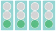 |
No change in therapy, Reevaluate in 6 months |
| 1 domain yellow | |
Reevaluate in ≤ 3 months |
| ≥ 2 domains yellow |  |
Consider optimization/change of therapy |
| or | or | |
| ≥ 1 domain red | |
|
Discussion
Several studies on the natural history of MS have suggested that the disease may be divided into two phases. The first phase is rather inflammatory driven and has a variable time until mild disability is reached (EDSS 3) while the second phase is more driven by degeneration with a uniform time course of several years until severe disability (EDSS 6) is reached [Leray et al. 2010]. This suggests that the window of opportunity to influence disease progression by therapy may be best during the first phase. Studies on the predictive value of the relapse rate on future disability support this concept since only the relapse rate in the first years of the disease correlates with future disability while relapses in later stages have no influence [Confavreux et al. 2000; Scalfari et al. 2010; Tremlett et al. 2009]. It is therefore of great importance to detect disease activity early in the disease course to reach the new treatment goal ‘NEDA’. However, the EDSS score that has been applied as a measure for disability in the past is known to be rather crude in patients with low disability (in the first phase of the disease) and is very much driven by walking disability, while factors like neuropsychology are underrepresented. Thus, new outcome disability measures need to be defined [Cohen et al. 2012]. The primary goal of the development of the MSDM was to propose a tool for a standardized examination that better reflects the many clinical faces of MS. Such a tool should detect clinical changes even during the early stages of the disease when disability is minimal. Furthermore, the tests should be practicable to be performed in everyday practice. The suggested tests can be performed within approximately 20 min, of which 13 min are required for instructions and analysis by assisting staff.
Why is such a rigorous follow-up examination of patients with MS necessary? As mentioned previously, therapeutic options are increasing and the new treatment goal should be NEDA. Nevertheless, this requires early detection of disease activity as there may be only a short therapeutic time window to achieve this goal. And this is probably also true for treatment escalation/optimization that should be implemented when relevant disease activity is observed. We suggest a treatment period of 6 months to reevaluate the treatment decision by assessing treatment success or failure. Such a rigorous short-term treatment optimization (after 3–6 months) has already been implemented successfully in other chronic autoimmune diseases like rheumatoid arthritis [Albrecht et al. 2014]. However, in rheumatoid arthritis the disease activity and the target to treat swollen joints and pain are easier to assess than the clinical activity in patients with MS. The MSDM is suggested as a tool to detect this activity in time.
The MSDM is proposed to be a compromise of good sensitivity considering the complexity of the various neurological functional systems and the practicability in routine practice. Other scores have been suggested in the past [Grand’Maison et al. 2013; Sormani et al. 2013] and some items are quite similar to the MSDM. However, the main difference is that we have omitted the EDSS and implemented a modified MSFC (PASAT supplemented by the SDMT and introduction of the LCSLC). Furthermore we have added the domain ‘neuropsychology’ since this aspect of the disease is regarded by many neurologists to be increasingly important, including fatigue, depression, quality of life, and employment status [Julian et al. 2008]. Neuropsychological examinations in particular can be time consuming and the MSDM is intended to provide a practicable set of tests for follow-up examinations.
Most of the domains proposed here are simple clinical tests that are independent of complicated technical resources. However, we acknowledge that timely and relatively frequent MRI investigations are not possible in every setting and depend on financial resources and the healthcare system. Nevertheless, the remaining domains, including relapse, disability progression, and neuropsychology, can even be assessed independently of the MRI. In this case, the model should be applied in a similar manner.
The practicability of the MSDM for the evaluation of progression in patients with MS must still be evaluated on a wide basis during everyday practice. Furthermore, prospective examinations are necessary to ascertain whether early detection of disease progression with these criteria, and fast optimization/change in treatment will indeed lead to NEDA and better long-term outcome. This is required in order to implement this aim in clinical trials as with regulatory agencies. We are convinced that the simple structure with four domains and the manageable conduction of the examinations in an everyday clinical setting might be useful in the treatment of patients with MS.
Footnotes
Funding: The project was initially supported by an unrestricted grant from Biogen Idec. The sponsor had no influence on the contents, results, and interpretation of results of this manuscript.
Conflict of interest statement: The authors have received honoraria for lecturing, consultation, and support for research from the following companies: MS: Biogen Idec GmbH, Baxter Deutschland GmbH, Bayer Vital GmbH, CSL Behring GmbH, Genzyme, Grifols, Merck-Serono GmbH, Novartis Pharma GmbH, Sanofi Aventis Deutschland GmbH and Teva GmbH. IKP: Actelion, Bayer Pharma AG, Biogen Idec, Genzyme, Merck Serono, Novartis and TEVA. BK: Biogen Idec GmbH, Bayer Vital GmbH, Genzyme, Merck-Serono GmbH, Novartis Pharma GmbH, Sanofi Aventis Deutschland GmbH and Teva GmbH
CL: Biogen Idec GmbH, Bayer Vital GmbH, Genzyme, Merck-Serono GmbH, Novartis Pharma GmbH, Sanofi Aventis Deutschland GmbH und Teva GmbH. BCK: Bayer Health Care, Biogen Idec, Genzyme/Sanofi Aventis, Grifols, Merck-Serono, Mitsubishi Europe, Novartis, Roche, Talecris and Teva.
Contributor Information
Martin Stangel, Clinical Neuroimmunology and Neurochemistry, Department of Neurology, Hannover Medical School, Carl-Neuberg-Str. 1, 30625 Hannover, Germany.
Iris Katharina Penner, Cognitive Psychology and Methodology, University of Basel, Basel, Switzerland.
Boris A. Kallmann, Multiple-Sclerosis-Center Franken, Bamberg, Germany
Carsten Lukas, Institute for Diagnostic and Interventional Radiology, St Josef Hospital, Ruhr-University Bochum, Germany.
Bernd C. Kieseier, Department of Neurology Medical Faculty, Heinrich-Heine University Düsseldorf, Germany
References
- Albrecht K., Kruger K., Wollenhaupt J., Alten R., Backhaus M., Baerwald C., et al. (2014) German guidelines for the sequential medical treatment of rheumatoid arthritis with traditional and biologic disease-modifying antirheumatic drugs. Rheumatol Int 34: 1–9. [DOI] [PubMed] [Google Scholar]
- Balcer L., Frohman E. (2010) Evaluating loss of visual function in multiple sclerosis as measured by low-contrast letter acuity. Neurology 74(Suppl. 3): S16–S23. [DOI] [PubMed] [Google Scholar]
- Barkhof F., Scheltens P., Frequin S., Nauta J., Tas M., Valk J., et al. (1992) Relapsing-remitting multiple sclerosis: sequential enhanced MR imaging vs clinical findings in determining disease activity. AJR Am J Roentgenol 159: 1041–1047. [DOI] [PubMed] [Google Scholar]
- Bevan C., Cree B. (2014) Disease activity free status: a new end point for a new era in multiple sclerosis clinical research? JAMA Neurol 71: 269–270. [DOI] [PubMed] [Google Scholar]
- Brex P., Ciccarelli O., O’Riordan J., Sailer M., Thompson A., Miller D. (2002) A longitudinal study of abnormalities on MRI and disability from multiple sclerosis. N Engl J Med 346: 158–164. [DOI] [PubMed] [Google Scholar]
- Cohen J., Krishnan A., Goodman A., Potts J., Wang P., Havrdova E., et al. (2014) The clinical meaning of walking speed as measured by the timed 25-foot walk in patients with multiple sclerosis. JAMA Neurol 71: 1386-1393. [DOI] [PubMed] [Google Scholar]
- Cohen J., Reingold S., Polman C., Wolinsky J. (2012) Disability outcome measures in multiple sclerosis clinical trials: current status and future prospects. Lancet Neurol 11: 467–476. [DOI] [PubMed] [Google Scholar]
- Confavreux C., Vukusic S., Moreau T., Adeleine P. (2000) Relapses and progression of disability in multiple sclerosis. N Engl J Med 343: 1430–1438. [DOI] [PubMed] [Google Scholar]
- Costelloe L., O’Rourke K., Kearney H., McGuigan C., Gribbin L., Duggan M., et al. (2007) The patient knows best: significant change in the physical component of the Multiple Sclerosis Impact Scale (MSIS-29 physical). J Neurol Neurosurg Psychiatry 78: 841–844. [DOI] [PMC free article] [PubMed] [Google Scholar]
- Debouverie M., Pittion-Vouyovitch S., Brissart H., Guillemin F. (2008) Physical dimension of fatigue correlated with disability change over time in patients with multiple sclerosis. J Neurol 255: 633–636. [DOI] [PubMed] [Google Scholar]
- Drake A., Weinstock-Guttman B., Morrow S., Hojnacki D., Munschauer F., Benedict R. (2010) Psychometrics and normative data for the Multiple Sclerosis Functional Composite: replacing the PASAT with the Symbol Digit Modalities Test. Mult Scler 16: 228–237. [DOI] [PubMed] [Google Scholar]
- Elbers R., Rietberg M., van Wegen E., Verhoef J., Kramer S., Terwee C., et al. (2012) Self-report fatigue questionnaires in multiple sclerosis, Parkinson’s disease and stroke: a systematic review of measurement properties. Qual Life Res 21: 925–944. [DOI] [PMC free article] [PubMed] [Google Scholar]
- Fisniku L., Brex P., Altmann D., Miszkiel K., Benton C., Lanyon R., et al. (2008) Disability and T2 MRI lesions: a 20-year follow-up of patients with relapse onset of multiple sclerosis. Brain 131: 808–817. [DOI] [PubMed] [Google Scholar]
- Freedman M., Patry D., Grand’Maison F., Myles M., Paty D., Selchen D., et al. (2004) Treatment optimization in mulitple sclerosis. Can J Neurol Sci 31: 157–168. [DOI] [PubMed] [Google Scholar]
- Freedman M., Selchen D., Arnold D., Prat A., Banwell B., Yeung M., et al. (2013) Treatment optimization in MS: Canadian MS Working Group updated recommendations. Can J Neurol Sci 40: 307–323. [DOI] [PubMed] [Google Scholar]
- Gold R., Hartung H., Stangel M., Wiendl H., Zipp F., Expertenmeetings T. (2012) Therapeutic goals of baseline and escalation therapy for relapsing-remitting multiple sclerosis. Akt Neurol 39: 342–350. [Google Scholar]
- Goldman M., Motl R., Rudick R. (2010) Possible clinical outcome measures for clinical trials in patients with multiple sclerosis. Ther Adv Neurol Disord 3: 229–239. [DOI] [PMC free article] [PubMed] [Google Scholar]
- Grand’Maison F., Bhan V., Freedman M., Myles M., Patry D., Selchen D., et al. (2013) Utility of the Canadian Treatment Optimization Recommendations (TOR) in MS care. Can J Neurol Sci 40: 527–535. [DOI] [PubMed] [Google Scholar]
- Havrdova E., Galetta S., Hutchinson M., Stefoski D., Bates D., Polman C., et al. (2009) Effect of natalizumab on clinical and radiological disease activity in multiple sclerosis: a retrospective analysis of the Natalizumab Safety and Efficacy in Relapsing-Remitting Multiple Sclerosis (AFFIRM) study. Lancet Neurol 8: 254–260. [DOI] [PubMed] [Google Scholar]
- Herrmann-Lingen C., Buss U., Snaith R. (1995) HADS-D – Hospital Anxiety and Depression Scale – Deutsche Version: Ein Fragebogen zur Erfassung von Angst und Depressivität in der somatischen Medizin. Bern, Huber Verlag [Google Scholar]
- Hirst C., Ingram G., Pearson O., Pickersgill T., Scolding N., Robertson N. (2008) Contribution of relapses to disability in multiple sclerosis. J Neurol 255: 280–287. [DOI] [PubMed] [Google Scholar]
- Hobart J., Lamping D., Fitzpatrick R., Riazi A., Thompson A. (2001) The Multiple Sclerosis Impact Scale (MSIS-29): a new patient-based outcome measure. Brain 124: 962–973. [DOI] [PubMed] [Google Scholar]
- Julian L., Vella L., Vollmer T., Hadjimichael O., Mohr D. (2008) Employment in multiple sclerosis. Exiting and re-entering the work force. J Neurol 255: 1354–1360. [DOI] [PMC free article] [PubMed] [Google Scholar]
- Leray E., Yaouanq J., Le Page E., Coustans M., Laplaud D., Oger J., et al. (2010) Evidence for a two-stage disability progression in multiple sclerosis. Brain 133: 1900–1913. [DOI] [PMC free article] [PubMed] [Google Scholar]
- Lovblad K., Anzalone N., Dorfler A., Essig M., Hurwitz B., Kappos L., et al. (2010) MR imaging in multiple sclerosis: review and recommendations for curren in relapsing-remitting multiple sclerosis patients. Mult Scler 14: 479–484. [DOI] [PMC free article] [PubMed] [Google Scholar]
- Rudick R., Lee J., Simon J., Ransohoff R., Fisher E. (2004) Defining interferon beta response status in multiple sclerosis patients. Ann Neurol 56: 548–555. [DOI] [PubMed] [Google Scholar]
- Rudick R., Polman C. (2009) Current approaches to the identification and management of breakthrough disease in patients with multiple sclerosis. Lancet Neurol 8: 545–559. [DOI] [PubMed] [Google Scholar]
- Rudick R., Polman C., Cohen J., Walton M., Miller A., Confavreux C., et al. (2009) Assessing disability progression with the Multiple Sclerosis Functional Composite. Mult Scler 15: 984–997. [DOI] [PubMed] [Google Scholar]
- Sailer M., Fazekas F., Gass A., Kappos L., Radue E., Rieckmann P., et al. (2008) Zerebrale und spinale MRT-Untersuchung bei Patienten mit klinisch isoliertem Syndrom oder gesicherter Multipler Sklerose. Rofo 180: 994–1001. [DOI] [PubMed] [Google Scholar]
- Scalfari A., Neuhaus A., Degenhardt A., Rice G., Muraro P., Daumer M, et al. (2010) The natural history of multiple sclerosis: a geographically based study 10: relapses and long-term disability. Brain 133: 1914–1929. [DOI] [PMC free article] [PubMed] [Google Scholar]
- Simon J., Li D., Traboulsee A., Coyle P., Arnold D., Barkhof F., et al. (2006) Standardized MR imaging protocol for multiple sclerosis: consortium of MS Centers consensus guidelines. AJNR Am J Neuroradiol 27: 455–461. [PMC free article] [PubMed] [Google Scholar]
- Sormani M., Bonzano L., Roccatagliata L., Cutter G., Mancardi G., Bruzzi P. (2009) Magnetic resonance imaging as a potential surrogate for relapses in multiple sclerosis: a meta-analytic approach. Ann Neurol 65: 268–275. [DOI] [PubMed] [Google Scholar]
- Sormani M., Bruzzi P. (2013) MRI lesions as a surrogate for relapses in multiple sclerosis: a meta-analysis of randomised trials. Lancet Neurol 12: 669–676. [DOI] [PubMed] [Google Scholar]
- Sormani M., De Stefano N. (2013) Defining and scoring response to IFN-beta in multiple sclerosis. Nat Rev Neurol 9: 504–512. [DOI] [PubMed] [Google Scholar]
- Sormani M., Li D., Bruzzi P., Stubinski B., Cornelisse P., Rocak S., et al. (2011) Combined MRI lesions and relapses as a surrogate for disability in multiple sclerosis. Neurology 77: 1684–1690. [DOI] [PubMed] [Google Scholar]
- Sormani M., Rio J., Tintore M., Signori A., Li D., Cornelisse P., et al. (2013) Scoring treatment response in patients with relapsing multiple sclerosis. Mult Scler 19: 605–612. [DOI] [PubMed] [Google Scholar]
- Tremlett H., Yousefi M., Devonshire V., Rieckmann P., Zhao Y. (2009) Impact of multiple sclerosis relapses on progression diminishes with time. Neurology 73: 1616–1623. [DOI] [PMC free article] [PubMed] [Google Scholar]
- Weiner H., Guttmann C., Khoury S., Orav E., Hohol M., Kikinis R., et al. (2000) Serial magnetic resonance imaging in multiple sclerosis: correlation with attacks, disability, and disease stage. J Neuroimmunol 104: 164–173. [DOI] [PubMed] [Google Scholar]
- Weinshenker B., Bass B., Rice G., Noseworthy J., Carriere W., Baskerville J., et al. (1989) The natural history of multiple sclerosis: a geographically based study. 2. Predictive value of the early clinical course. Brain 112(Pt 6): 1419–1428. [DOI] [PubMed] [Google Scholar]
- Yong V. (2002) Differential mechanisms of action of interferon-beta and glatiramer acetate in MS. Neurology 59: 802–808. [DOI] [PubMed] [Google Scholar]