



Abstract
Malnutrition in utero (IU) could alter pancreatic development. Reported here are the effects of high fat diet (HFD) during pregnancy on fetal growth and pancreatic morphology in an “At Risk” animal model of metabolic disease, the glucose transporter 4 heterozygous mouse (G4+/−).
Wild type (WT) female mice mated with G4+/− males were fed HFD or control (CD) diet for 2 weeks prior to mating and throughout pregnancy. At embryonic day18.5 fetuses were sacrificed and pancreata isolated for analysis of morphology and expression of genes involved in insulin-cell development, proliferation, apoptosis, glucose transport and function.
Compared to WT CD, WT HFD fetal pancreata had a 2.4 fold increase in the number of glucagon cells (p=0.023). HFD also increased glucagon cell size by 18% in WT pancreata compared to WT CD. Compared to WT CD, G4+/− CD had an increased number of insulin cells, and decreased insulin and glucagon cell size. Compared to G4+/− CD, G4+/− HFD fetuses had increased pancreatic gene expression of Igf2, a mitogen and inhibitor of apoptosis. Expression of genes involved in proliferation, apoptosis, glucose transport and insulin secretion were not altered in WT HFD compared with G4+/− HFD pancreata.
In contrast to WT HFD pancreata, HFD exposure did not alter pancreatic islet morphology in fetuses with GLUT4 haploinsufficiency; this may be mediated in part by increased Igf2 expression. Thus, interactions between IU diet and fetal genetics may play a critical role in the developmental origins of health and disease.
Keywords: high fat diet, pancreas, fetus, programming, glucagon cells
Introduction
Metabolic Syndrome (MetS) is a cluster of risk factors including obesity, dyslipidemia, insulin resistance and hypertension. MetS increases the risk for type 2 diabetes mellitus (T2D) which is characterized by peripheral insulin resistance and insulin cell dysfunction (Grundy, et al. 2004).
In Western Societies, fat and carbohydrate-dense foods have become increasingly abundant and easily accessible (Cordain, et al. 2005) contributing to the increased prevalence of MetS in adults in the United States (Mozumdar and Liguori 2011). Increased globalization, associated with a nutritional transition towards Western diets, is thought to be a contributing factor to the increasing prevalence of obesity and T2D globally (Popkin 2006)). In addition to poor diet and genetics, evidence suggests that an altered intrauterine environment (IU) plays a key role in the development of MetS (Vuguin, et al. 2013) and that interactions between the IU environment and lifestyle can increase risk of MetS and T2D in people who are genetically susceptible (Hu 2011).
Poor nutrition during pregnancy impacts fetal growth and development particularly that of the endocrine pancreas (Snoeck, et al. 1990; Vuguin, et al. 2013). Specifically, alterations in the IU environment caused by a low calorie, low protein or a high fat diet (HFD) affects the function of the endocrine pancreas by altering islet size, islet vascularization, number of insulin-cells (INS), insulin content, function, and parasympathetic innervation (Cerf, et al. 2005; Dahri, et al. 1991; Ford, et al. 2009; Garofano, et al. 1997; Ng, et al. 2010; Rodriguez-Trejo, et al. 2012; Vogt, et al. 2014). In addition, it has been suggested that HFD during fetal development induced glucagon-cell (GLU) hypertrophy and hyperplasia, resulting in an increase in GLU-cell number and volume in the neonatal offspring (Cerf et al. 2005).
These findings could be partially explained by altered expression of growth factors, such as insulin like growth factors (Igfs) and regulatory proteins involved in endocrine cell differentiation, such as the transcription factor pancreatic and duodenal homeobox 1 (Pdx1) (Chen, et al. 2012; Park, et al. 2008). Specifically, HFD exposure during development reduced Pdx1 immunoreactivity in a rodent model suggesting that Pdx1 is susceptible to IU environment (Cerf, et al. 2009).
The insulin growth factor (Igf) system, an important metabolic and mitogenic factor, is the major regulator of fetal growth and development. Igf2 mRNA is highly expressed in islet cells and some ductal epithelial cells in late fetal life (Hill, et al. 1999) and co-localizes with INS- and GLU-cells in human fetal pancreas (Portela-Gomes and Hoog 2000). Igf2 is mitogenic for INS cells (Calderari, et al. 2007; Hill et al. 1999), and inhibits INS cell apoptosis (Cornu, et al. 2009; Hill et al. 1999; Raile, et al. 2003). Fetal expression of Igf2 has been shown to be increased by a HFD IU (Zhang, et al. 2009), suggesting that changes in Igf2 expression, in response to the altered IU environment, may play a role in the programming of the endocrine pancreas.
To develop T2D, pancreatic dysfunction has to be accompanied by a state of peripheral insulin resistance. Peripheral insulin resistance can be defined as a reduction in the ability of target tissues such as skeletal muscle, white adipose tissue and heart to respond to insulin. One response of insulin resistance is a reduction in insulin stimulated glucose uptake, mediated via the glucose transporter 4 (GLUT4) (Bryant, et al. 2002; Zierath, et al. 1996). In animal models, GLUT4 haploinsufficiency (G4+/−) results in peripheral insulin resistance and T2D (Charron and Kahn 1990; Li, et al. 2000; Rossetti, et al. 1997; Stenbit, et al. 1997). In addition, as G4+/− mice age, they develop islet cell hyperplasia due to an increase in INS cell number (Brissova, et al. 2005)
Studies have demonstrated that GLUT4 mRNA and GLUT4 protein is expressed in the GLU and INS cells of mouse, rat and human endocrine pancreas (Bahr, et al. 2012; Kobayashi, et al. 2004). GLUT4 expression in pancreatic endocrine cells seems to be regulated by glucose and insulin (Bahr et al. 2012). Specifically, in GLU cells, high glucose levels decrease and high insulin levels increase GLUT4 expression. In contrast, the opposite occurs in INS cells. In addition, pancreatic GLUT4 expression is elevated in T2D patients and decreased in animal models of type 1 diabetes suggesting that alterations in GLUT4 expression in the endocrine pancreas may play a role in the regulation of pancreatic cell function during disease states (Bahr et al. 2012).
While effects of maternal nutrition on INS cell development and function have been extensively studied, very few studies have examined the effects of HFD IU on GLU cell development and function (Cerf et al. 2005). We have previously demonstrated that exposure to a HFD IU and during lactation leads to development of MetS in G4+/− and WT offspring (Hartil, et al. 2009; Vuguin et al. 2013). Although both animal models developed features of metabolic disease, genotype-dependent differences were observed. Specifically, HFD IU WT fetuses have significantly higher glucose levels compared to HFD IU G4+/− fetuses. We hypothesize that GLUT4 haploinsufficiency protects the pancreatic endocrine cell function from the effects of HFD thus protecting the G4+/− fetus from becoming hyperglycemic.
The animal model used in this study provides a unique opportunity to study the influence of genotype and diet on fetal pancreatic islet development (Vuguin et al. 2013). The results of this study suggest that the interaction between a HFD with genotype during gestation ‘programs’ the fetus for increased susceptibility to T2D in part by altering pancreatic islet cell composition leading to an impaired ability to optimize glucose homeostasis.
Materials and Methods
Animal protocols were approved by the Institute for Animal Care and Use Committee at the Albert Einstein College of Medicine. As previously described (Hartil et al. 2009), age and body weight (BW) matched female WT mice (CD1 background) were maintained on control PicoLab® Mouse Diet #5058 (CD: 9% fat as soybean oil and animal fat, 20% protein, 53% carbohydrate, 3.59 kcal/g), or switched to high fat Bio-Serv Product #F3282 (HFD: 35.5% fat as lard, 20% protein, 36.3% carbohydrate, 5.29 kcal/g) 2 weeks prior to mating and throughout pregnancy (IU). Females were bred to non-littermate G4+/− males. Pregnancy was determined by the detection of a copulatory plug and defined as embryonic day (e) 0.5. Pregnant mice were sacrificed on e18.5.
A total of 196 fetuses were euthanized by immediate decapitation following dissection from the uterine horn. Fifty-six fetuses were exposed to CD and 140 were exposed to HFD IU. This cohort was produced from a total of 4 WT females exposed to CD and 11 WT females exposed to HFD. The number of fetuses per litter and fetus weight were recorded at sacrifice. The numbers of abnormal and dead pups were also noted. Genotyping and sex determination of fetuses were performed as previously described (Hartil et al. 2009; Vuguin et al. 2013).
Tissue processing for immunostaining
These techniques have been previously described (Kedees, et al. 2007; Vuguin, et al. 2006). Embryos were placed in ice cold PBS and decapitated. The dorsal pancreas was removed by dissection and fixed overnight by immersion in a fixative solution of 4% paraformadehyde in 0.1 M phosphate buffer. Fixed tissues were infiltrated in 30% sucrose and mounted in embedding matrix (Lipshaw Co. Pittsburgh, PA). Cryosections (10–20 micron) were mounted onto glass slides coated with a solution of 1% gelatin containing 0.05% chromium potassium sulfate.
Immunoflourescence
As previously described (Kedees et al. 2007; Vuguin et al. 2006), sections were incubated sequentially in empirically derived optimal dilutions of control serum or primary antibody overnight at 4°C and with a 1:200 dilution of the secondary antibody (in 0.01M PBS). After completion of the staining procedure, sections were covered with 2–3 drops of Vectashield Solution (Vector Labs, Inc., Burlingame, CA) (Vuguin et al. 2006).
Source of Antibodies
Guinea pig anti-bovine INS antibodies were purchased from Linco Research, Inc. (Eureka, MO). Rabbit anti-human GLU was purchased from Calbiochem, Inc. (San Diego, CA). Anti-rabbit GLUT2 sera was purchased from Chemicon, Inc. (Temecula, CA). Rabbit antiserum to Pdx1 was a generous gift from C.V.E. Wright (Vanderbilt University, Nashville, TN). Antibodies were used at the following dilutions: anti-insulin (INS) at 1:400, anti-glucagon (GLU) (1:4,000), anti-Pdx-1 (1:5,000), and anti-GLUT2 (1:1,000). Secondary antibodies: Alexafluor 488 anti-mouse, anti-rat and anti-rabbit IgG, Alexafluor 594 anti-guinea pig, anti-rabbit and anti-mouse IgG were purchased from Molecular Probes, Inc. (Eugene, OR).
Confocal Microscopy
Confocal images were obtained using a Radiance 2000 confocal microscope (BioRad, Inc., Hercules, CA) attached to a Zeiss Axioskop microscope (Carl Zeiss, Inc.). Images of 540 X 540 pixels were obtained and processed using Adobe Photoshop 6.0 (Adobe Systems, Mountain View, CA) (Vuguin et al. 2006).
Morphometric Analysis
The number of islets, size of individual endocrine cells, and cell number ratio for cells expressing insulin (INS) and glucagon (GLU) were calculated as previously described (Kedees et al. 2007; Vuguin et al. 2006). Consecutive 10 to 20 μm sections were obtained from each pancreas (n=4–8 per fetal genotype/diet from at least 4 litters/diet) Number of slides examined: 12 to 15 sections/pancreas/genotype/diet were examined at 200–300μm apart. Sections were projected on the screen of a video monitor and cell counting and area measurements were made using the National Institute of Health Image J software (http://rsb.info.nih.gov/ij/).
Definitions of Parameters Measured
Total Endocrine Area (μm2/mm2)
Total endocrine area was defined as the sum of INS and GLU expressing areas normalized to the total pancreatic area.
INS and GLU Expressing Areas (μm2)
INS and GLU expressing areas were calculated as the product of the cell number by the cell size for either INS or GLU expressing cells.
Total Pancreatic Area (mm2)
Total pancreatic area was determined by manually defining the perimeter of the exocrine and endocrine pancreatic tissue in each section examined.
Islet Number (#islets/mm2)
Islet number was defined as aggregates of at least 5 INS or GLU expressing cells (Garofano, et al. 2000). The islet numbers were normalized to total pancreatic area.
INS and GLU cell number (#cells/mm2)
The relative number of INS and GLU cells per islet was determined by the point sampling method and normalized to total pancreatic area in each section as previously described (Garofano et al. 2000). At least 5000 points were scored in 30 islets/genotype/diet.
INS: GLU cell ratio
INS:GLU cell ratio was determined by dividing the total number of INS expressing cells by the total number of GLU expressing cells.
INS and GLU cell size (μm2)
The area of individual INS or GLU expressing cells was determined by manually defining the perimeter of the cells immunostained by either INS or GLU. More than 50 cells/pancreas/genotype/diet from multiple litters were measured and averaged.
RNA Isolation and Quantitative Real-time PCR Analysis
Total RNA was prepared from fetal pancreata, liver (GLUT 4 expression negative control), gastrocnemius and adipose tissue (GLUT 4 expression positive controls) (n=6–10 diet/genotype, n=4–8 litters) as previously described (Hartil et al. 2009; Vuguin et al. 2013). The RNA was checked for DNA contamination, using PCR with control primers as described previously (Ouhilal, et al. 2012).
Quantitative real-time PCR (qRT-PCR) was the method of choice to determine the expression of genes of interest (Ouhilal et al. 2012). Four commonly used housekeeping genes, ubiquitin, β-actin, hypoxanthine guanine ribotransferase, and Cyclophilin B were used for normalization, as described previously (Ouhilal et al. 2012). For quantitative analysis, all samples were normalized to the genes described above using the ΔΔCT value method. Each sample was measured in triplicate to assess technical variability (Ouhilal et al. 2012).
Data Analysis
Data are expressed as the mean ± SEM. Statistical analyses were performed using JMP IN 5.1 software (SAS Institute, Cary, NC). ANOVA was used to test the difference between the means of two (t-test) or more groups. Significance was defined as p< 0.05.
Results
Our previous studies demonstrated that maternal exposure to HFD decreased fetal body weight at e18.5 and increased blood glucose levels without altering plasma insulin levels (Hartil et al. 2009; Vuguin et al. 2013). Consistent with those studies, HFD IU decreased fetal growth by 10% independent of fetal genotype (Table 1). To determine whether HFD exposure affected pancreatic morphology or islet cell composition, we examined e18.5 WT and G4+/− pancreata. No significant differences were seen between the groups when analyzed based on fetal sex with regards to pancreatic morphology or gene expression.
Table 1.
Metabolic Parameters and Pancreatic Morphology seen in WT and G4+/− Fetuses Exposed to either CD or HFD During Development.
| WT fetus | G4 +/− fetus | |||
|---|---|---|---|---|
| CD | HFD | CD | HFD | |
| Body Weight (g) | 1.32±0.019A | 1.18±0.015B | 1.34±0.017A | 1.17±0.019B |
| Total Endocrine Area (μm2/mm2) | 0.0104±0.001A | 0.0204±0.009B | 0.0124±0.001A | 0.0106±0.003A |
| Islet Number (#islets/mm2) | 6.2±0.4 A | 6.2±0.6A | 5.8±0.7A | 7.5±0.9A |
| GLU Cell Number (#cells/mm2) | 38±7A | 91±20B | 64±13AB | 54±17AB |
| INS Cell Number (#cells/mm2) | 88±10 A | 110±20 AB | 153±15B | 123±37AB |
| INS:GLU Cell Number | 2.6±0.3 A | 1.3±0.2B | 2.5±0.2A | 2.3±0.2A |
| INS Cell Size (μm2) | 79±7 A | 91±11 A | 58±1B | 65±4AB |
| GLU Cell Size (μm2) | 81±1A | 96±7B | 61±2C | 72±6ABC |
Data is expressed as mean ±SE. n=5–19/genotype. Values that do not share the same letter are significantly different from each other p<0.05.
Pancreatic Morphology
Total endocrine area (the sum of INS and GLU expressing areas normalized to the total pancreatic area) was increased almost two-fold in WT HFD fetal pancreata (Table 1) compared to WT CD (p=0.0015). In contrast, there was no statistical difference in total endocrine area in G4+/− HFD compared to G4+/− CD fetal pancreata. Despite a two-fold increase in total endocrine area in WT HFD fetal pancreata, HFD did not increase the number of islets (defined as aggregates of at least 5 INS or GLU expressing cells) in either genotype (Table 1). Islets, independent of the genotype and diet, were comprised of a mantle of GLU cells surrounded by a core of INS cells (Figure 1A to D).
Figure 1.
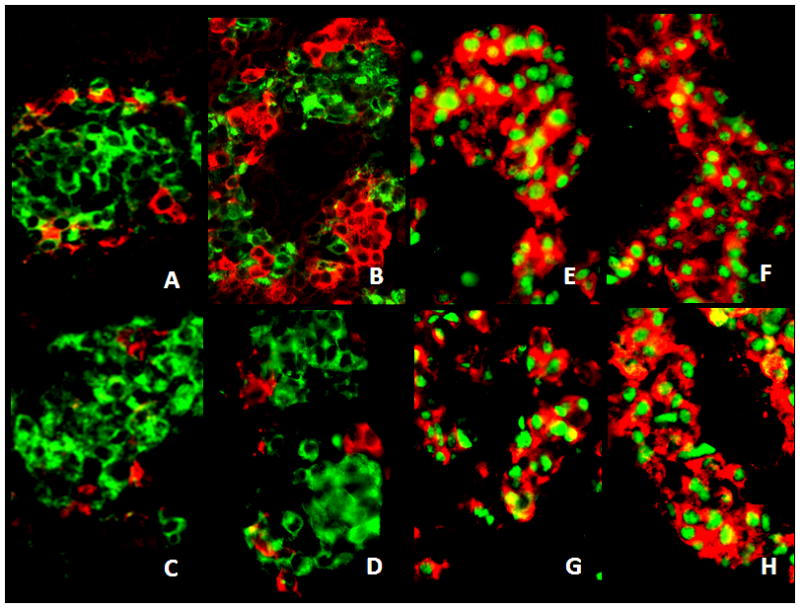
Photomicrographs of confocal images of representative islets from embryos at e-18.5 immunostained for insulin (green) (A–D), glucagon (red) (A–D), and insulin (red) (E–H) and Pdx1 (green) (E–H) cells in WT C (A, E), WT HFD (B, F), G4+/− CD (C, G) and G4+/− HFD (D, H). Pancreata from WT HFD (B) fetuses exhibit an increased number of GLU cells (red) as compared to WT CD (A), G4+/− CD (C) and G4+/− HFD (D) fetuses. HFD did not alter the expression or the localization of Pdx1 (green) in WT (E, F) or G4+/− (G, H) fetal pancreata compared to those from CD pancreata. Scale = 40 μm
To determine whether the increase in total endocrine area in WT HFD pancreata was associated with an increase in the relative abundance or size of endocrine cells within the islets, the number and area of GLU and INS immunoreactive cells was quantified.
Morphometric analysis revealed that the mean number of GLU cells per pancreata was 2.4-fold higher in WT HFD when compared to WT CD (p=0.023) (Figure 1A and B). In contrast, no change was observed in the mean number of GLU cells per pancreata between G4+/− CD and G4+/− HFD (Figure 1C and D). Similarly, no change in the relative number of INS cells was observed when each genotype was compared to the opposite diet. However, the mean number of INS cells per pancreata was 1.7-fold higher in G4+/− CD when compared to WT CD (p=0.007) (Table 1 and Figure 1C).
G4+/− HFD and G4+/− CD fetal pancreata had a significantly higher INS:GLU expressing cell number when compared to WT HFD, however, this was not statistically different when compared to WT CD fetal pancreata (Table 1 and Figure 1A to D).
Exposure to HFD did not affect INS cell size in WT or G4+/− fetal pancreas when compared to the fetuses exposed to CD. However, G4+/− CD pancreata had smaller INS cells compared to WT independent of the IU diet (p=0.03 vs. WT HFD and p<0.002 vs. WT CD) but was not different when compared to G4+/− HFD (Table 1).
Exposure to HFD increased GLU cell size by 18% in both WT and G4+/− fetal pancreata, although this only reached statistical significance in WT HFD vs. WT CD (p=0.03) (Table 1). Interestingly, similar to reduced INS cell size, G4+/− CD had smaller GLU cells compared to WT independent of diet (p=0.0033 vs. WT HFD and p<0.0001 vs. WT CD) (Table 1).
GLUT2 and Pdx1 localization
To determine whether HFD IU regulates factors involved in the regulation of INS cell development, differentiation and function, we determined Pdx1 and GLUT2 localization in islets of fetuses exposed to both diets. HFD IU did not alter the localization of Pdx1 in WT or G4+/−fetal pancreata compared to a CD (Figure 1E to H). Similarly, HFD did not alter the GLUT2 localization in WT or G4+/− fetuses (data not shown).
Gene Expression
To determine whether HFD IU regulates factors involved in INS cell differentiation, proliferation, survival, apoptosis and function, we determined mRNA expression of pancreatic and duodenal homeobox1 (Pdx1), SRY (Sex Determining Region Y)-Box 9 (Sox9), baculoviral IAP repeat containing 5 (BIRC5-Survivin), B-Cell CLL/Lymphoma 2 (BCL2), and apoptosis-related cysteine peptidase (CASP 3) (Table 2). HFD IU did not alter mRNA expression of Pdx1, Sox9, BIRC5, BCL2 and CASP 3 in WT or G4+/− fetal pancreata compared to a CD.
Table 2.
Effect of HFD IU Exposure on Fetal Pancreatic Gene Expression
| Gene Name | Gene Symbol | Gene Sequence (Forward and Reverse) | WT HFD IU vs. WT C IU | G4+/− HFD vs. G4+/− C |
|---|---|---|---|---|
| A. Cell Differentiation | ||||
| Pancreatic and duodenal homeobox 1 | Pdx1 | CGCGTCCAGCTCCCTTT CCTGCCCACTGGCCTTT |
NS | NS |
| SRY (Sex Determining Region Y)-Box 9 | Sox9 | GTACCCGCATCTGCACAAC CTCCTCCACGAAGGGTCTCT |
NS | NS |
| B. Cell Survival and Apoptosis | ||||
| Baculoviral IAP repeat containing 5 | BIRC5-Survivin | CCCGATGACAACCCGATA CATCTGCTTCTTGACAGTGAGG |
NS | NS |
| B-Cell CLL/Lymphoma 2 | BCL2 | AGTACCTGAACCGGCATCTG GGGGCCATATAGTTCCACAAA |
NS | NS |
| Apoptosis-related cysteine peptidase | CASP 3 | GAGGCTGACTTCCTGTATGCTT AACCACGACCCGTCCTTT |
NS | NS |
| D. Cortisol Receptor and Cell Function | ||||
| Nuclear receptor subfamily 3, group C, member 1 | NR3C1 | TGGAGCTACAGTCAAGGTTTCT GCTTGGAATCTGCCTGAGA |
NS | NS |
| ATP-binding cassette, sub-family C, member 8− sulfonylurea receptor subunit | SUR1 | GACGGCTGGGCAGATCTG GAGGTTTGGGCATAAGAAGAAAAA |
NS | NS |
| Potassium Inwardly-Rectifying Channel, Subfamily J, Member 86.1 | KCNJ8; Kir6.1 | AGCCGCCATGCTGTGATT CCCACCCGGAACATGAAG |
NS | NS |
| C. Glucose Transport | ||||
| Solute Carrier Family 2 (Facilitated Glucose Transporter), Member 4 | GLUT4 | CTGCAAAGCGTAGGTACCAA CCTCCCGCCCTTAGTTG |
ND | ND |
| Solute carrier family 2 (facilitated glucose transporter), member 2 | GLUT2 | TTGACTGGAGCCCTCTTGATG CACTTCGTCCAGCAATGATGA |
NS | NS |
| E. Growth Factors | ||||
| Insulin like growth factor | Igf1 | AACAAGCCCACAGGCTATGG AAGCAACACTCATCCACAATGC |
NS | NS |
| Insulin like growth factor | Igf2 | CATCGTCCCCTGATCGTGTT CACTGATGGTTGCTGGACATCT |
NS | 5* |
Gene expression was determined for genes involved in: (A) cell differentiation, (B) cell survival and apoptosis, (C) glucose transport, (D) cortisol receptor and cell function and (E) growth factors. Fold change indicates the increase in mRNA measured by qRT-PCR in HF IU compared to C IU in WT and G4+/− fetal pancreata (n=6–10/genotype diet at e18.5)
p<0.001 in HF IU vs. C IU diet; NS =non significant, ND= no detected
To determine whether HFD IU regulates expression of genes involved in insulin secretion, pancreatic growth and (whatever NR3C1 does), we determined mRNA expression of the potassium inwardly-rectifying channel (Kir6.1), sulfonylurea receptor (SUR1), glucocorticoid receptor (NR3C1) and the insulin like growth factors, Igf1 and Igf2. HFD IU did not alter mRNA expression of Kir6.1, SUR1, NR3C1 or Igf1 in the pancreas of WT and G4+/− fetuses.
HFD significantly increased expression of Igf2 mRNA, a potent INS cell growth and anti-apoptotic factor, in G4+/− HFD pancreas compared to G4+/− CD. In contrast, no significant difference in Igf2 expression was observed in WT HFD fetal pancreata compared to WT CD. To determine whether HFD IU regulates expression of genes involved in facilitated glucose transport in pancreatic islets, mRNA expression of the main facilitated glucose transporter expressed in pancreatic islets, GLUT 2 was measured. In addition, GLUT 4 mRNA expression was determined. HFD did not alter mRNA expression of GLUT 2 in WT or G4+/− fetuses. GLUT 4 mRNA expression was not detected in fetal pancreata at e18.5.
Discussion
Diabetes mellitus is a complex disease resulting from the interplay between genetics and the environment. It results from dysfunctional pancreatic INS cells that cannot compensate for the metabolic demands imposed by peripheral tissues such as skeletal muscle and adipose tissue. Due to the role of both genetics and the IU environment, and the importance of the pancreas on the development of diabetes, we sought to determine the effect of maternal HFD on pancreatic development in a mouse model genetically predisposed to develop features of diabetes, the G4+/− mouse (Charron and Kahn 1990; Li et al. 2000; Rossetti et al. 1997; Stenbit et al. 1997).
Consistent with our previous studies, HFD exposure decreased fetal body weight (Vuguin et al. 2013). Similar to a rat model of HFD IU that resulted in poor fetal growth, exposure to HFD during development was accompanied by an increase in GLU cell volume and number in WT, but not G4+/−, pancreata (Cerf et al. 2009).
HFD did not alter the islet morphology in G4+/− fetuses despite an increase in RNA expression of the mitogenic growth factor Igf2 when compared to CD. These data suggest that increased Igf2 could be a compensatory response to maintain normal islet morphology.
There were no discernible structural differences in the islets of fetuses exposed to HFD. INS cells were present at the core and GLU cells were present at the mantle, suggesting normal regulation of islet formation. In contrast to other models of IU programming, HFD IU did not alter gene expression and or localization of the transcription factor, Pdx1, thought to be relevant for INS cell adaptation to an altered IU environment (Chen et al. 2012; Gesina, et al. 2006; Rodriguez-Trejo et al. 2012). Additionally, the expression of genes implicated in pancreatic growth (Igf1) or insulin secretory capacity (Kir6.1 and SUR1) were not altered by maternal HFD.
In animal models, the most profound changes in islet cell morphology seen in response to a HFD IU were the number and the volume of INS cells, which were significantly reduced in neonatal rats (Cerf et al. 2009). In contrast, no detectable change in INS cell number was observed. These findings may represent differences in the species or differences in the INS cell adaptation to a different metabolic environment.
INS cell mass adapts to changes in metabolic homeostasis. These adaptations can occur through increases or decreases in INS cell number, through changes in proliferation, neogenesis, apoptosis or cell size. INS cell number was not significantly altered by diet, but was increased 1.7-fold in G4+/− CD compared to WT CD. Based on gene expression at e18.5 data we did not find evidence of increased proliferation and/ or apoptosis. Future studies done at earlier developmental stages may be necessary to confirm these findings.
INS cell size was significantly reduced in G4+/− CD fetuses compared to WT CD. Adaptations in INS cell size have been observed during pregnancy (Dhawan, et al. 2007) and in response to persistent hyperinsulinemia (Anlauf, et al. 2005). Larger INS cells containing more protein per cell are known to be highly glucose responsive (Martens, et al. 2010). Our finding that G4+/− CD fetuses exhibit smaller but more INS cells suggests an adaptation to the metabolic environment associated with GLUT4 haploinsufficiency.
HFD decreased the INS:GLU expressing cell number in WT HFD fetuses. Further analysis revealed a significant increase in GLU cell number and size in WT HFD pancreata. When the cell number was multiplied by the individual cell surface area, to approximate the endocrine cell mass, an increase in the total endocrine area was observed.
The mechanism that leads to GLU cell hyperplasia in WT HFD fetal pancreas is not clear. GLU cell hyperplasia has been seen in models with impaired GLU synthesis/signaling (Vuguin et al. 2006), glucagon receptor expression (Chen, et al. 2011) and GLU action (Chen, et al. 2005); inactivation of Pax4 (Sosa-Pineda, et al. 1997); circulating low insulin levels (Rahier, et al. 1983; Sosa-Pineda et al. 1997; Thyssen, et al. 2006); mild hypoglycemia and chronic HFD (Fiori, et al. 2013). Since circulating insulin levels were normal and glucose levels were elevated in our HFD IU model, other factors must be responsible for the increase in GLU cell number (Jiang and Zhang 2003).
In addition to the GLU cell hyperplasia, WT fetuses exposed to a HFD displayed an increased GLU cell size that could potentially lead to increased GLU levels (Huang, et al. 2013). Increased GLU levels could explain the increase in expression of genes involved in gluconeogenesis and the increased glucose levels, which we previously reported in the livers of WT HFD fetuses (Vuguin, et al. 2013). Unfortunately, we were not able to determine serum GLU levels. Accurate determination of fetal GLU serum levels usually requires pooling serum from several fetuses to collect at least 50–100 μl of serum per sample (www.biotrend.com/download/gl-32k.pdf). Because of the technical difficulties associated with the adequate determination of serum GLU levels, alterations in pancreatic morphology were assessed instead. Thus, further studies are needed to confirm the role of GLU in the GLU cell phenotype observed in WT fetal pancreata exposed to HFD.
Increased GLU cell number and cell size could also occur in response to lower glucose levels during fetal development in a HFD IU. Decreased glucose levels would signal the central nervous system to increase GLU cell number and secretion leading to GLU-cell hyperplasia that may result in hyperglycemia (Brunicardi, et al. 1995; Furuta, et al. 1997). Alternatively, high glucose levels and GLU cell hyperplasia could be the result of cell transformation of GLU expressing cells into INS cells (Thorel, et al. 2010). GLU cells are the earliest identifiable cells of the pancreatic endocrine lineage, capable of becoming INS expressing cells (Gromada, et al. 2007). It is possible that the GLU cells in our model are incapable of being transformed into INS cells. This seems unlikely, since a decrease in INS cell number or size as seen in another models of GLU cell hyperplasia, is not a feature found in this model (Cerf et al. 2009).
Increased INS:GLU endocrine cell ratio was seen only in WT HFD fetal pancreas while increased Igf2 gene expression was seen in HFD G4+/− fetal pancreas which had normal islet architecture. It is possible that different signals regulate GLU cell number in islets of WT HFD and G4+/− mice.
GLUT4 has previously been found to be expressed at low levels in pancreatic GLU and INS cells of adult mouse, rat and human endocrine pancreas (Bahr et al. 2012; Kobayashi et al. 2004) and its expression is regulated by glucose and insulin levels (Bahr et al. 2012). Unfortunately, GLUT4 mRNA expression was not detected in fetal pancreata at e18.5. GLUT4 expression is turned on in brown adipose tissue, heart and skeletal muscle towards the end of pregnancy (Santalucia, et al. 1992). Its expression increases progressively after birth, and it has been found to be sensitive to alterations in maternal nutrient intake (Gardner, et al. 2005; Thamotharan, et al. 2005). Thus, the developmental timing of its expression in the pancreas could explain why we were not able to detect the presence of GLUT4 mRNA.
The exact role of Igf2 expression in the fetal pancreas is unclear. In genetic models, decreased Igf2 expression in fetal pancreata lead to lower INS cell proliferation rates and increased INS cell apoptosis (Kulkarni 2005). Overexpression of Igf2 in INS cells is associated with an increase in INS cell mass (Devedjian, et al. 2000), while global overexpression of Igf2 gene causes islet GLU cell hyperplasia with an abnormal INS:GLU ratio (Petrik, et al. 1999).
In one model, maternal under-nutrition decreased pancreatic Igf2 expression and was associated with increased INS cell mass (Martin, et al. 2005). In contrast, another study demonstrated that maternal under-nutrition increased pancreatic Igf2 expression with no alteration in INS cell mass (de Miguel-Santos, et al. 2010).
In rats, high carbohydrate diet during the neonatal period decreased Igf2 mRNA expression and GLU cell number (Petrik, et al. 2001). Thus, the lack of consistency between studies makes it difficult to determine the precise role of Igf2 in the pancreatic phenotype and determination of the INS:GLU cell number. Some differences may be related to compensatory increases in Igf1 expression that could potentially lead to increased INS cell mass (Martin et al. 2005). Igf1 expression was not different in our study. In addition, gene expression was determined in whole fetal pancreatic tissue and it may be that expression levels are regulated differently in response to genotype and/or HFD IU in different (pancreatic) islet cell types.
In conclusion, nature (genotype) modifies the effects of nurture (HFD) on fetal pancreatic development. We propose that changes in endocrine cell ratio in islets induced by IU nutritional and genetic manipulation may affect islet cell responsiveness to physiological or pathological stimuli. Further investigation is needed to confirm whether adaptations that occur in WT HFD islets IU are maladaptive in later life and whether developing in a HFD IU milieu affects the capacity of GLU cells to respond to insulin resistance with increasing age and/or progressive metabolic deterioration. Additionally, it is unknown whether the lack of GLU cell hyperplasia in G4+/− pancreata in response to HFD might be detrimental to development during times when glucose delivery is interrupted, such as during time of delivery. Understanding the molecular basis of an altered INS:GLU expressing cell number is an attractive objective particularly in light of new treatments that may directly act on these pathogenic mechanisms of T2D.
Acknowledgments
Funding:
This study was supported by grants from the National Institute of Health DK47435 (MJC) and K08-HD042172 (PV), R21 DK081194 (to M.J.C. and P.M.V.), and F31 DK093332 to (LW) and an ADA Mentor based postdoctoral fellowship (MJC). Additional support was generously provided by the Diabetes and Liver Research and Training and Cancer Centers of Albert Einstein College of Medicine.
We are grateful to Mr. Carlos Vargas, Ms. Amy Sorvino and Ms. Goutami Sayal for their technical expertise.
Footnotes
Declaration of Interest:
The authors declare that there is no conflict of interest that could be perceived as prejudicing the impartiality of the research reported.
Bibliography
- Anlauf M, Wieben D, Perren A, Sipos B, Komminoth P, Raffel A, Kruse ML, Fottner C, Knoefel WT, Monig H, et al. Persistent hyperinsulinemic hypoglycemia in 15 adults with diffuse nesidioblastosis: diagnostic criteria, incidence, and characterization of beta-cell changes. Am J Surg Pathol. 2005;29:524–533. doi: 10.1097/01.pas.0000151617.14598.ae. [DOI] [PubMed] [Google Scholar]
- Bahr I, Bazwinsky-Wutschke I, Wolgast S, Hofmann K, Streck S, Muhlbauer E, Wedekind D, Peschke E. GLUT4 in the endocrine pancreas--indicating an impact in pancreatic islet cell physiology? Horm Metab Res. 2012;44:442–450. doi: 10.1055/s-0032-1306335. [DOI] [PubMed] [Google Scholar]
- Brissova M, Blaha M, Spear C, Nicholson W, Radhika A, Shiota M, Charron MJ, Wright CV, Powers AC. Reduced PDX-1 expression impairs islet response to insulin resistance and worsens glucose homeostasis. Am J Physiol Endocrinol Metab. 2005;288:E707–714. doi: 10.1152/ajpendo.00252.2004. [DOI] [PubMed] [Google Scholar]
- Brunicardi FC, Shavelle DM, Andersen DK. Neural regulation of the endocrine pancreas. Int J Pancreatol. 1995;18:177–195. doi: 10.1007/BF02784941. [DOI] [PubMed] [Google Scholar]
- Bryant NJ, Govers R, James DE. Regulated transport of the glucose transporter GLUT4. Nat Rev Mol Cell Biol. 2002;3:267–277. doi: 10.1038/nrm782. [DOI] [PubMed] [Google Scholar]
- Calderari S, Gangnerau MN, Thibault M, Meile MJ, Kassis N, Alvarez C, Portha B, Serradas P. Defective IGF2 and IGF1R protein production in embryonic pancreas precedes beta cell mass anomaly in the Goto-Kakizaki rat model of type 2 diabetes. Diabetologia. 2007;50:1463–1471. doi: 10.1007/s00125-007-0676-2. [DOI] [PubMed] [Google Scholar]
- Cerf ME, Chapman CS, Muller CJ, Louw J. Gestational high-fat programming impairs insulin release and reduces Pdx-1 and glucokinase immunoreactivity in neonatal Wistar rats. Metabolism. 2009;58:1787–1792. doi: 10.1016/j.metabol.2009.06.007. [DOI] [PubMed] [Google Scholar]
- Cerf ME, Williams K, Nkomo XI, Muller CJ, Du Toit DF, Louw J, Wolfe-Coote SA. Islet cell response in the neonatal rat after exposure to a high-fat diet during pregnancy. Am J Physiol Regul Integr Comp Physiol. 2005;288:R1122–1128. doi: 10.1152/ajpregu.00335.2004. [DOI] [PubMed] [Google Scholar]
- Charron MJ, Kahn BB. Divergent molecular mechanisms for insulin-resistant glucose transport in muscle and adipose cells in vivo. J Biol Chem. 1990;265:7994–8000. [PubMed] [Google Scholar]
- Chen M, Gavrilova O, Zhao WQ, Nguyen A, Lorenzo J, Shen L, Nackers L, Pack S, Jou W, Weinstein LS. Increased glucose tolerance and reduced adiposity in the absence of fasting hypoglycemia in mice with liver-specific Gs alpha deficiency. J Clin Invest. 2005;115:3217–3227. doi: 10.1172/JCI24196. [DOI] [PMC free article] [PubMed] [Google Scholar]
- Chen M, Mema E, Kelleher J, Nemechek N, Berger A, Wang J, Xie T, Gavrilova O, Drucker DJ, Weinstein LS. Absence of the glucagon-like peptide-1 receptor does not affect the metabolic phenotype of mice with liver-specific G(s)alpha deficiency. Endocrinology. 2011;152:3343–3350. doi: 10.1210/en.2011-0012. [DOI] [PMC free article] [PubMed] [Google Scholar]
- Chen X, Rozance PJ, Hay WW, Jr, Limesand SW. Insulin-like growth factor and fibroblast growth factor expression profiles in growth-restricted fetal sheep pancreas. Exp Biol Med (Maywood) 2012;237:524–529. doi: 10.1258/ebm.2012.011375. [DOI] [PMC free article] [PubMed] [Google Scholar]
- Cordain L, Eaton SB, Sebastian A, Mann N, Lindeberg S, Watkins BA, O’Keefe JH, Brand-Miller J. Origins and evolution of the Western diet: health implications for the 21st century. Am J Clin Nutr. 2005;81:341–354. doi: 10.1093/ajcn.81.2.341. [DOI] [PubMed] [Google Scholar]
- Cornu M, Yang JY, Jaccard E, Poussin C, Widmann C, Thorens B. Glucagon-like peptide-1 protects beta-cells against apoptosis by increasing the activity of an IGF-2/IGF-1 receptor autocrine loop. Diabetes. 2009;58:1816–1825. doi: 10.2337/db09-0063. [DOI] [PMC free article] [PubMed] [Google Scholar]
- Dahri S, Snoeck A, Reusens-Billen B, Remacle C, Hoet JJ. Islet function in offspring of mothers on low-protein diet during gestation. Diabetes. 1991;40(Suppl 2):115–120. doi: 10.2337/diab.40.2.s115. [DOI] [PubMed] [Google Scholar]
- de Miguel-Santos L, Fernandez-Millan E, Angeles Martin M, Escriva F, Alvarez C. Maternal undernutrition increases pancreatic IGF-2 and partially suppresses the physiological wave of {beta}-cell apoptosis during the neonatal period. J Mol Endocrinol. 2010;44:25–36. doi: 10.1677/JME-09-0045. [DOI] [PubMed] [Google Scholar]
- Devedjian JC, George M, Casellas A, Pujol A, Visa J, Pelegrin M, Gros L, Bosch F. Transgenic mice overexpressing insulin-like growth factor-II in beta cells develop type 2 diabetes. J Clin Invest. 2000;105:731–740. doi: 10.1172/JCI5656. [DOI] [PMC free article] [PubMed] [Google Scholar]
- Dhawan S, Georgia S, Bhushan A. Formation and regeneration of the endocrine pancreas. Curr Opin Cell Biol. 2007;19:634–645. doi: 10.1016/j.ceb.2007.09.015. [DOI] [PMC free article] [PubMed] [Google Scholar]
- Fiori JL, Shin YK, Kim W, Krzysik-Walker SM, Gonzalez-Mariscal I, Carlson OD, Sanghvi M, Moaddel R, Farhang K, Gadkaree SK, et al. Resveratrol prevents beta-cell dedifferentiation in nonhuman primates given a high-fat/high-sugar diet. Diabetes. 2013;62:3500–3513. doi: 10.2337/db13-0266. [DOI] [PMC free article] [PubMed] [Google Scholar]
- Ford SP, Zhang L, Zhu M, Miller MM, Smith DT, Hess BW, Moss GE, Nathanielsz PW, Nijland MJ. Maternal obesity accelerates fetal pancreatic beta-cell but not alpha-cell development in sheep: prenatal consequences. Am J Physiol Regul Integr Comp Physiol. 2009;297:R835–843. doi: 10.1152/ajpregu.00072.2009. [DOI] [PMC free article] [PubMed] [Google Scholar]
- Furuta M, Yano H, Zhou A, Rouille Y, Holst JJ, Carroll R, Ravazzola M, Orci L, Furuta H, Steiner DF. Defective prohormone processing and altered pancreatic islet morphology in mice lacking active SPC2. Proc Natl Acad Sci U S A. 1997;94:6646–6651. doi: 10.1073/pnas.94.13.6646. [DOI] [PMC free article] [PubMed] [Google Scholar]
- Gardner DS, Tingey K, Van Bon BW, Ozanne SE, Wilson V, Dandrea J, Keisler DH, Stephenson T, Symonds ME. Programming of glucose-insulin metabolism in adult sheep after maternal undernutrition. Am J Physiol Regul Integr Comp Physiol. 2005;289:R947–954. doi: 10.1152/ajpregu.00120.2005. [DOI] [PubMed] [Google Scholar]
- Garofano A, Czernichow P, Breant B. In utero undernutrition impairs rat beta-cell development. Diabetologia. 1997;40:1231–1234. doi: 10.1007/s001250050812. [DOI] [PubMed] [Google Scholar]
- Garofano A, Czernichow P, Breant B. Impaired beta-cell regeneration in perinatally malnourished rats: a study with STZ. FASEB J. 2000;14:2611–2617. doi: 10.1096/fj.00-0015com. [DOI] [PubMed] [Google Scholar]
- Gesina E, Blondeau B, Milet A, Le Nin I, Duchene B, Czernichow P, Scharfmann R, Tronche F, Breant B. Glucocorticoid signalling affects pancreatic development through both direct and indirect effects. Diabetologia. 2006;49:2939–2947. doi: 10.1007/s00125-006-0449-3. [DOI] [PMC free article] [PubMed] [Google Scholar]
- Gromada J, Franklin I, Wollheim CB. Alpha-cells of the endocrine pancreas: 35 years of research but the enigma remains. Endocr Rev. 2007;28:84–116. doi: 10.1210/er.2006-0007. [DOI] [PubMed] [Google Scholar]
- Grundy SM, Brewer HB, Jr, Cleeman JI, Smith SC, Jr, Lenfant C. Definition of metabolic syndrome: Report of the National Heart, Lung, and Blood Institute/American Heart Association conference on scientific issues related to definition. Circulation. 2004;109:433–438. doi: 10.1161/01.CIR.0000111245.75752.C6. [DOI] [PubMed] [Google Scholar]
- Hartil K, Vuguin PM, Kruse M, Schmuel E, Fiallo A, Vargas C, Warner MJ, Durand JL, Jelicks LA, Charron MJ. Maternal substrate utilization programs the development of the metabolic syndrome in male mice exposed to high fat in utero. Pediatr Res. 2009;66:368–373. doi: 10.1203/PDR.0b013e3181b33375. [DOI] [PMC free article] [PubMed] [Google Scholar]
- Hill DJ, Hogg J, Petrik J, Arany E, Han VK. Cellular distribution and ontogeny of insulin-like growth factors (IGFs) and IGF binding protein messenger RNAs and peptides in developing rat pancreas. J Endocrinol. 1999;160:305–317. doi: 10.1677/joe.0.1600305. [DOI] [PubMed] [Google Scholar]
- Hu FB. Globalization of diabetes: the role of diet, lifestyle, and genes. Diabetes Care. 2011;34:1249–1257. doi: 10.2337/dc11-0442. [DOI] [PMC free article] [PubMed] [Google Scholar]
- Huang YC, Rupnik MS, Karimian N, Herrera PL, Gilon P, Feng ZP, Gaisano HY. In situ electrophysiological examination of pancreatic alpha cells in the streptozotocin-induced diabetes model, revealing the cellular basis of glucagon hypersecretion. Diabetes. 2013;62:519–530. doi: 10.2337/db11-0786. [DOI] [PMC free article] [PubMed] [Google Scholar]
- Jiang G, Zhang BB. Glucagon and regulation of glucose metabolism. Am J Physiol Endocrinol Metab. 2003;284:E671–678. doi: 10.1152/ajpendo.00492.2002. [DOI] [PubMed] [Google Scholar]
- Kedees MH, Guz Y, Vuguin PM, Vargas C, Cui L, Steiner DF, Charron MJ, Teitelman G. Nestin expression in pancreatic endocrine and exocrine cells of mice lacking glucagon signaling. Dev Dyn. 2007;236:1126–1133. doi: 10.1002/dvdy.21112. [DOI] [PMC free article] [PubMed] [Google Scholar]
- Kobayashi H, Mitsui T, Nomura S, Ohno Y, Kadomatsu K, Muramatsu T, Nagasaka T, Mizutani S. Expression of glucose transporter 4 in the human pancreatic islet of Langerhans. Biochem Biophys Res Commun. 2004;314:1121–1125. doi: 10.1016/j.bbrc.2004.01.010. [DOI] [PubMed] [Google Scholar]
- Kulkarni RN. New insights into the roles of insulin/IGF-I in the development and maintenance of beta-cell mass. Rev Endocr Metab Disord. 2005;6:199–210. doi: 10.1007/s11154-005-3051-y. [DOI] [PubMed] [Google Scholar]
- Li J, Houseknecht KL, Stenbit AE, Katz EB, Charron MJ. Reduced glucose uptake precedes insulin signaling defects in adipocytes from heterozygous GLUT4 knockout mice. FASEB J. 2000;14:1117–1125. doi: 10.1096/fasebj.14.9.1117. [DOI] [PubMed] [Google Scholar]
- Martens GA, Jiang L, Verhaeghen K, Connolly JB, Geromanos SG, Stange G, Van Oudenhove L, Devreese B, Hellemans KH, Ling Z, et al. Protein markers for insulin-producing beta cells with higher glucose sensitivity. Plos ONE. 2010;5:e14214. doi: 10.1371/journal.pone.0014214. [DOI] [PMC free article] [PubMed] [Google Scholar]
- Martin MA, Serradas P, Ramos S, Fernandez E, Goya L, Gangnerau MN, Lacorne M, Pascual-Leone AM, Escriva F, Portha B, et al. Protein-caloric food restriction affects insulin-like growth factor system in fetal Wistar rat. Endocrinology. 2005;146:1364–1371. doi: 10.1210/en.2004-0665. [DOI] [PubMed] [Google Scholar]
- Mozumdar A, Liguori G. Persistent increase of prevalence of metabolic syndrome among U.S. adults: NHANES III to NHANES 1999–2006. Diabetes Care. 2011;34:216–219. doi: 10.2337/dc10-0879. [DOI] [PMC free article] [PubMed] [Google Scholar]
- Ng SF, Lin RC, Laybutt DR, Barres R, Owens JA, Morris MJ. Chronic high-fat diet in fathers programs beta-cell dysfunction in female rat offspring. Nature. 2010;467:963–966. doi: 10.1038/nature09491. [DOI] [PubMed] [Google Scholar]
- Ouhilal S, Vuguin P, Cui L, Du XQ, Gelling RW, Reznik SE, Russell R, Parlow AF, Karpovsky C, Santoro N, et al. Hypoglycemia, hyperglucagonemia, and fetoplacental defects in glucagon receptor knockout mice: a role for glucagon action in pregnancy maintenance. Am J Physiol Endocrinol Metab. 2012;302:E522–531. doi: 10.1152/ajpendo.00420.2011. [DOI] [PMC free article] [PubMed] [Google Scholar]
- Park JH, Stoffers DA, Nicholls RD, Simmons RA. Development of type 2 diabetes following intrauterine growth retardation in rats is associated with progressive epigenetic silencing of Pdx1. J Clin Invest. 2008;118:2316–2324. doi: 10.1172/JCI33655. [DOI] [PMC free article] [PubMed] [Google Scholar]
- Petrik J, Pell JM, Arany E, McDonald TJ, Dean WL, Reik W, Hill DJ. Overexpression of insulin-like growth factor-II in transgenic mice is associated with pancreatic islet cell hyperplasia. Endocrinology. 1999;140:2353–2363. doi: 10.1210/endo.140.5.6732. [DOI] [PubMed] [Google Scholar]
- Petrik J, Srinivasan M, Aalinkeel R, Coukell S, Arany E, Patel MS, Hill DJ. A long-term high-carbohydrate diet causes an altered ontogeny of pancreatic islets of Langerhans in the neonatal rat. Pediatr Res. 2001;49:84–92. doi: 10.1203/00006450-200101000-00019. [DOI] [PubMed] [Google Scholar]
- Popkin BM. Global nutrition dynamics: the world is shifting rapidly toward a diet linked with noncommunicable diseases. Am J Clin Nutr. 2006;84:289–298. doi: 10.1093/ajcn/84.1.289. [DOI] [PubMed] [Google Scholar]
- Portela-Gomes GM, Hoog A. Insulin-like growth factor II in human fetal pancreas and its co-localization with the major islet hormones: comparison with adult pancreas. J Endocrinol. 2000;165:245–251. doi: 10.1677/joe.0.1650245. [DOI] [PubMed] [Google Scholar]
- Rahier J, Goebbels RM, Henquin JC. Cellular composition of the human diabetic pancreas. Diabetologia. 1983;24:366–371. doi: 10.1007/BF00251826. [DOI] [PubMed] [Google Scholar]
- Raile K, Berthold A, Banning U, Horn F, Pfeiffer G, Kiess W. IGFs, basic FGF, and glucose modulate proliferation and apoptosis induced by IFNgamma but not by IL-1beta in rat INS-1E beta-cells. Horm Metab Res. 2003;35:407–414. doi: 10.1055/s-2003-41621. [DOI] [PubMed] [Google Scholar]
- Rodriguez-Trejo A, Ortiz-Lopez MG, Zambrano E, de Granados-Silvestre ML, Mendez C, Blondeau B, Breant B, Nathanielsz PW, Menjivar M. Developmental programming of neonatal pancreatic beta-cells by a maternal low-protein diet in rats involves a switch from proliferation to differentiation. Am J Physiol Endocrinol Metab. 2012;302:E1431–1439. doi: 10.1152/ajpendo.00619.2011. [DOI] [PMC free article] [PubMed] [Google Scholar]
- Rossetti L, Stenbit AE, Chen W, Hu M, Barzilai N, Katz EB, Charron MJ. Peripheral but not hepatic insulin resistance in mice with one disrupted allele of the glucose transporter type 4 (GLUT4) gene. J Clin Invest. 1997;100:1831–1839. doi: 10.1172/JCI119711. [DOI] [PMC free article] [PubMed] [Google Scholar]
- Santalucia T, Camps M, Castello A, Munoz P, Nuel A, Testar X, Palacin M, Zorzano A. Developmental regulation of GLUT-1 (erythroid/Hep G2) and GLUT-4 (muscle/fat) glucose transporter expression in rat heart, skeletal muscle, and brown adipose tissue. Endocrinology. 1992;130:837–846. doi: 10.1210/endo.130.2.1370797. [DOI] [PubMed] [Google Scholar]
- Snoeck A, Remacle C, Reusens B, Hoet JJ. Effect of a low protein diet during pregnancy on the fetal rat endocrine pancreas. Biol Neonate. 1990;57:107–118. doi: 10.1159/000243170. [DOI] [PubMed] [Google Scholar]
- Sosa-Pineda B, Chowdhury K, Torres M, Oliver G, Gruss P. The Pax4 gene is essential for differentiation of insulin-producing beta cells in the mammalian pancreas. Nature. 1997;386:399–402. doi: 10.1038/386399a0. [DOI] [PubMed] [Google Scholar]
- Stenbit AE, Tsao TS, Li J, Burcelin R, Geenen DL, Factor SM, Houseknecht K, Katz EB, Charron MJ. GLUT4 heterozygous knockout mice develop muscle insulin resistance and diabetes. Nat Med. 1997;3:1096–1101. doi: 10.1038/nm1097-1096. [DOI] [PubMed] [Google Scholar]
- Thamotharan M, Shin BC, Suddirikku DT, Thamotharan S, Garg M, Devaskar SU. GLUT4 expression and subcellular localization in the intrauterine growth-restricted adult rat female offspring. Am J Physiol Endocrinol Metab. 2005;288:E935–947. doi: 10.1152/ajpendo.00342.2004. [DOI] [PubMed] [Google Scholar]
- Thorel F, Nepote V, Avril I, Kohno K, Desgraz R, Chera S, Herrera PL. Conversion of adult pancreatic alpha-cells to beta-cells after extreme beta–cell loss. Nature. 2010 doi: 10.1038/nature08894. [DOI] [PMC free article] [PubMed] [Google Scholar]
- Thyssen S, Arany E, Hill DJ. Ontogeny of regeneration of beta-cells in the neonatal rat after treatment with streptozotocin. Endocrinology. 2006;147:2346–2356. doi: 10.1210/en.2005-0396. [DOI] [PubMed] [Google Scholar]
- Vogt MC, Paeger L, Hess S, Steculorum SM, Awazawa M, Hampel B, Neupert S, Nicholls HT, Mauer J, Hausen AC, et al. Neonatal insulin action impairs hypothalamic neurocircuit formation in response to maternal high-fat feeding. Cell. 2014;156:495–509. doi: 10.1016/j.cell.2014.01.008. [DOI] [PMC free article] [PubMed] [Google Scholar]
- Vuguin PM, Hartil K, Kruse M, Kaur H, Lin CL, Fiallo A, Glenn AS, Patel A, Williams L, Seki Y, et al. Shared effects of genetic and intrauterine and perinatal environment on the development of metabolic syndrome. PLoS One. 2013;8:e63021. doi: 10.1371/journal.pone.0063021. [DOI] [PMC free article] [PubMed] [Google Scholar]
- Vuguin PM, Kedees MH, Cui L, Guz Y, Gelling RW, Nejathaim M, Charron MJ, Teitelman G. Ablation of the glucagon receptor gene increases fetal lethality and produces alterations in islet development and maturation. Endocrinology. 2006;147:3995–4006. doi: 10.1210/en.2005-1410. [DOI] [PMC free article] [PubMed] [Google Scholar]
- Zhang J, Zhang F, Didelot X, Bruce KD, Cagampang FR, Vatish M, Hanson M, Lehnert H, Ceriello A, Byrne CD. Maternal high fat diet during pregnancy and lactation alters hepatic expression of insulin like growth factor-2 and key microRNAs in the adult offspring. BMC Genomics. 2009;10:478. doi: 10.1186/1471-2164-10-478. [DOI] [PMC free article] [PubMed] [Google Scholar]
- Zierath JR, He L, Guma A, Odegoard Wahlstrom E, Klip A, Wallberg-Henriksson H. Insulin action on glucose transport and plasma membrane GLUT4 content in skeletal muscle from patients with NIDDM. Diabetologia. 1996;39:1180–1189. doi: 10.1007/BF02658504. [DOI] [PubMed] [Google Scholar]