Abstract
Background:
Spinal cord herniation was first described in 1974. It generally occurs in middle-aged adults in the thoracic spine. Symptoms typically include back pain and progressive paraparesis characterized by Brown-Séquard syndrome. Surgical reduction of the hernia improves the attendant symptoms and signs, even in patients with longstanding deficits.
Case Description:
A 66-year-old female with back pain for 7 years, accompanied by paresthesias and a progressive paraparesis, underwent a thoracic MRI which documented a ventral spinal cord herniation at the T4 level. Following a laminectomy, with reduction of the hernia and ventral dural repair, the patient improved.
Conclusion:
Herniation of the thoracic cord, documented on MR, may produce symptomatic paraparesis which may resolve following laminectomy with ventral dural repair.
Keywords: Laminectomy, magnetic resonance imaging, microsurgery, neurosurgical procedures, spinal cord diseases
INTRODUCTION
Idiopathic spinal cord herniation (ISCH), most commonly found in the thoracic spine, was first described in 1974.[12] On thoracic MR studies, the spinal cord is typically pushed through a ventral defect/tear in the dura.[10] The ISCH is commonly found in middle-aged adult females and is characterized by progressive lower extremity sensoriomotor deficit, or Brown-Séquard syndrome.[7,10,11] Although as yet there is no clear explanation of the etiology/pathology of this lesion, the surgical management is relatively straightforward and consists of reduction of the herniated spinal cord with ventral dural repair. Of interest, patients go on to improve even with longstanding preoperative deficits.[10] The case reported here is a 66-year-old female with a T4 ISCH, and a literature review of the topic is also provided.
CASE REPORT
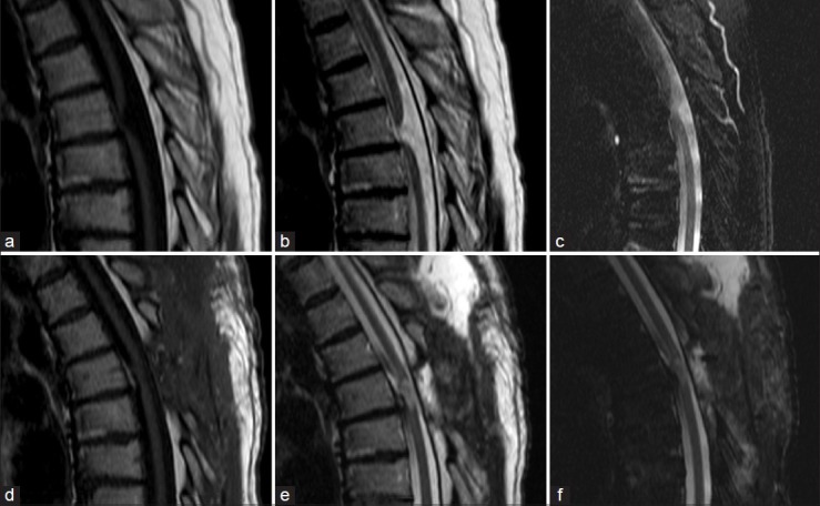
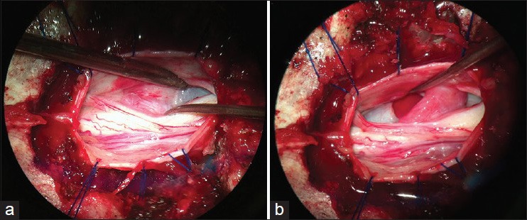
A 66-year-old female farm worker presented with 7 years of progressive back pain and impaired motor and sphincter function. On neurological examination, she exhibited a sensory level at T4 with a spastic paraparesis and bilateral (right greater than left) muscle atrophy.[1] The MR revealed a ventral dural gap at the T4 level [Figure 1a–c]. She underwent a T4 laminectomy at which time the ventral dural gap was repaired (e.g. ventral dura mater defect repaired utilizing a 5-0 prolene suture to attach a muscle/fascia patch to the surrounding healthy dura mater) and the cord was reduced [Figure 2a] at surgery. The herniated cord appeared “violaceous/pale” in color and was hardened [Figure 2b]. Postoperatively, the patient showed improvement in muscle strength in the lower limbs, allowing her to walk with assistance (with a cane or walker).
Figure 1.

Magnetic resonance of the dorsal spine [sagittal acquisition – (a and d) T1-weighted images; (b and e) T2-weighted images; (c and f) SPIR (Selective partial inversion recover) T2-weighted images] demonstrating the spinal cord herniation (a-c) and the radiological result following surgical treatment (d–f)
Figure 2.

Intraoperative photograph. (a) Here, the herniated spinal cord content is demonstrated (instrument tip to the right). (b) The dural defect (indicated by the instrument), the reduced spinal cord area, and the tapering of the spinal cord at the herniation level are shown
DISCUSSION
Clinical and diagnostic studies
Utilizing MRI, ISCH has been increasingly recognized. In one series, it was reported to occur in 0.08% of cases.[6,7] It typically occurs in middle-aged females (range 22-71 years of age) versus males.[1,7,9,10,11] It most commonly presents as a Brown-Séquard syndrome or a non-focal deficit.[1,6,9] Although postoperative improvement is commonly reported, the more severe the preoperative deficit, the worse is the prognosis (e.g. 80% of the cases with spastic paraparesis regain motor function).[1,4,8,9]
Pathophysiology
Spontaneous ventral cord herniation usually involves the mid-thoracic spine, where the cord is naturally anteriorly situated due to the natural kyphosis.[5] This facilitates ventral cord herniation through an anterior/anteromedial dural defect that may have resulted from a ventral disc protrusion (causing weakness of dural fibers), an arachnoid cyst (pushing the cord to an anterior sleeve), or other ventral lesions.[1,3,4,5]
One author[8] suggested that two factors account for these lesions. They include an anterior dural defect leading to an extradural arachnoid cyst (congenital), or pseudomeningocele (iatrogenic), and the concave defect situated on the site of the spinal curvature (dorsally in the cervical spine and ventrolaterally or ventrally in the thoracic spine).
Other etiologies of spinal cord herniation
There are also multiple other etiological theories as to why ventral thoracic cord herniations occur. The predominant one is that the ventral position/adherence of the thoracic cord leads to compression and extrusion through ventral dural defects attributable to cardiac, pulmonary, and/or CSF pulsations.[1,2,6] Other theories include chronic inflammation, lytic substances, and inflammatory conditions.
Value of MR in diagnosing spinal cord herniation
Although MRI is the most accurate tool for diagnosing spinal cord herniation, myelo-CT studies also prove useful.[9] They typically show thinning of the spinal cord and adherence to the anterior spinal canal. T2-weighted images can further directly demonstrate an anterior dural “gap.”
Classification of sagittal and axial MR types of cord herniation
Sagittal MR: In the sagittal plane, three types of thoracic MR cord herniations are identified: Type K shows an obvious ventral spinal kink; Type D, the discontinuous type, is characterized by the spinal cord completely disappearing at the herniation site; and Type P is the protrusion type wherein the anterior subarachnoid space ventral to the thoracic cord disappears, almost without a focal “kink.”[7]
Axial MR: Using axial images, the location of cord herniations is classified as central (Type C) and lateral (Type L) types. Furthermore, the laterality of the herniated spinal cord is classified based on its correspondence (same; Type S) or non-correspondence (opposite; Type O) with the location of the herniation.[7]
Surgery and outcome
Surgery for ISCH is appropriate when a progressive and/or severe neurological deficit occurs.[9] At first, reduction and repair of the dural defect through thoracotomy and partial corpectomy was described; however, this involved prolonged recovery for most patients.[12] Most authors, therefore, prefer a laminectomy or laminoplasty often accompanied by looking for and repair of an attendant arachnoid cyst.[5,6,8] In this case, a laminectomy at T4 with the resection of the dentate ligament permitted gentle rotation of the spinal cord and its reduction through the ventral dural defect that was then repaired with prolene 5-0 sutures.
Prognosis is better if the preoperative deficit is limited.[1,4,8,9] Nevertheless, when the neurological symptoms are already very serious, with paraplegia and spasticity,[1,9] as in our case, significant improvement is not likely to occur.
CONCLUSION
Patients who present with slowly progressive paraparesis or Brown-Séquard syndrome may suffer from a ventral thoracic spinal cord herniation (ISCH). Surgical decompression, utilizing a laminectomy or laminoplasty, may allow for reduction of the cord herniation while facilitating ventral dural repair.
Footnotes
Available FREE in open access from: http://www.surgicalneurologyint.com/text.asp?2014/5/16/564/148042
Contributor Information
Rodrigo Becco De Souza, Email: rodrigobecco@yahoo.com.br.
Guilherme Brasileiro De Aguiar, Email: guilhermebraguiar@yahoo.com.br.
Jefferson Walter Daniel, Email: dolldani@uol.com.br.
José Carlos Esteves Veiga, Email: jcemveiga@uol.com.br.
REFERENCES
- 1.Ammar KN, Pritchard PR, Matz PG, Hadley MN. Spontaneous thoracic spinal cord herniation: Three cases with long-term follow-up. Neurosurgery. 2005;57:E1067. doi: 10.1227/01.neu.0000180016.69507.e0. [DOI] [PubMed] [Google Scholar]
- 2.Dix JE, Griffitt W, Yates C, Johnson B. Spontaneous thoracic spinal cord herniation through an anterior dural defect. AJNR Am J Neuroradiol. 1998;19:1345–8. [PMC free article] [PubMed] [Google Scholar]
- 3.Groen RJ, Middel B. Idiopathic cord herniation. J Neurosurg Spine. 2010;12:714–6. doi: 10.3171/2009.10.SPINE09829. [DOI] [PubMed] [Google Scholar]
- 4.Groen RJ, Middel B, Meilof JF, de Vos-van de Biezenbos JB, Enting RH, Coppes MH, et al. Operative treatment of anterior thoracic spinal cord herniation: Three new cases and an individual patient data meta-analysis of 126 case reports. Neurosurgery. 2009;64(3 Suppl):S145–59. doi: 10.1227/01.NEU.0000327686.99072.E7. [DOI] [PubMed] [Google Scholar]
- 5.Gwinn R, Henderson F. Transdural herniation of the thoracic spinal cord: Untethering via a posterolateral transpedicular approach. Report of three cases. J Neurosurg Spine. 2004;1:223–7. doi: 10.3171/spi.2004.1.2.0223. [DOI] [PubMed] [Google Scholar]
- 6.Hassler W, Al-Kahlout E, Schick U. Spontaneous herniation of the spinal cord: Operative technique and follow-up in 10 cases. J Neurosurg Spine. 2008;9:438–43. doi: 10.3171/SPI.2008.9.11.438. [DOI] [PubMed] [Google Scholar]
- 7.Imagama S, Matsuyama Y, Sakai Y, Nakamura H, Katayama Y, Ito Z, et al. Image classification of idiopathic spinal cord herniation based on symptom severity and surgical outcome: A multicenter study. J Neurosurg Spine. 2009;11:310–9. doi: 10.3171/2009.4.SPINE08691. [DOI] [PubMed] [Google Scholar]
- 8.Kumar R, Taha J, Greiner AL. Herniation of the spinal cord. Case report. J Neurosurg. 1995;82:131–6. doi: 10.3171/jns.1995.82.1.0131. [DOI] [PubMed] [Google Scholar]
- 9.Maira G, Denaro L, Doglietto F, Mangiola A, Colosimo C. Idiopathic spinal cord herniation: Diagnostic, surgical, and follow-up data obtained in five cases. J Neurosurg Spine. 2006;4:10–9. doi: 10.3171/spi.2006.4.1.10. [DOI] [PubMed] [Google Scholar]
- 10.Miyake S, Tamaki N, Nagashima T, Kurata H, Eguchi T, Kimura H. Idiopathic spinal cord herniation. Report of two cases and review of the literature. Neurosurg. 1998;88:331–5. doi: 10.3171/jns.1998.88.2.0331. [DOI] [PubMed] [Google Scholar]
- 11.Srinivasan A, Bourque P, Goyal M. Spontaneous thoracic spinal cord herniation. Neurology. 2004;63:2187. doi: 10.1212/01.wnl.0000140620.32658.0b. [DOI] [PubMed] [Google Scholar]
- 12.Wortzman G, Tasker RR, Rewcastle NB, Richardson JC, Pearson FG. Spontaneous incarcerated herniation of the spinal cord into a vertebral body: A unique cause of paraplegia. Case report. J Neurosurg. 1974;41:631–5. doi: 10.3171/jns.1974.41.5.0631. [DOI] [PubMed] [Google Scholar]


