Abstract
Purpose.
Because human bone marrow (BM) CD34+ stem cells home into damaged tissue and may play an important role in tissue repair, this pilot clinical trial explored the safety and feasibility of intravitreal autologous CD34+ BM cells as potential therapy for ischemic or degenerative retinal conditions.
Methods.
This prospective study enrolled six subjects (six eyes) with irreversible vision loss from retinal vascular occlusion, hereditary or nonexudative age-related macular degeneration, or retinitis pigmentosa. CD34+ cells were isolated under Good Manufacturing Practice conditions from the mononuclear cellular fraction of the BM aspirate using a CliniMACs magnetic cell sorter. After intravitreal CD34+ cell injection, serial ophthalmic examinations, microperimetry/perimetry, fluorescein angiography, electroretinography (ERG), optical coherence tomography (OCT), and adaptive optics OCT were performed during the 6-month follow-up.
Results.
A mean of 3.4 million (range, 1–7 million) CD34+ cells were isolated and injected per eye. The therapy was well tolerated with no intraocular inflammation or hyperproliferation. Best-corrected visual acuity and full-field ERG showed no worsening after 6 months. Clinical examination also showed no worsening during follow-up except among age-related macular degeneration subjects in whom mild progression of geographic atrophy was noted in both the study eye and contralateral eye at 6-month follow-up, concurrent with some possible decline on multifocal ERG and microperimetry. Cellular in vivo imaging using adaptive optics OCT showed changes suggestive of new cellular incorporation into the macula of the hereditary macular degeneration study eye.
Conclusions.
Intravitreal autologous BM CD34+ cell therapy appears feasible and well tolerated in eyes with ischemic or degenerative retinal conditions and merits further exploration. (ClinicalTrials.gov number, NCT01736059.)
Keywords: stem cells, retinal degeneration, retinal imaging, retinal vein occlusion, OCT
This preliminary phase 1 clinical trial reports no safety or feasibility issues in exploring the use of intravitreal autologous bone marrow CD34+ stem cells as potential therapy for ischemic or degenerative retinal disorders.
Introduction
Degenerative and ischemic retinal conditions remain leading causes of irreversible blindness, despite advances in drug and laser therapies for complications associated with these conditions. Currently, age-related macular degeneration (AMD) remains a leading cause of irreversible legal blindness among the elderly, and Stargardt's disease remains a leading cause of hereditary macular degeneration leading to legal blindness.1,2 When the degenerative or ischemic retinal condition is diffuse and extends beyond the macula, almost total blindness can result. This can occur in patients with advanced hereditary retinal degeneration, such as retinitis pigmentosa, or severe retinal vascular occlusion, a leading vascular cause of blindness in the elderly.3
Tissue regeneration is potentially possible with cellular therapy. Partially differentiated retinal pigment epithelial cells derived from embryonic stem cells and inducible pluripotent stem cells have been developed.4–6 Preclinical studies suggest that these cells may slow progression of retinal degeneration when injected into the subretinal space.4 Clinical trials have been initiated to explore these cells as a potential treatment for macular degeneration.7 However, the allogeneic therapy requires systemic immunosuppressive therapy to minimize rejection.7 Furthermore, these stem cells are potentially teratogenic, raising long-term safety concerns.4–7
Bone marrow (BM) contains a high concentration of lineage-negative adult stem cells that appear to play an important role in tissue maintenance and repair. Among the cells in human BM, much attention has been on CD34+ cells. These cells have been used safely for BM transplantation to reconstitute the hematopoietic system and in clinical trials of coronary heart disease and peripheral limb ischemia for revascularization.8–15 These cells were explored in animal models as potential therapy for degenerative or ischemic retinal conditions since they are multipotent and can have local trophic effects.15–19 Intravitreally injected CD34+ cells migrate into the retina and home into the damaged retinal vasculature or neuronal tissue.18–20 These human cells are detected in the mouse retina as long as 6 months following injection with no associated safety issues.19
This report describes the preliminary observations of the first clinical trial exploring the safety and feasibility of using intravitreally injected autologous BM CD34+ cells to treat degenerative or ischemic retinal conditions.
Methods
This single-center, prospective, open-labeled, phase 1 study enrolled patients with irreversible vision loss from degenerative or ischemic retinal conditions seen in the Retina Service Eye Clinic at the University of California-Davis Eye Center between November 2012 and August 2014. The study was based on an Investigational New Drug (IND) approved by the Food and Drug Administration (FDA; IND 13307) and conducted according to a protocol approved by the University of California-Davis (UCD) School of Medicine Office of Human Research Protection and Stem Cell Research Oversight Committee. All subjects provided written informed consent before study initiation. The UCD Eye Center Clinical Trials Unit was responsible for the day-to-day conduct of the trial. The study was overseen independently by the UCD Clinical and Translational Science Center. It was conducted in accordance with the tenets of the Declaration of Helsinki. The study was registered with www.clinicaltrials.gov prior to initiation (NCT01736059).
Study subjects were 18 years of age or older with irreversible vision loss for over 6 months in the study eye from hereditary or nonexudative (dry) AMD, retinitis pigmentosa, or retinal vascular occlusion. Best-corrected visual acuity (BCVA) was measured by a certified visual acuity examiner using the Early Treatment Diabetic Retinopathy Study (ETDRS) visual acuity charts with appropriate refraction during each study visit. Enrollment BCVA was 20/100 to counting fingers in the study eye, with equal or better BCVA in the contralateral eye. Exclusion criteria included any concurrent condition causing vision loss, coagulopathy or other hematologic disorders, concurrent use of coumadin or systemic immunosuppressive therapy, significant media opacity, and history of neovascularization or macular edema in the study eye requiring therapy within 3 months of enrollment or anticipated during the study period.
A comprehensive eye examination including a BCVA was performed at baseline, 1 day, 1 week, 2 weeks, 1 month, 3 months, and 6 months following cell injection. In addition, macular spectral-domain optical coherence tomography (OCT; Cirrus; Carl Zeiss Meditec, Inc., Dublin, CA, USA) and microperimetry (Ellex Macular Integrity Assessment Microperimeter, CenterVue S.p.A., Padova, Italy) were performed on both eyes at baseline and at 2 weeks, 1 month, 3 months, and 6 months following cellular therapy. If the subject had unstable fixation, Octopus Goldmann perimetry was performed instead. Fundus photography and fluorescein angiography were performed at baseline and at 1-, 3-, and 6-month visits. Electroretinography (ERG; Epsion Visual Electrophyiology System, model D300; Diagnosys LLC, Littleton, MA, USA) was performed on both eyes at baseline and at 1- and 6-month follow-up. Both full-field and multifocal ERGs were performed under light-adapted (photopic) conditions. Full-field ERG was performed also under dark-adapted (scotopic) conditions after 20 minutes of dark adaptation.
The primary outcome measures were (1) the incidence and severity of ocular and systemic adverse events associated with study treatment and (2) the number of CD34+ cells isolated and injected intravitreally from a single BM aspirate.
Bone Marrow Aspiration and CD34+ Cell Isolation
Bone marrow aspiration from the iliac crest was performed under local anesthesia (1% lidocaine intramuscular) without sedation by a licensed experienced hematologist (MA). Subjects received lorazepam (1 mg) and hydromorphone (2 mg) orally, approximately 1 hour before the procedure as antianxiety and analgesic medication, respectively. Approximately 40 to 50 mL BM was obtained sterilely with a single aspiration. The aspirate was transported to the UCD Good Manufacturing Practice facility in heparinized syringes (with no preservative) within 30 minutes for CD34+ cell isolation. A Ficoll gradient separation of the mononuclear cellular fraction followed by a CliniMACs (Miltenyi Biotec, Inc., Auburn, CA, USA) magnetic cell sorter for CD34+ cell isolation was performed. The release criteria for the final cellular product were passing the stat viability cell count, gram stain, and endotoxin assay. A 14-day sterility test was also set up but was not used as a release criterion since results would not be immediately available. An approved positive culture action plan, however, was in place for the eventuality of a positive culture. The final cellular product was resuspended in balanced saline solution (0.1 mL final volume, BSS Plus; Alcon Laboratories, Fort Worth, TX, USA) for intravitreal injection, and all isolated CD34+ cells were injected intravitreally. The BM aspiration site was visually inspected for bleeding or infection at 1-day and 1-week visits.
Intravitreal Injection
The study eye was dilated and manually massaged gently for 5 minutes prior to injection to soften the eye. The eye was anesthetized with topical proparacaine and subconjunctival injection of 2% lidocaine, cleansed with 5% betadine, and kept open with a sterile lid speculum. The entire 0.1-mL volume of isolated CD34+ cells was injected intravitreally 4 mm posterior to the infratemporal limbus by an experienced retinal specialist (SSP) using a 30-gauge short needle. The lid speculum was removed and balanced saline solution was used to rinse the eye. Visual acuity of at least counting fingers was confirmed after the injection. Indirect ophthalmoscopy was performed to confirm optic nerve perfusion.
Adaptive Optics OCT (AO-OCT) Imaging of the Macula
The OCT utilized a broadband superluminescent diode at 836 nm with 112-nm spectral bandwidth (Broadlighter; Superlum LTD, Cork, Ireland) and a spectrometer consisting of a diffraction grating and a linear charge-coupled device detector (AViiVA, e2v; Chelmsford, England). A portion of the OCT light was reflected into a Hartmann-Shack wavefront sensor (Northrop Grumman Corp., Falls Church, VA, USA) to measure the study eye's aberrations, and a high-stroke 97-actuator membrane magnetic deformable mirror (ALPAO S.A.S., St. Martin, France) corrected them to achieve diffraction-limited resolution. This configuration allowed a measured axial resolution of 3.5 μm at the retina and thus a ~3- × 3- × 3.5-μm3 volumetric resolution. A bite bar and a forehead rest assembly mounted on an X-Y-Z remote-controlled motorized translation stage was used to reduce head motion and allow precise positioning of the eye's pupil in the center of the imaging system's entrance pupil. The study eye was dilated with 1% tropicamide. The raw cross-sectional AO-OCT images consisted of 1000 axial scans each, acquired at a rate of 18 kHz. The AO-OCT images shown in this report are intensity averages of five consecutive raw images, motion corrected using the ImageJ StackReg plugin.21,22 More technical details about the AO-OCT system have been published previously.23
Results
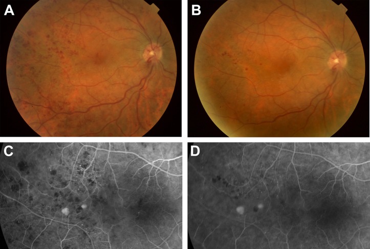
Table 1 summarizes the demographic and clinical features of the six study subjects who enrolled in the study. All completed the 6-month follow-up period. All were young adults except for the two AMD subjects (age range, 23–85 years; mean, 48 years). All had advanced vision loss in the study eye with baseline BCVA of 20/200 to 20/800. Subject 1 had unilateral irreversible vision loss of 9-month duration from a combined central retinal artery and vein occlusion with persistent retinal hemorrhages but no macular edema or retinal neovascularization at the time of enrollment (Fig. 1A). The remaining five subjects had progressive vision loss in both eyes of 5 years or longer duration (range, 5–20 years) from various degenerative retinal conditions. Subjects 2 and 3 had bilateral symmetrical loss of central vision from Stargardt's disease, which was diagnosed clinically by the presence of fleck retinopathy and “dark choroid” on fluorescein angiography. Subjects 4 and 6 had asymmetric but bilateral loss of central vision from atrophic nonexudative AMD. Subject 5 had advanced central and peripheral vision loss in both eyes from retinitis pigmentosa, which was diagnosed by the presence of bone spicules on fundus examination and a flat unrecordable ERG in both eyes.
Table 1.
Demographic and Clinical Characteristics of Enrolled Subjects
|
Age/Sex/Eye |
Diagnosis |
Baseline BCVA* |
Best Follow-Up BCVA* (No. Letters Gained) |
6-Month BCVA* (No. Letters Gained) |
| 1. 39/male/OD | CRAO/CRVO | 20/800 | 20/40 @ 3 mo | 20/63 |
| (65) | (55) | |||
| 2. 31/male/OS | Stargardt's | 20/200+2 | 20/100−2 @ 3 mo | 20/125+2 |
| (11) | (10) | |||
| 3. 23/male/OD | Stargardt's | 20/200 | 20/160+2 @ 4 wk | 20/160 |
| (9) | (7) | |||
| 4. 76/female/OS | AMD | 20/200 | 20/80−2 @ 4 wk | 20/80−2 |
| (18) | (18) | |||
| 5. 36/male/OS | RP | 20/640 | 20/250 @ 2 wk | 20/400−2 |
| (20) | (8) | |||
| 6. 85/female/OS | AMD | 20/200−1 | 20/160−2 @ 4 wk | 20/200−2 |
| (3) | (−1) |
Based on visual acuity with refraction obtained with ETDRS chart by a certified visual acuity tester. CRAO/CRVO, combined central retinal artery and vein occlusion; RP, retinitis pigmentosa; OS, left eye; OD, right eye.
Figure 1.
Serial fundus photographs and fluorescein angiograms of subject 1 with a history of combined central retinal artery and vein occlusion diagnosed 9 months prior to study enrollment. (A) Fundus photograph of the right eye taken at time of enrollment showing persistent extensive retinal hemorrhages especially temporally with some retinal venous congestion. (B) Fundus photography of the study eye taken 3 months following intravitreal CD34+ cell injection showing marked resolution of retinal hemorrhages compared to enrollment. (C) Fluorescein angiogram of the study eye at enrollment showing an area temporal to the macula with microaneurysmal and telangiectatic retinal vascular changes with some patches of retinal ischemia. Patches of blocked fluorescence from retinal hemorrhages are also noted along with two small patches of fluorescein pooling from small focal areas of pigment epithelial detachment. (D) Fluorescein angiogram of the study eye taken 3 months following CD34+ cell therapy shows partial resolution of the microaneurysmal and telangiectatic retinal vascular changes and blocked fluorescence from the retinal hemorrhages noted at enrollment. The changes are more noteworthy inferior to the two small areas of fluorescein pooling from pigment epithelial detachment that remain unchanged.
Bone Marrow Aspiration and CD34+ Isolation/Injection
All subjects tolerated the BM aspiration and intravitreal injection of CD34+ cells and completed the 6-month study follow-up without any adverse effects except for grade 1 local pain immediately following BM aspiration. All the products passed the initial release tests, and all the cultures remained negative during the 14-day sterility test. The total number of CD34+ cells isolated for intravitreal injection from a single BM aspiration ranged from 1.0 to 7 million cells per eye (mean 3.4 million cells). All isolated CD34+ cells were injected intravitreally. There was no obvious correlation between the age of the subject and the number of CD34+ cells isolated in this small pilot study.
Follow-Up Visual Acuity and Eye Examination
Improvement in BCVA ranged from 0 to 11 lines in the ETDRS visual acuity chart during the 6-month follow-up (mean improvement of three lines). Improvement of two or more lines of BCVA (i.e., 10 letters or more) was noted in four of six subjects during the course of the study. The improvement in BCVA was larger in subject 1 with retinal vascular occlusion when compared to the other subjects who had retinal degenerative conditions. The time course of improvement varied among the subjects. However, some improvement in measurement of BCVA was noted in all subjects by 1-month follow-up. Most of the improvement was sustained during the 6-month follow-up period, with some fluctuations. However, the final BCVA at 6-month follow-up usually was within one line of best-measured visual acuity during the follow-up period (Table 1).
Most subjects noted some mild transient floaters during the first few days following cell injection. The injected cells were visible in the anterior vitreous just after the injection in some eyes but not visible after 1 week in all eyes. There was no evidence of intraocular inflammation or hyperproliferation of cells in the study eye on serial eye examination. Subject 4 developed a new midperipheral choroidal neovascular membrane in the nonstudy eye 2 weeks after cellular therapy, which was successfully treated with photodynamic therapy. Some mild extrafoveal enlargement of geographic atrophy was noted in both eyes in both AMD subjects (4 and 6) after 6-month follow-up as accessed by fluorescein angiography. No other changes were noted on follow-up examination, fluorescein angiography, or OCT of the macula of the study eye of any of the five subjects with retinal degenerative conditions. In contrast, subject 1 with the retinal vascular occlusion had marked resolution of the retinal hemorrhages at 3-month follow-up, which was sustained at the 6-month follow-up (Figs. 1A, 1B). Fluorescein angiography showed concurrent partial resolution of the microaneurysmal and telangiectatic retinal vascular changes (Figs. 1C, 1D). No new retinal ischemia, ocular neovascularization, or macular edema was noted during the follow-up period in any of the study eyes.
Microperimetry and Perimetry
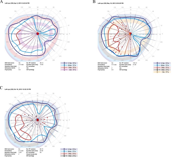
Macular function in the study eye was evaluated by microperimetry during the course of the study in the five of the six subjects with reliable fixation. The findings in the study eyes are summarized in Table 2. Subject 5 had poor fixation, and Octopus Goldmann perimetry was performed instead (Fig. 2).
Table 2.
Macular Microperimetry Findings of the Study Eyes
|
Baseline* Mean, % Reduced |
1 Month* Mean, % Reduced |
6 Months* Mean, % Reduced |
|
| 1 | 16.8, | 18.4, | 19.2, |
| 70.3 | 59.5 | 45.9 | |
| 2 | 27.1, | 28.3, | 26.6, |
| 24.3 | 10.8 | 13.5 | |
| 3 | 18.9, | 18.9, | 23.5, |
| 25.6 | 26.0 | 45.9 | |
| 4 | 16.9, | 17.1, | 14.7, |
| 86.5 | 75.7 | 94.7 | |
| 6 | 16.9, | 17.7, | 14.6, |
| 75.5 | 64.9 | 83.5 |
Values are expressed as mean sensitivity threshold (dB), percentage reduced sensitivity. Subject 5 had Goldmann perimetry as shown in Figure 2.
Figure 2.
Serial Goldmann perimetry of the study eye of subject 5 with advanced retinitis pigmentosa. (A) Baseline perimetry. (B) Perimetry at 1 month following intravitreal CD34+ cell therapy showing improved sensitivity in the nasal visual field to III4e stimulus (blue). (C) Perimetry at final follow-up at 6 months following enrollment shows sustained improvement in nasal visual field noted at 1-month follow-up perimetry.
Subject 1 with retinal vascular occlusion showed progressive improvement in macular function on microperimetry starting at 1-month follow-up. Macular mean sensitivity threshold improved from 16.8 to 19.2 dB, and the percentage reduced sensitivity decreased from 70.3% to 45.9% by 6-month follow-up.
Subject 2 with Stargardt's disease had mean macular sensitivity in the study eye, which remained relatively stable during the course of the study, ranging from 28.3 to 26.6 dB; but the percentage reduced sensitivity improved from 24.3% at baseline to 10.8% and 13.5% at 1- and 6-month follow-up visits, respectively. Subject 3 with more advanced Stargardt's disease had improvement in mean macular sensitivity from 18.9 to 23.5 dB by 6-month follow-up, although the percentage reduced sensitivity worsened from 25.6% to 45.9% during the study period.
Both subjects with AMD (subjects 4 and 6) had some improvement in the percentage reduced sensitivity at the 1-month visit, but by 6-month follow-up, some further reduction in mean macular sensitivity and increase in percentage reduced sensitivity were noted in both study eyes. The areas of the macula with worsening macular function on microperimetry corresponded to the areas of slight worsening of geographic atrophy noted on fluorescein angiography, observed in both the study eye and the contralateral eye of both AMD subjects by 6-month follow-up (Fig. 3).
Figure 3.
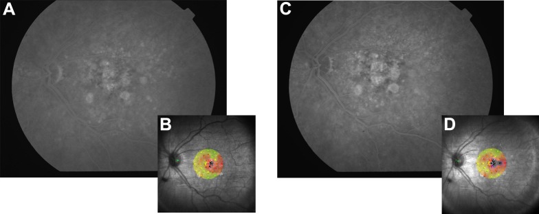
Fluorescein angiogram and microperimetry of the study eye of subject 4 with age-related macular degeneration showing mild extrafoveal enlargement of geographic atrophy during the study follow-up period with corresponding area of worsening macular function on microperimetry. (A) Late transit fluorescein angiogram of the study left eye at baseline showing patches of geographic atrophy involving the fovea. (B) Corresponding macular sensitivity map on microperimetry of the left eye at baseline. (C) Late transit fluorescein angiogram of the study left eye at 6-month study follow-up showing some enlargement of geographic atrophy temporal to the fovea. (D) Corresponding macular sensitivity map on microperimetry of the left eye at 6-month study follow-up showing an increase in scotoma temporally. Color coding of macular sensitivity map: green, normal function; red/orange, decreased function; black, absolute scotoma.
Subject 5 with retinitis pigmentosa was noted to have improvement in peripheral visual function on Goldmann perimetry at 1-month follow-up examination, which appeared sustained at the final 6-month follow-up visit (Fig. 2).
Electroretinography
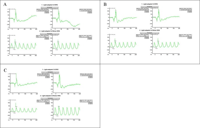
All subjects had reduced but recordable full-field ERG at baseline in the study eye except subject 5, who had flat unrecordable ERG readings in both eyes at baseline and at follow-up consistent with the diagnosis of retinitis pigmentosa. Among subjects with detectable full-field ERG signal at baseline, there was no worsening of signal amplitude during the follow-up in the study eye. Subject 1 had a 50% reduction in amplitude in the study eye at baseline, which normalized at the 1-month visit and returned to baseline level at 6-month follow-up. Subject 2 had bilateral symmetric decrease in photopic and flicker amplitude at baseline and at 1-month follow-up consistent with the diagnosis of Stargardt's disease (Figs. 4A, 4B). This subject was noted to have a 30% to 40% increase in the photopic and flicker ERG amplitude only in the study eye at the final 6-month follow-up study visit (Fig. 4C).
Figure 4.
Full-field photopic electroretinogram of subject 2 with Stargardt's disease at baseline (A), 1 month (B), and 6 months (C) following intravitreal CD34+ cell injection in the left eye (left eye recording is depicted in the right recording panel) showing an increase in the b-wave and flicker signal amplitude in the study eye at 6-month follow-up visit relative to the untreated contralateral eye and baseline recordings. The rectangular boxes within the recording represent the age-matched normal b-wave and flicker amplitude range for each recording, as follows: normal mean flicker amplitude ± SD 106 ± 32 μV; normal mean b-wave amplitude 128 ± 46 μV.
Macular multifocal ERG was abnormal at baseline in all the study eyes. During the course of the study, the number of abnormal hexagons and mean variance on multifocal ERG remained relatively stable in the study eye compared to baseline (Table 3) except among some study eyes with bilateral degenerative macular conditions (subjects 2, 4, and 6), where some possible mild worsening of macular function was noted in both eyes that might be attributed to progression of the degenerative condition during the course of the study.
Table 3.
Macular Multifocal Electroretinography Findings of the Study Subjects
|
Subject No. |
Multifocal Electroretinography, No. Abnormal Hexagons*/Mean Variance† |
|||
|
Baseline |
At 6-Month Follow-Up |
|||
|
Study Eye |
Contralateral Eye |
Study Eye |
Contralateral Eye |
|
| 1 CRAO/CRVO | 25/NA | 0/NA | 24/NA | 0/NA |
| 2 Stargardt's | 31/−1.7 | 24/−1.8 | 37/−2.2 | 51/−2.9 |
| 3 Stargardt's | 56/−3.4 | 56/−3.1 | 51/−3.0 | 55/−3.2 |
| 4 AMD | 1/−0.9 | 3/−1.1 | 6/−1.2 | 13/−1.4 |
| 5 RP | 61/−4.0 | 61/−4.1 | 60/−3.9 | 61/−3.5 |
| 6 AMD | 1/−0.9 | 3/−0.3 | 8/−1.2 | 4/−1.1 |
Hexagons with 2 or more standard deviation decrease in amplitude from normal values from a total of 61 hexagons covering the macula.
Mean variance refers to the mean recorded standard deviation in amplitude from normal values for the 61 hexagons covering the macula.
Adaptive Optics OCT Macular Imaging
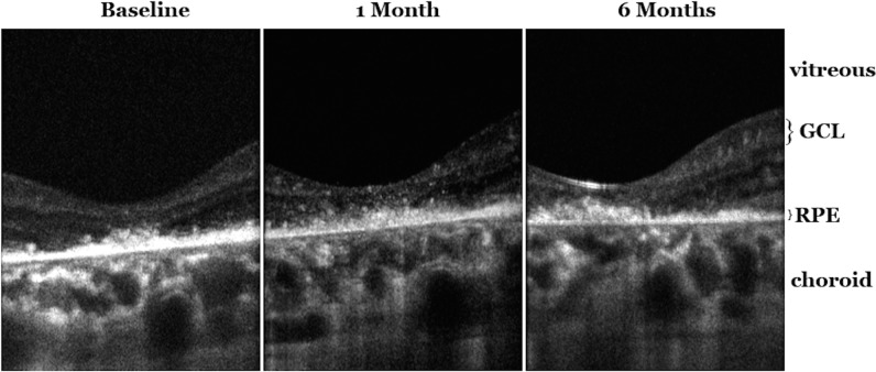
In order to visualize possible changes in the macula at a cellular level during the course of the study, AO-OCT imaging was performed. High-quality AO-OCT imaging of the macula was obtained only on subject 2 since the remaining study subjects had media opacity, atypical optical aberrations, or fixation problems that compromised image quality for AO-OCT imaging. Multiple punctate hyperreflective (white) deposits were noted within the macula 1 month after intravitreal CD34+ cell injection (Fig. 5). The size of these deposits was approximately the size of the CD34+ cells, that is, 10 μm. At 6-month follow-up, these deposits became less visible within the retinal layers, and the deeper retinal pigment epithelial layer appeared somewhat more hyperreflective, suggestive of migration of these deposits or cells into this layer.
Figure 5.
Adaptive optics optical coherence tomography (AO-OCT) imaging of the central macula of the study eye of subject with Stargardt's disease. Left: B-scan cross-sectional images of the central macula at study enrollment showing the irregular hyperreflective retinal pigment epithelial layer with overlying photoreceptor inner segment–outer segment layer not well visualized. Middle: Cross-sectional B-scan images of the central macula 1 month after intravitreal CD34+ cell injection showing new multiple hyperreflective (white) deposits within the retinal layers. Right: Cross-sectional B-scan image taken 6 months after CD34+ cell injection showing less prominent hyperreflective deposits within the retinal layers and a hint of an increase in hyperreflectivity of the irregular retinal pigment epithelial layer centrally under the fovea. GCL, ganglion cell layer; RPE, retinal pigment epithelium.
Discussion
This report summarizes the initial findings of the first clinical trial exploring the use of intravitreal autologous BM CD34+ cells as potential therapy for ischemic or degenerative retinal conditions that are blinding and currently untreatable. This study is based on the first IND approved by the FDA for this route of CD34+ cell administration.19 The findings are preliminary but demonstrate that the treatment was feasible, without any major safety concerns for the duration of the study follow-up. The number of CD34+ cells isolated and injected intravitreally among our subjects was close to the upper limit specified by the study protocol based on available preclinical safety data, that is, 10 million CD34+ cells per eye.19 Both the BM aspiration and intravitreal cell injection were well tolerated and not associated with any ocular or systemic adverse effects during the 6-month follow-up. Three of the study subjects have been examined as part of standard of care after exiting from the study. No long-term ocular or systemic adverse effects have been noted among these patients with follow-up ranging from 12 to 18 months (Park SS, unpublished data, 2014).
Our findings are consistent with preclinical observations in NOD-SCID mice showing no long-term ocular or systemic safety concerns associated with intravitreally injected BM CD34+ cells.19 Two previous small pilot clinical trials using intravitreal autologous BM mononuclear cells also reported no safety concerns over 6-month follow-up, but visual benefit was minimal to none.24,25 The BM mononuclear cells used in these previous clinical trials were obtained by Ficoll gradient separation of the BM aspirate, the first step used in our study to isolate CD34+ cells. Thus the BM mononuclear cellular fraction contained CD34+ cells, but in very low concentration.
The observations of our pilot clinical trial using purified BM CD34+ cells are similar to those in previous studies using mononuclear cells in that no safety or feasibility issues were noted, but they differ in that four of six study subjects had two or more lines of BCVA improvement following cell therapy. Furthermore, possible improvement in microperimetry/perimetry, ERG, and/or fluorescein angiography was noted in some of the study eyes. However, since some possible worsening of macular function also was noted by microperimetry and multifocal ERG in some study eyes with macular degeneration, it is difficult to be certain whether the observed clinical signs of possible improvement following cell therapy represent true treatment effects. Nonetheless, the possible improvements in visual function are encouraging observations among these subjects with a history of longstanding irreversible advanced vision loss, which had been progressive among five of our six subjects with retinal degeneration.
Adult BM contains stem cells that are believed to play an important role in normal tissue maintenance and repair. Bone marrow lineage-negative stem cells have been shown in preclinical studies to slow progression of retinal degeneration after intravitreal administration.17 Among BM lineage-negative cells, human CD34+ cells have been explored in clinical trials for their ability to home into and repair damaged tissue.8,14,26,27 CD34+ cells are often called “hematopoietic stem/progenitor cells” because they can be stimulated to differentiate into various blood cell lineages and fully reconstitute the blood-forming system.27,28 However, the full function and potential of these cells are still being investigated. Endothelial progenitors also express CD34 on their surface, for instance, so a CD34+ population from BM will include the revascularization population.13,28,29 Intravitreal injection of CD34+ cells resulted in homing of these cells into the damaged retinal tissue within days in preclinical studies.18,20 Transretinal migration of these CD34+ cells into the outer retina has been demonstrated following intravitreal injection in an animal model of laser retinal injury.20
In this clinical study, histopathologic analysis to confirm the intraretinal incorporation of these CD34+ cells following intravitreal injection was not possible. However, AO-OCT imaging allows cellular macular imaging in vivo.23 Serial high-quality AO-OCT imaging of the macula of subject 2 showed new fine hyperreflective deposits within the retinal layers 1 month after cellular therapy (Fig. 5). These deposits were approximately the size of CD34+ cells (~10 μm) and too small to be visualized using the clinical-grade spectral-domain OCT instrument with lower transverse resolution. These AO-OCT findings appear to be unique to our study eye and have not been observed in over 50 human eyes with various degenerative or acquired maculopathy imaged with this AO-OCT imaging system (Panorgias A, Jonnal R, Zawadzki RJ, et al., personal observation, 2014). Further studies are needed to validate whether these observed AO-OCT changes indeed represent direct intraretinal incorporation of viable CD34+ cells. However, the findings are consistent with observations made in preclinical studies20 and demonstrate for the first time the potential usefulness with high-resolution AO-OCT imaging in studying the treatment effect of cellular therapy.
In summary, this preliminary report of a phase 1 study noted no safety or feasibility issues associated with intravitreal autologous BM CD34+ cell therapy. Although the study was small, with limited follow-up, and enrolled subjects with various ischemic and degenerative retinal conditions, the primary endpoints of our study were achieved in that a desired number of CD34+ cells were purified consistently for intravitreal administration and no adverse local or systemic effects were observed associated with this cellular therapy. Although BCVA improved in most eyes during the course of the study, improvement in visual function on subjective testing, such as BCVA or perimetry, may be attributed to learning or placebo effects. Furthermore, spontaneous improvement in vision can occur in eyes with less severe retinal vascular occlusion, although such improvements are more commonly seen in eyes with better initial visual acuity or commonly occur within the first few months following the ischemic event.30 This phase 1 study was not specifically designed to measure efficacy. Nonetheless, possible changes noted on objective testing, such as AO-OCT, ERG, and fluorescein angiography, among some of our study subjects following cellular therapy are findings that warrant further investigation. Since mild progression of geographic atrophy was observed the study eye and contralateral eye of subjects with AMD by 6-month follow-up, this cellular therapy may not stop the progression of degenerative retinal conditions, especially in advanced stages. A larger prospective study with longer follow-up is planned to further explore the safety and potential efficacy of this cellular therapy.
Acknowledgments
The authors thank members of the University of California (UC)-Davis Eye Center Clinical Trials Unit, in particular Marisa Salvador, Idalow Good, and Barbara Holderreed, for assessment of patients and submitting local regulatory paperwork. They also thank members of the UC-Davis Good Manufacturing Practice (GMP) facility for their technical support and Tracy Hysong, MS, CCRP, of the UC-Davis Clinical and Translational Science Center for clinical data monitoring.
Presented in part at the 46th annual meeting of the Retina Society, Beverly Hills, California, United States, September 29, 2013; at the 37th annual meeting of the Macula Society, Key Largo, Florida, United States, February 20, 2014; and at the annual meeting of the Association for Research in Vision and Ophthalmology, Orlando, Florida, United States, May 6, 2014.
Supported by Foundation Fighting Blindness Grant CD-CDBT 0808-0471-UCD (JN), National Eye Institute Grant NIH EY024239 (JSW), a Research to Prevent Blindness unrestricted departmental grant, and the UC-Davis Retina Research Fund (SSP). JN and GB are partially supported by the California Institute for Regenerative Medicine (CIRM). The UC-Davis GMP facility was built with funds from CIRM Major Facilities Grant FA-00611.
Disclosure: S.S. Park, None; G. Bauer, None; M. Abedi, None; S. Pontow, None; A. Panorgias, None; R. Jonnal, None; R.J. Zawadzki, None; J.S. Werner, None; J. Nolta, None
References
- 1. Bressler NM. Age-related macular degeneration is the leading cause of blindness. JAMA. 2004; 291: 1900–1901. [DOI] [PubMed] [Google Scholar]
- 2. Yanuzzi LA. The Retinal Atlas. United Kingdom: Elsevier Saunders; 2010: 63–68. [Google Scholar]
- 3. Mitchell P, Smithe W, Chang A. Prevalence and association of retinal vein occlusion in Australia: the Blue Mountain Eye Study. Arch Ophthalmol. 1996; 114: 1243–1247. [DOI] [PubMed] [Google Scholar]
- 4. Lu B, Malcuit C, Wang S, et al. Long-term safety and function of RPE from human embryonic stem cells in preclinical models of macular degeneration. Stem Cells. 2009; 27: 2126–2135. [DOI] [PubMed] [Google Scholar]
- 5. Hirami Y, Osakada F, Takahashi K, et al. Generation of retinal cells from mouse and human induced pluripotent stem cells. Neurosci Lett. 2009; 458: 126–131. [DOI] [PubMed] [Google Scholar]
- 6. Cyranoski D. iPS cells in humans. Nat Biotechnol. 2013; 31: 775. 24022140 [Google Scholar]
- 7. Schwartz SD, Hubschman JP, Heilwell G, et al. Embryonic stem cell trials for macular degeneration: a preliminary report. Lancet. 2012; 379: 713–720. [DOI] [PubMed] [Google Scholar]
- 8. Booth C, Veys P. T cell depletion in paediatric stem cell transplantation. Clin Exp Immunol. 2013; 172: 139–147. [DOI] [PMC free article] [PubMed] [Google Scholar]
- 9. Jakob P, Landmesser U. Current status of cell-based therapy for heart failure. Curr Heart Fail Rep. 2013; 10: 165–176. [DOI] [PubMed] [Google Scholar]
- 10. Vrtovec B, Poglajen G, Sever M, et al. CD34+ stem cell therapy in nonischemic dilated cardiomyopathy patients. Clin Pharmacol Ther. 2013; 94: 452–458. [DOI] [PubMed] [Google Scholar]
- 11. Patel AN, Geffner L, Vina RF, et al. Surgical treatment for congestive heart failure with autologous adult stem cell transplantation: a prospective randomized study. J Thorac Cardiovasc Surg. 2005; 130: 1631–1638. [DOI] [PubMed] [Google Scholar]
- 12. Rafii S, Meeus S, Dias S, et al. Contribution of marrow-derived progenitors to vascular and cardiac regeneration. Semin Cell Dev Biol. 2002; 13: 61–67. [DOI] [PubMed] [Google Scholar]
- 13. Schmeisser A, Strasser RH. Phenotypic overlap between hematopoietic cells with suggested angioblastic potential and vascular endothelial cells. J Hematother Stem Cell Res. 2002; 11: 69–79. [DOI] [PubMed] [Google Scholar]
- 14. Zhang QH, She MP. Biological behaviour and role of endothelial progenitor cells in vascular diseases. Chin Med J (Engl). 2007; 120: 2297–2303. [PubMed] [Google Scholar]
- 15. Sheridan CM, Pate S, Hiscott P, Wong D, Pattwell DM, Kent D. Expression of hypoxia-inducible factor-1alpha and -2alpha in human choroidal neovascular membranes. Graefes Arch Clin Exp Ophthalmol. 2009; 247: 1361–1367. [DOI] [PubMed] [Google Scholar]
- 16. Chang KH, Chan-Ling T, McFarland EL, et al. IGF binding protein-3 regulates hematopoietic stem cell and endothelial precursor cell function during vascular development. Proc Natl Acad Sci U S A. 2007; 104: 10595–10600. [DOI] [PMC free article] [PubMed] [Google Scholar]
- 17. Otani A, Dorrell MI, Kinder K, et al. Rescue of retinal degeneration by intravitreally injected adult bone marrow-derived lineage-negative hematopoietic stem cells. J Clin Invest. 2004; 114: 765–774. [DOI] [PMC free article] [PubMed] [Google Scholar]
- 18. Caballero S, Sengupta N, Afzal A, et al. Ischemic vascular damage can be repaired by healthy, but not diabetic, endothelial progenitor cells. Diabetes. 2007; 56: 960–967. [DOI] [PMC free article] [PubMed] [Google Scholar]
- 19. Park SS, Caballero S, Bauer G, et al. Long-term effects of intravitreal injection of GMP-grade bone marrow derived CD34+ Cells in NOD-SCID mice with acute ischemia-reperfusion injury. Invest Ophthalmol Vis Sci. 2012; 53: 986–994. [DOI] [PMC free article] [PubMed] [Google Scholar]
- 20. Calzi L, Kent DL, Change KH, et al. Labeling of stem cells with monocrystalline iron oxide for tracking and localization by magnetic resonance imaging. Microvasc Res. 2009; 78: 132–139. [DOI] [PMC free article] [PubMed] [Google Scholar]
- 21. Schneider CA, Rasband WS, Eliceiri KW. NIH. Image to ImageJ: 25 years of image analysis. Nat Methods. 2012; 9: 671–675. [DOI] [PMC free article] [PubMed] [Google Scholar]
- 22. Thevenaz P, Ruttimann UE, Unser M. A pyramid approach to subpixel registration based on intensity. IEEE Trans Image Process. 1998; 7: 27–41. [DOI] [PubMed] [Google Scholar]
- 23. Zawadzki RJ, Choi SS, Fuller AR, Evans JW, Hamann B, Werner JS. Cellular resolution volumetric in vivo retinal imaging with adaptive optics-optical coherence tomography. Opt Express. 2009; 17: 4084–4094. [DOI] [PMC free article] [PubMed] [Google Scholar]
- 24. Siqueira RC, Messias A, Voltarelli JC, Scott IU, Jorge R. Intravitreal injection of autologous bone marrow-derived mononuclear cells for hereditary retinal dystrophy: a phase I trial. Retina. 2011; 31: 1207–1214. [DOI] [PubMed] [Google Scholar]
- 25. Jonas JB, Witzens-Harig M, Arseniev L, Ho AD. Intravitreal autologous bone-marrow-derived mononuclear cell transplantation. Acta Ophthalmologica. 2010; 88: e131. [DOI] [PubMed] [Google Scholar]
- 26. Kuroda R, Matsumoto T, Kawakami Y, Fukui T, Mifune Y, Kurosaka M. Clinical impact of circulating CD34-positive cells on bone regeneration and healing. Tissue Eng Part B Rev. 2014; 20: 190–199. [DOI] [PMC free article] [PubMed] [Google Scholar]
- 27. Sidney LE, Branch MJ, Dunphy SE, Dua HS, Hopkinson A. Concise review: evidence for CD34 as a common marker for diverse progenitors. Stem Cells. 2014; 32: 380–389. [DOI] [PMC free article] [PubMed] [Google Scholar]
- 28. Masuda H, Asahara T. Clonogenic assay of endothelial progenitor cells. Trends Cardiovasc Med. 2013; 23: 99–103. [DOI] [PubMed] [Google Scholar]
- 29. Lin CS, Lue TF. Defining vascular stem cells. Stem Cells Dev. 2013; 22: 1018–1026. [DOI] [PMC free article] [PubMed] [Google Scholar]
- 30. McIntosh RL, Rogers SL, Lim L, et al. Natural history of central retinal vein occlusion: an evidence-based systematic review. Ophthalmology. 2010; 117: 1113–1123. [DOI] [PubMed] [Google Scholar]


