F1000Research logoLink to F1000Research
. 2014 Aug 26;3:74. Originally published 2014 Mar 18. [Version 2] doi: 10.12688/f1000research.3718.2

Role of bacteriophages in STEC infections: new implications for the design of prophylactic and treatment approaches

Jaime H Amorim 1, Manuel E Del Cogliano 2, Romina J Fernandez-Brando 4, Marcos F Bilen 2, Monica R Jesus 1, Wilson B Luiz 1, Marina S Palermo 4, Rita CC Ferreira 1, Esteban G Servat 3, Pablo D Ghiringhelli 2, Luis CS Ferreira 1, Leticia V Bentancor 2,a
PMCID: PMC4288416  PMID: 25580222

Version Changes

Revised. Amendments from Version 1

In this version we have addressed the reviewers’ questions, have slightly modified Figures 1 and 2, and have added additional authors as they performed the new experiments

Abstract

Shiga toxin (Stx) is considered the main virulence factor in Shiga toxin-producing Escherichia coli (STEC) infections. Previously we reported the expression of biologically active Stx by eukaryotic cells in vitro and in vivo following transfection with plasmids encoding Stx under control of the native bacterial promoter 1,2. Since stx genes are present in the genome of lysogenic bacteriophages, here we evaluated the relevance of bacteriophages during STEC infection. We used the non-pathogenic E. coli C600 strain carrying a lysogenic 933W mutant bacteriophage in which the stx operon was replaced by a gene encoding the green fluorescent protein (GFP). Tracking GFP expression using an In Vivo Imaging System (IVIS), we detected fluorescence in liver, kidney, and intestine of mice infected with the recombinant E. coli strain after treatment with ciprofloxacin, which induces the lytic replication and release of bacteriophages. In addition, we showed that chitosan, a linear polysaccharide composed of d-glucosamine residues and with a number of commercial and biomedical uses, had strong anti-bacteriophage effects, as demonstrated at in vitro and in vivo conditions. These findings bring promising perspectives for the prevention and treatment of haemolytic uremic syndrome (HUS) cases.

Introduction

Infections by Shiga toxin-producing Escherichia coli (STEC) strains are a serious public health concern, resulting in diarrhea, hemorrhagic colitis, and haemolytic uremic syndrome (HUS).

Stx is the main virulence factor in STEC strains. The stx gene is present in the genome of prophages, which are similar to the bacteriophage lambda found in the lysogenic form of various E. coli strains. Previously we reported that the native promoter of the Stx-encoding gene can drive expression of the toxin in eukaryotic cells in both in vivo and in vitro conditions 1, 2.

Many questions remain unanswered with regard to the mechanism by which STEC infection causes HUS. In particular, we are interested in understanding how Stx enters the systemic circulation and why only very small numbers of bacteria are sufficient to induce HUS in humans 3.

Based on our previous observations that the native stx gene promoter is active in host cells, we seek to understand the role bacteriophages play in the pathogenesis of STEC strains. Recently, it was reported that bacteriophages carrying the stx gene are required for the development of HUS in the murine model 4. Our hypothesis is that eukaryotic host cells are transduced with and/or infected by Stx-encoding bacteriophages, leading to in vivo dissemination after entry in. This would also explain why very small numbers of bacteria are sufficient to cause HUS.

In order to test whether bacteriophages are responsible for the induction of HUS, we used an anti-bacteriophage agent to inactivate them. Chitosan, a linear polysaccharide polymer obtained after the deacetylation of chitin, the structural element in the exoskeleton of crustaceans, possesses strong antimicrobial activity against several pathogenic microorganisms 5. Its antiviral activity was reported on the bacteriophage c2, which infects Lactococcus strains, and on bacteriophage MS2, which infects E. coli 6, without significantly affecting the growth of the bacterial strains 7. In order to test our hypothesis, which would make Stx-encoding bacteriophages a new target for prevention and treatment of STEC infections; we used chitosan as an anti-bacteriophage agent both in vitro and in vivo.

For that purpose we employed recombinant phages in which the Stx-encoding genes were replaced by the gene encoding the green fluorescent protein (GFP). The results demonstrated that STEC phages can systemically disseminate in different mouse tissues and organs after delivery directly into the stomach of mice. In addition, with the present results we demonstrated that chitosan has strong inhibitory effects on STEC bacteriophage as demonstrated under in vitro and in vivo conditions.

Materials and methods

Strains

The E. coli C600ΔTOX:GFP strain is a lysogenized C600 strain carrying the 933W bacteriophage in which the stx gene was replaced by the gfp sequence (ϕΔTOX:GFP) 8. The bacterial strain was generously provided by Dr. Alison Weiss. The enterohemorrhagic E. coli (EHEC) EDL933W strain (ATCC 43895) is lysogenic for the wild-type bacteriophage from which ϕΔTOX:GFP was obtained. E. coli Y 1090 strain was used in the bacteriophage titration assay (ATCC 37197).

Transduction of eukaryotic cells

BHK-21 cells (Syrian hamster kidney fibroblasts from the American Type Culture Collection) cells were grown on 12-well plates (Nunc) in complete medium (10% fetal bovine serum in DMEM medium, Gibco, USA) for use in the transduction assay. Phages (ϕΔTOX:GFP), at a multiplicity of infection (M.O.I) equal to 1, were added to BHK-21 cells spread the day before on 12 wells plate (Nunc). BHK-21 cells were counted with a Neubauer camera, and the bacteriophage titers were measured as described below. Transduction of BHK-21 cells was enhanced by centrifugation at 1,000 × g for 10 min at room temperature as previously reported 1. After incubation at 37°C for 3 hours, the phage-containing medium was removed. Cells were washed twice with phosphate buffered saline (PBS) and then incubated in complete medium (DMEM, Gibco, USA). Twenty four hours post-transduction, cells were washed with PBS, harvested by Trypsin-EDTA incubation and centrifuged at 2,655 × g for 15 minutes. DNA was harvested from pellets after incubation for 5 minutes at 98°C in lysis solution (Tris pH8 50 mM, SDS 2%, Triton-X100 5%) and the harvested DNA was used for PCR. Primers: Up-R 5´CCGCTCGAGACTAGTGCAAAAGCGAGCCTGGTAAATAAATATG3´; Up-D 5´GGAATTCCATATGCTCGTTGAGGCATATGAAAATCAGAC3´. The reaction was run in a Eppendorf Termocycler at an initial 92°C for 120 seconds and then at 92°C for 20 seconds and 60°C for 20 seconds and 72°C for 120 seconds for 35 cycles using primers giving a fragment of 1310 bp on the upstream region of gfp gene into the bacteriophage genome.

Bacteriophage induction

The E. coli C600ΔTOX:GFP strain was grown in Luria Broth (LB) plus 10 mM CaCl 2 and chloroamphenicol (Sigma) (15 μg/ml final concentration) overnight (ON) at 37°C under agitation. The ON culture was diluted to OD 600nm = 0.1 in LB plus 10 mM CaCl 2 and chloramphenicol (Sigma) (15 μg/ml final concentration). Induction was carried out by adding ciprofloxacin to a final concentration of 40 ng/ml 9. Bacteria were incubated for 6 hours at 37°C under agitation. Cultures were then centrifuged at 5000 rpm for 15 minutes. The bacteriophage-containing supernatant was filtered with 0.2 μm filters and kept at 4°C until the titration assay was performed.

Titration assay

E. coli strain (ATCC 37197) was grown in LB plus ampicillin overnight at 37°C under agitation. The culture was diluted 1:100 in LB plus ampicillin and incubated for 2 additional hours at 37°C under agitation. At the end of the incubation, 500 μl samples of the E. coli strain were incubated with 5, 50 and 100 μl of a suspension containing bacteriophages for 30 minutes at room temperature. At the end of this incubation, 3 ml of Top Agar (Tryptone 1%; NaCl 0.5%; Agar 0.7%) plus CaCl 2 (10 mM final concentration) was added, and plated on LB-Amp agar plates. Plates were incubated at 37°C and lysis plaques were visually counted.

Bacteriophage inactivation assay

The ϕΔTOX:GFP phage was incubated with chitosan (Sigma 448877) at a final concentration of 5 mg/ml in phosphate buffer 10 mM, at Ph = 7 for 10 minutes at room temperature, and the bacteriophage titers were measured as described in titration assay section. Chitosan was also used in the bacteriophage induction assay described above. Chitosan was added 2 and 4 hours post-induction and bacteriophage titers were analyzed at 6 hours post-induction.

Mice

BALB/c and DBA-2 mice were bred in-house at the animal facility of the Microbiology Department of the Sao Paulo University, Brazil. The protocol was approved by the Ethics Committee on Animal Experiments of the Institute of Biomedical Sciences (Protocol number 106), University of São Paulo. Male mice aged 6 weeks (18 to 20 g) were used for the In Vivo Imaging System (IVIS). Immature male and female DBA-2 mice (17–21 days of age, approximately 8–11 g body weight) were used immediately after weaning for the infection assays with EDL933W strain (n = 4). Mice were maintained under a 12-hour light-dark cycle at 22 ± 2°C and fed a standard diet and water ad libitum.

Ethics statement

The experimental protocol of this study followed the ethical principles for animal experimentation adopted by the Brazilian College of Animal Experimentation (COBEA) and was approved by the Ethics Committee on Animal Experiments of the Institute of Biomedical Sciences (Protocol number 106), University of São Paulo, in accordance with the principles set forth in the Guide for the Care and Use of Laboratory Animals (National Institutes of Health, 1985).

EHEC infection

Immature male and female DBA-2 mice (17–21 days of age, approximately 8–11 g body weight) were used immediately after weaning for the infection assays (n = 4). E. coli EDL933W strain was used for the mouse infection experiments following the protocol previously reported by Brando and collaborators 9. Briefly, the E. coli EDL933W strain was grown in Tryptic Soy Broth (TSB, DIFCO, BD) overnight at 37°C. The culture was centrifuged at 14,000 rpm for 15 minutes and the bacterial pellet washed twice in phosphate buffered saline (PBS). Bacterial cells were suspended to a final concentration of 3 × 10 13 CFU/ml. The bacterial suspension (100 μl) was delivered directly into the stomach of mice after 8 hours of food starvation, via a gavage needle. After 4 hours of ingesting the bacterial suspension, mice were given food and water. Control animals received 100 μl of sterile PBS. Survival was observed for one week. Both groups were composed by 4 animals.

In vivo chitosan protective effects

Immature male and female DBA-2 were infected with the E. coli EDL933W strain, as described above, and treated with 100 μl of a chitosan solution at a concentration of 5 mg/ml (500 μg of chitosan per mouse) orally administered 2 hours after infection and survival was recorded. Chitosan effects were also measured. -month old BALB/c mice orally infected with the E. coli C600ϕΔTOX:GFP strain. In vivo bacteriophage induction was carried out with ciprofloxacin as previously described by Zhang and collaborators 9. Two 2 hours after induction with ciprofloxacin, 100 μl of the chitosan solution was administered orally to the mice and GFP dissemination by IVIS was analyzed.

In Vivo Imaging System (IVIS)

Two-month old BALB/c mice were orally infected with the E. coli C600:ϕΔTOX-GFP strain. Briefly, bacterial cells cultivated overnight in LB medium were washed with phosphate buffered saline (PBS), centrifuged again and suspended in a 20% sucrose to have a concentration of 1 × 10 10 CFU. Mice were inoculated orally with 10 9 bacterial cells and in vivo bacteriophage excision was carried out as described by Zhang and collaborators 9. Mice were submitted to euthanasia with CO 2 inhalation 24 hours later. Blood, spleens, kidneys, lungs, brains, intestines, hearts and livers were harvested by surgical removal and kept in PBS solution for evaluation of GFP expression. GFP was excited at 465nm and detected at 510nm. Mice were analysed in a living Imaging 4.3.1 Calipter model (Life Sciences).

Statistical analysis

Statistical significance between treatments and controls was analyzed using the Prism 5.0 software (GraphPad Software), and the corresponding P values are indicated in the figures. Data correspond to means ± standard errors of the means (SEM) for individual mice. Statistical differences were determined using the one-way analysis of variance (ANOVA).

Results

Induction of ϕΔTOX:GFP by ciprofloxacin and chitosan anti-bacteriophage effects

Lytic induction was triggered in the E. coli C600ΔTOX:GFP strain using ciprofloxacin 9. We observed a significant decrease in the optical density of the bacterial culture after addition of the antibiotic and the release of phages into the culture supernatant ( Figure 1, panel A and B). The bacteriophage titers were determined at different time points after lytic induction and a significant increase in the number of viable bacteriophages was observed after induction ( Figure 1, panel B). The effect of chitosan as an anti-bacteriophage agent was also examined. To this aim, we added chitosan to the bacterial culture 2 or 4 hours post-induction and we observed the complete inactivation of the ϕΔTOX:GFP without measurable toxic effects to the bacterial strain ( Figure 1, panels A and B).

Figure 1. Induction of ϕΔTOX:GFP by ciprofloxacin and effect of chitosan in vitro.

Figure 1.

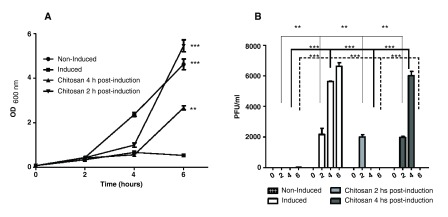
A. Growth curve: the E. coli C600ΔTOX:GFP strain was induced with ciprofloxacin and the optical density was measured at 600nm at 0, 2, 4 and 6 hours after induction. Non-induced culture of E. coli C600ΔTOX:GFP strain was used as control. Chitosan was added at 2 or 4 hours after induction. B. Bacteriophage ϕΔTOX:GFP titers: bacteriophage titers were determined at 0, 2, 4 and 6 hours post-induction. Chitosan was added at 2 or 4 h post-induction. * p<0.05.

Transduction of mammalian cells with ϕΔTOX:GFP

We previously reported the capacity of ϕΔTOX:GFP to transduce macrophages in vitro. To further evaluate the ability of chitosan to inhibit bacteriophage transduction, BHK-21 cells were transduced for 3 hours with ϕΔTOX:GFP, ϕΔTOX:GFP plus chitosan or ϕΔTOX:GFP previously treated with DNAse. Addition of DNAse would eliminate any free bacteriophage DNA in the bacterial lysates. As shown in Figure 2, ϕΔTOX:GFP DNA was detected by PCR in exposed mammalian cells, confirming that the virus was proficient to transduce this cell line. Similar results were also obtained in cells exposed to bacteriophages treated with DNAse ( Figure 2). However, no phage DNA was detected when BHK-21 cells were infected with ϕΔTOX:GFP incubated with chitosan, confirming the inactivating action of chitosan on ϕΔTOX:GFP ( Figure 2).

Figure 2. Detection of ϕΔTOX:GFP DNA in transfected mammalian cells.

Figure 2.

A. PCR on DNA extracted from BHK-21 cells: 24 hours after transduction, BHK-21 cells were washed and treated with Trypsin-EDTA solution. DNA was extracted and PCR was performed. Lane 1: Cells transfected with ϕΔTOX:GFP. Lane 2: Cells transfected with ϕΔTOX:GFP previously treated with chitosan. Lane 3: Cells transfected with ϕΔTOX:GFP previously treated with DNAse. Lane 4. Untreated cells. Lane 5. Positive PCR control (ϕΔTOX:GFP DNA). Lane 6. Negative PCR control. Lane 7. 1 kb ladder (Invitrogen).

GFP detection in mice inoculated with the lysogenic E. coli C600Δ TOX:GFP strain

To demonstrate the in vivo dissemination of ϕΔTOX:GFP, mice were infected with the lysogenic E. coli C600ΔTOX:GFP strain followed by gastrointestinal administration of ciprofloxacin. In order to evaluate the effect of chitosan in vivo, a group of mice was administered with chitosan 2 hours post-induction and a control group of uninfected mice was evaluated for auto-fluorescence background control in each organ. One day after infection, organs were harvested and examined for GFP expression. As shown in Figure 3, GFP was detected in the intestine, liver and, to a lesser extent, kidney of mice orally infected with the lysogenic E. coli C600ΔTOX:GFP strain and treated with ciprofloxacin. Remarkably, administration of chitosan 2 hours after infection caused a sharp decrease in GFP detection in organs of orally infected mice ( Figure 3, panels A and B). Moreover, positive detection of phages was observed in intestine homogenates and blood samples of infected mice after ciprofloxacin induction (data not shown). These results indirectly demonstrate that ϕΔTOX:GFP is released by the lysogenic bacterial E. coli strain and systemically spread and transduce cells in different mouse organs and tissues after oral infection and lytic induction. Another possibility is that, the bacteriophage could be taken by pinocytosis by eukaryotic cells, and, once inside the cell, GFP or Stx2 are expressed.

Figure 3. Detection of in vivo GFP expression in mice infected with the lysogenic E. coli C600ΔTOX:GFP strain using In Vivo Imaging System (IVIS).

Figure 3.

A. IVIS Representative image: ciprofloxacin was administered 2 hours post-infection to induce ϕΔTOX:GFP in vivo. Mice were treated with chitosan 2 hours after bacteriophage induction. All mice were sacrificed 24 hours post-infection and brains, hearts, lungs, livers, spleens, kidneys and intestines were harvested and analyzed by IVIS. Fluorescence intensity was recorded as photons/sec/cm 2, and the signal intensity represents the amount of GFP present. B. Graphic of fluorescence intensity on GFP-positive organs. Four animals per group were analyzed and the fluorescence intensity was quantified using Living Imaging 4.3.1 in Calipter Life Sciences.

Effect of chitosan on the mortality of mice orally inoculated with the EHEC EDL933W strain

In order to evaluate the in vivo effect of chitosan during the infection process, mice were intragastrically infected with a wild-type EHEC EDL933W strain, based on the model described by Brando and collaborators 10. Another mouse group was also treated with chitosan, intragastrically administered 2 hours post-infection, and survival was followed for one week. Partial protection was observed in mice treated with chitosan as demonstrated by the delay in the death time ( Figure 4). Mice infected with the EHEC EDL933W strain died 72 hours post-infection while mice infected with the same strain and subsequently treated with chitosan died 168 hours after infection.

Figure 4. Treatment with chitosan delays death of mice infected with the EHEC EDL933W strain.

Figure 4.

Mice were infected orally with the EHEC EDL933W strain. Controls did not receive chitosan (dots and broken line) and the experimental group received chitosan 2 hours post-infection (square and fill line). Survival rates were followed for one week.

Induction of ϕΔTOX:GFP strain by ciprofloxacin and chitosan effect

Induction of ϕΔTOX:GFP by ciprofloxacin and effect of chitosan in vitro. A. Growth curve: the E. coli C600DTOX:GFP strain was induced with ciprofloxacin and the optical density was measured at 600 nm at 0, 2, 4 and 6 hours after induction. Non-induced culture of E. coli C600DTOX:GFP strain was used as control. Chitosan was added at 2 or 4 hours after induction. B. Bacteriophage ϕΔTOX:GFP titers: bacteriophage titers were determined at 0, 2, 4 and 6 hours post-induction. Chitosan was added at 2 or 4 h post-induction. *p<0.05.

Discussion

Lambda bacteriophages are used as carriers in gene transfer and vaccine delivery experiments based on the capacity to in vivo transduce mammalian cells 11. Tyler and collaborators recently showed that prophage induction is required for renal disease and lethality in the EHEC mouse model, suggesting that free bacteriophages encoding Stx may play a direct role in the disease 4. Our results give a further support to that hypothesis and help understand why only small numbers of bacteria are usually capable to induce HUS in humans 3. If bacteriophages are induced in the gastrointestinal tract, infect different host cells, and promote Stx expression, a reduced number of bacteria would suffice to cause significant damage.

In previous reports, we showed that the native phage promoter controlling Stx expression is active in eukaryotic cells both in vitro 1 and in vivo 2 conditions. Based on these results, we sought to evaluate whether bacteriophages could be considered a target for treating STEC infections. To this aim, we used GFP as an in vitro and in vivo reporter of phage dissemination based on a lysogenic E. coli C600ΔTOX:GFP strain and following bacteriophage induction. GFP expression was observed in liver, intestine and kidney of mice orally infected with the lysogenic strain and subsequently exposed to phage inducing conditions by oral administration with ciprofloxacin. Of particular relevance was the observation that chitosan, a natural polysaccharide polycationic polymer, exerted a direct inactivation effect on ϕΔTOX:GFP in vitro and drastically reduced the detection of fluorescence in mice orally infected with the lysogenic E. coli C600ΔTOX:GFP strain. Our findings indicate that chitosan possesses strong anti-bacteriophage effects in vitro and probably also in vivo, as demonstrated with the lysogenic E. coli C600ΔTOX:GFP strain. This positively charged polymeric polysaccharide has been reported to inhibit other bacteriophages and probably acts through electrostatic interactions with negatively charged capsid proteins 6. Based on these effects we propose that chitosan could be a viable alternative for the treatment of STEC infections. Chitosan is already used in food and medicine, and it is harmless to humans, making it a cheap and safe option for this application.

The same chitosan protective effects were also observed in vivo based on mice infected with the wild-type EHEC EDL933W strain which is lysogenic for the same bacteriophage used to generate the E. coli C600ΔTOX:GFP strain. The fact that only partial protection was observed in mice infected with the E. coli C600ΔTOX:GFP strain and subsequently treated with chitosan may be due to the short half-life of the compound 12.

Altogether, these findings suggest a paradigm change on the role of bacteriophages in STEC infections, indicating that these bacteriophages have a pivotal role on the development of HUS. The present observations further suggest that prophylaxis and treatment of human bacterial infections carrying virulence factors on lysogenic bacteriophages could require targeting of the bacteriophages instead of, or as well as, the bacteria and toxins involved.

Data availability

F1000Research: Dataset 1. Induction of ϕΔTOX:GFP strain by ciprofloxacin and chitosan effect, 10.5256/f1000research.3718.d34269 13

Acknowledgments

We would like to acknowledge Dr. Alison A. Weiss for providing strain E. coli C600: ΔTOX-GFP.

Funding Statement

This work was supported by PICT 2411 from the Agencia Nacional de Promoción Científica y Tecnológica, Argentina (to L.V.B) and Fundação de Amparo à Pesquisa do Estado de São Paulo (FAPESP), Brazil (to L.C.S.F). LVB and PDG are members of the Research Career of CONICET (Consejo Nacional de Ciencia y Tecnología).

The funders had no role in study design, data collection and analysis, decision to publish, or preparation of the manuscript.

v2; ref status: indexed

References

  • 1.Bentancor LV, Bilen MF, Mejías MP, et al. : Functional capacity of Shiga-toxin promoter sequences in eukaryotic cells. PLoS One. 2013;8(2):e57128. 10.1371/journal.pone.0057128 [DOI] [PMC free article] [PubMed] [Google Scholar]
  • 2.Bentancor LV, Mejías MP, Pinto A, et al. : Promoter sequence of Shiga toxin 2 (Stx2) is recognized in vivo leading to production of biologically active Stx2. MBio. 2013;4(5):e00501–13. 10.1128/mBio.00501-13 [DOI] [PMC free article] [PubMed] [Google Scholar]
  • 3.Petruzziello-Pellegrini TN, Marsden PA: Shiga toxin-associated hemolytic uremic syndrome: advances in pathogenesis and therapeutics. Curr Opin Nephrol Hypertens. 2012;21(4):433–40. 10.1097/MNH.0b013e328354a62e [DOI] [PMC free article] [PubMed] [Google Scholar]
  • 4.Tyler JS, Beeri K, Reynolds JL, et al. : Prophage induction is enhanced and required for renal disease and lethality in an EHEC mouse model. PLoS Pathog. 2013;9(3):e1003236. 10.1371/journal.ppat.1003236 [DOI] [PMC free article] [PubMed] [Google Scholar]
  • 5.Kong M, Chen XG, Xing K, et al. : Antimicrobial properties of chitosan and mode of action: a state of the art review. Int J Food Microbiol. 2010;144(1):51–63. 10.1016/j.ijfoodmicro.2010.09.012 [DOI] [PubMed] [Google Scholar]
  • 6.Ly-Chatain MH, Moussaoui S, Vera A, et al. : Antiviral effect of cationic compounds on bacteriophages. Front Microbiol. 2013;4:46. eCollection 2013. 10.3389/fmicb.2013.00046 [DOI] [PMC free article] [PubMed] [Google Scholar]
  • 7.Kochkina ZM, Pospeshny G, Chirkov SN: [Inhibition by chitosan of productive infection of T-series bacteriophages in the Escherichia coli culture]. Mikrobiologiia. 1995;64(2):211–5. [PubMed] [Google Scholar]
  • 8.Gamage SD, Strasser JE, Chalk CL, et al. : Nonpathogenic Escherichia coli can contribute to the production of Shiga toxin. Infect Immun. 2003;71(6):3107–3115. 10.1128/IAI.71.6.3107-3115.2003 [DOI] [PMC free article] [PubMed] [Google Scholar]
  • 9.Zhang X, McDaniel AD, Wolf LE, et al. : Quinolone antibiotics induce Shiga toxin-encoding bacteriophages, toxin production, and death in mice. J Infect Dis. 2000;181(2):664–70. 10.1086/315239 [DOI] [PubMed] [Google Scholar]
  • 10.Brando RJ, Miliwebsky E, Bentancor L, et al. : Renal damage and death in weaned mice after oral infection with Shiga toxin 2-producing Escherichia coli strains. Clin Exp Immunol. 2008;153(2):297–306. 10.1111/j.1365-2249.2008.03698.x [DOI] [PMC free article] [PubMed] [Google Scholar]
  • 11.Lankes HA, Zanghi CN, Santos K, et al. : In vivo gene delivery and expression by bacteriophage lambda vectors. J Appl Microbiol. 2007;102(5):1337–49. 10.1111/j.1365-2672.2006.03182.x [DOI] [PMC free article] [PubMed] [Google Scholar]
  • 12.Soane RJ, Frier M, Perkins AC, et al. : Evaluation of the clearance characteristics of bioadhesive systems in humans. Int J Pharm. 1999;178(1):55–65. 10.1016/S0378-5173(98)00367-6 [DOI] [PubMed] [Google Scholar]
  • 13.Bentancor L, Ferreira LCS, Amorim JH: Induction of ϕΔTOX:GFP strain by ciprofloxacin and chitosan effect. F1000Research. 2014. Data Source [Google Scholar]
F1000Res. 2015 Jan 5. doi: 10.5256/f1000research.5299.r5927

Referee response for version 2

Mikael Skurnik 1

First of all I am sorry for the long delay in my getting back to this evaluation. While the authors have replied adequately to some of my comments there are some that are not. I will deal them below.

Major points:

Point 1. It is not enough to write the data of the dose response experiments in the reply. The results of the dose response experiments should be integrated into the main article.

Point 2. Inclusion of the mouse experiment (Fig. 4) in the article is not acceptable and should be deleted completely. In addition to having relatively small number of mice per group, the use of both male and female mice in the experiments is not appropriate as the responses can vary between the sexes. This and the small numbers of mice could be the reason for non-consistent killing results. In addition, it is not clear either from the authors' reply of from the text and the figure, how many mice were actually in the experiment. The figure 4 indicates that 50 % of both mouse groups died. How many mice was 50%, one or two? I have hard time to believe that two non-treated mice both died after 72 hr and two chitosan-treated both after 168 hr after infection and at the same time two mice in both groups survived. Therefore the mouse experiments in my opinion do not have any significance to one or other direction and should not be included in the present article.

Point 3. While the both controls the authors used are OK, they don't control the effect of ciprofloxacin in the experiment. Chitosan could also neutralize ciprofloxacin and thereby rescue the bacterial growth. The E. coli C600 strain is very widely used strain and should be easy to get. Preferentially it should be from the same source from where the lysogenic strains were obtained. I have the strain also in my strain collection and I can send it to the authors if they don't find it elsewhere.

Point 4. OK

Point 5. While the response seems OK, I don't find anywhere in the article the results and the additional figure. These should be integrated to the article.

Point 6.  Even though the C600 strain is not invasive, bacteria may be taken up by endocytosis and thereby enter the organs. The authors should exclude this possibility. While it would be difficult to detect the bacteria from intestinal tissue, other organs should be sterile.If the authors cannot see any bacterial growth from the sterile organs, that would be a sufficient control.

Point 7. See point 2 above.

All previous Minor points were all addressed adequately.

Please check carefully the text on page 4 right column top paragraph.

I have read this submission. I believe that I have an appropriate level of expertise to confirm that it is of an acceptable scientific standard, however I have significant reservations, as outlined above.

F1000Res. 2014 Dec 29. doi: 10.5256/f1000research.5299.r6885

Referee response for version 2

Cristina Ibarra 1

This is an interesting paper where the authors have examined the hypothesis that  the stx-carrying prophage upon induction in vivo are required for the development of HUS in the murine model. Eukaryotic host cells would transduce these phages, leading to increase of Stx production in the target organs. This would explain why very small numbers of bacteria are sufficient to cause HUS. The authors have also examined the anti-bacteriophage effects of chitosan in vivo and in vitro and demonstrated that chitosan has strong inhibitory effects on STEC bacteriophage and suggest that it could be a prophylactic for the prevention and treatment of HUS patients.

In general, the presented study is conducted straight forward, is written concisely and the applied methodology is adequate.

I recommend indexation of this article without changes.

I have read this submission. I believe that I have an appropriate level of expertise to confirm that it is of an acceptable scientific standard.

F1000Res. 2014 Sep 9. doi: 10.5256/f1000research.5299.r6090

Referee response for version 2

Raúl Raya 1

I consider the authors have satisfactorily answered all my comments and that the manuscript is acceptable.

Should the phrase:  “T he fact that only partial protection was observed in mice infected with the E. coli C600ΔTOX:GFP strain and subsequently treated with chitosan may be due to the short half-life of the compound.” instead be: “ The fact that only partial protection was observed in mice infected with the wild-type EHEC EDL933W strain and subsequently treated with chitosan may be due to the short half-life of the compound”?

The authors should indicate the concentration of DNAase used.

I have read this submission. I believe that I have an appropriate level of expertise to confirm that it is of an acceptable scientific standard.

F1000Res. 2014 Apr 17. doi: 10.5256/f1000research.3984.r4510

Referee response for version 1

Raúl Raya 1

The article written by Amorim et al. describes the anti-phage activity of chitosan on two variants (wild type and a derivative where the stx gene was replaced by the gfp gene) of the temperate Shiga-toxin producing phage EDL933W. The anti-phage activity was evaluated both in vivo and in vitro. The authors suggest that chitosan could be a viable alternative for the treatment of STEC infections.

Major:

Phage Induction/anti-phage effects of chitosan/Figure 1:

It seems that chitosan not only sequesters free-phage particles, but also stimulates the growth of uninduced cells (see induced cells treated with chitosan 2 hours post-induction reached higher final OD values). So, does chitosan inhibit the induction process of the temperate phage? Or, does chitosan also adsorb/inactivate ciprofloxacin? Even though in the Materials and Methods a “Bacteriophage inactivation assay” is described, no data is presented. A dose-response curve should be presented, to determine the phage binding (inactivation) capacity of chitosan.

In vivo experiments:

If the authors suggest that Stx phages, rather than bacterial cells, may be responsible for the development of the STEC infections, why they did not use purified phage in the vivo experiments? Does chitosan adsorb/inactivate the Shiga-toxins? If so, may it explain the delayed response observed in Figure 4 (“EDL933W plus chitosan”)?

Minors:

Abstract:

  • Provide a reference after “… plasmids encoding Stx under control of the native bacterial promoter.”

  • Change “ E. coli K12 strain” to “ E. coli C600 strain”.


Materials and Methods:

  • Delete “… was generously provided by Dr. Luis Carlos de Souza Ferreira, LDV-USP, Brazil.”, since Dr. Ferreira is one of the authors of the manuscript.

  • Check the sentence “…was generously provided by Dr. Alison Weiss”; it is repeated twice in the Materials and Methods, and also in the Acknowledgments.

  • Change “This is a non-pathogenic phage…” to “This is a non-pathogenic cell…”

  • Change “…complete DMEM medium” to “DMEM medium”. Or, if the “complete DMEM medium” contains 10% fetal bovine serum, change “…complete DMEM medium” for “…complete medium”.

  • Please, indicate how DNA was harvested.

  • Change “Tris pH8 50mM” to “Tris pH8 50 mM”.

  • Change “BALB/c mice were bred…” to ““BALB/c and DBA-2 mice were bred…”

  • Change “…under a 12-h light-dark…” to “…under a 12-hour light-dark…”.

  • Delete the sentence “Two-month old BALB/c mice … and GFP dissemination by IVIS was analyzed”. It is redundant.


Results:

  • Figure 1B: Should “Bacteriophage/ml” be “PFU/ml”? Why phage titers are so low?

  • Figure 1A: Change “hs” for “hours” or “h”.

  • Figure 2: Lanes 5 and 6 should read: “positive PCR control” and “negative PCR control”, respectively. In lane 78, indicate in the figure the kb values of the ladder.

  • Delete “viable” in “viable phages”. Were phages transduced or adsorbed to mammalian cells?

  • The sentence “Mice were orally challenged with a wild-type EDL933W” is not correct, since there was a direct delivery of bacterial cells into the stomach of mice.

Discussion:


  • Check “deliv-ery”

I have read this submission. I believe that I have an appropriate level of expertise to confirm that it is of an acceptable scientific standard, however I have significant reservations, as outlined above.

F1000Res. 2014 Aug 1.
Leticia Bentancor 1

Phage Induction/anti-phage effects of chitosan/Figure 1:

The higher final OD values determined on the cells treated with chitosan 2 hours post-induction, versus the OD value determined on un-induced cells, is not statistically significant. However, we tested if chitosan is capable of inactivating ciprofloxacin. Ciprofloxacin and chitosan were incubated for 10 minutes at room temperature with chitosan at 5mg/ml. After pre-incubation, the mix was used to induce bacteriophage excision. To see a more significant effect on bacteriophage excision, the induction was incubated overnight. The OD value measured showed a non-significant difference between non-induced culture and induced culture with the antibiotic pre-incubated with chitosan (the experiment was performed in triplicate).

On the other hand, purified bacteriophages were incubated with chitosan, and bacteriophage inactivation was observed with a lysis plaques assay. Bacteriophages were incubated for 10 minutes at room temperature with chitosan at 5mg/ml. After incubation, bacteriophage inactivation was evaluated. The bacteriophage solution containing a titer of 4x10 3pfu/ml was 100% inactivated after chitosan incubation. The assay was performed in triplicate. This result showed the capacity of chitosan to inactivate bacteriophage  in vitro.

Also, we did a dose-response curve of chitosan. We used 5mg/ml, 2.5mg/ml, and 1mg/ml of chitosan on purified bacteriophage solution. To evaluate it, the bacteriophage solution was incubated for 10 minutes at room temperature with the different doses of Chitosan and the bacteriophage inactivation was evaluated with a lysis plaques assay. Chitosan at 1mg/ml lost the inactivation activity on the bacteriophages. Chitosan at 5mg/ml and 2.5 mg/ml showed a 100% efficiency on bacteriophage inactivation, however, 1mg/ml of chitosan showed a loss of inactivation, showing between 5-10% of bacteriophage inactivation. This experiment was performed in triplicate.

In vivo  experiments:

Chitosan was analyzed  in vitro and  in vivo on fDTOX:GFP. Inactivation of fDTOX:GFP was observed  in vitro with a lysis plaques assay. On the other hand, a decrease of GFP was observed  in vivo by IVIS. These results shown that chitosan has the capacity to inactive bacteriophages in absence of Shiga-toxins. A direct action of chitosan on Shiga toxin was not evaluated in this work since we do not have purified Stx2 for such experiments. The authors are working on murine infection with fStx2, but the results obtained will be part of a new publication.

Abstract:

  • Provide a reference after “… plasmids encoding Stx under control of the native bacterial promoter.”

    The reference was provided. 

  • Change “ E. coli K12 strain” to “ E. coli C600 strain”.

    The change was made.

Materials and Methods:

  • Delete “… was generously provided by Dr. Luis Carlos de Souza Ferreira, LDV-USP, Brazil.”, since Dr. Ferreira is one of the authors of the manuscript.

    Answer:  We deleted “… was generously provided by Dr. Luis Carlos de Souza Ferreira, LDV-USP, Brazil.”

  • Check the sentence “…was generously provided by Dr. Alison Weiss”; it is repeated twice in the Materials and Methods, and also in the Acknowledgments.

     We deleted the sentence in the sub-section “ Transduction of eukaryotic cells” of material and methods section. 

  • Change “This is a non-pathogenic phage…” to “This is a non-pathogenic cell…”

    We changed “This is a non-pathogenic phage…” to “This is a non-pathogenic cell…”. However, this non-pathogenic cell produces the excision of a non-pathogenic phage.

  • Change “…complete DMEM medium” to “DMEM medium”. Or, if the “complete DMEM medium” contains 10% fetal bovine serum, change “…complete DMEM medium” for “…complete medium”.

    Complete DMEM medium contains 10% fetal bovine serum, so, we changed for the second option “…complete medium”.

  • Please, indicate how DNA was harvested.

    Cells were harvested using Trypsin-EDTA solution. After that, DNA was harvested from pellets by incubation with lysis solution described in material and methods. We included “by Trypsin-EDTA incubation” to clarify the procedure.

  • Change “Tris pH8 50mM” to “Tris pH8 50 mM”.

    The change was made.

  • Change “BALB/c mice were bred…” to ““BALB/c and DBA-2 mice were bred…”

     We changed “BALB/c mice were bred…”  for ““BALB/c and DBA-2 mice were bred…”

  • Change “…under a 12-h light-dark…” to “…under a 12-hour light-dark…”.

    The change was made.

  • Delete the sentence “Two-month old BALB/c mice … and GFP dissemination by IVIS was analyzed”. It is redundant.

    We have two different mouse models. First, we have the model used to analyze GFP dissemination in which we used two months old mice. Second, we have the model used to analyze protection effect in which we used immature mice. For this reason we clarify the model every time. Let me know if you consider that we need to delete the sentence   “Two-month old BALB/c mice … and GFP dissemination by IVIS was analyzed”.

Results:

  • Figure 1B: Should “Bacteriophage/ml” be “PFU/ml”? Why phage titers are so low?

    Bacteriophage/ml was changed to PFU/ml as reviewer suggested. See below. It is true that bacteriophage titers are low. An optimization for bacteriophage purification was done to obtain a higher titer of bacteriophage. The antibiotics used to induce C600DTOX:GFP was selected as an alternative for mitomycin C. The efficiency of bacteriophage induction is strain dependent.  Zhang and collaborators reported a titer equal to 1,3x10 5 pfu/ml using ciprofloxacin but they used pathogenic strain  E. coli O157:H7.  We also observed a higher titer inducing the EDL933W strain, for this reason we suppose that the low titer observed is dependent on the strain used.

  • Figure 1A: Change “hs” for “hours” or “h”.

     The change was made.

  • Figure 2: Lanes 5 and 6 should read: “positive PCR control” and “negative PCR control”, respectively. In lane 78, indicate in the figure the kb values of the ladder.

    The changes were made.

  • Delete “viable” in “viable phages”. Were phages transduced or adsorbed to mammalian cells?

    We deleted “viable” in “viable phages”. In this context, phages purified from tissue were detected by lysis plaque assay.

  • The sentence “Mice were orally challenged with a wild-type EDL933W” is not correct, since there was a direct delivery of bacterial cells into the stomach of mice.

     The sentence “Mice were orally challenged with a wild-type EDL933W” was change by “Mice were intragastrically infected with a wild-type EDL933W”.

Discussion:

  • Check “deliv-ery”

    We did not find deliv-ery in the Discussion section.

F1000Res. 2014 Mar 31. doi: 10.5256/f1000research.3984.r4170

Referee response for version 1

Mikael Skurnik 1

The paper by Amorim et al. deals with the role of bacteriophages in STEC-infections. The authors have earlier demonstrated that the stx genes can be expressed within eukaryotic cells, provided the prophage-carried stx-DNA is introduced there in naked form, i.e., in transfected plasmids. In the present work, the authors wanted to test/prove the hypothesis that the stx-carrying prophage upon induction in vivo could contribute to the toxin production. They also tested whether the polysaccharide chitosan has anti- stx phage effect. I have some major and minor points:

Major

  1. Bacteriophage inactivation assay: The experimental design of the bacteriophage inactivation assay uses only one concentration of chitosan. To demonstrate specificity, dose dependence should be demonstrated. In addition, the in vivo dose of chitosan was not indicated in the methods section (100 µl/mouse of 5 ml/ml chitosan was given orally to mice as indicated in the Effect of chitosan in vivo section.

  2. The mouse experiments were performed with too small a number of mice.

  3. Figure 1A of growth curves is missing a crucial control. What happens to E. coli C600 under the ciprofloxacin treatment?

  4. Figure 1B: The lack of the 4 hr column in chitosan 4h post-induction does not seem logical to me. There should be a ca 6000 PFU/ml column similar to that one in the induced 4hr sample. This discrepancy should be explained.

  5. Figure 2: the PCR experiment does not provide evidence of transduction. The definition of transduction is that DNA moves from one cell to another. PCR detects the phage DNA either free in the cell cytoplasm or packed in endocytosed phage particles. Therefore, the authors need to demonstrate that infective phage particles disappear from infected cells. The experiment also does not exclude the possibility that phage particles are just adsorbed on the cell surface.

  6. The experiment reported in figure 3 should also include bacterial counts from the organs as it is very likely that live E. coli bacteria, after a massive dose of 10 13 bacteria per mouse, end up in the organs. Therefore the authors should demonstrate that the GFP response is not from bacteria infected by the GFP-phages.

  7. The Figure 4 experiment was performed with only 4 mice. Such an experiment should not be shown in a publication. In addition, different chitosan doses should be tested here also.

Minor

  1. Introduction, paragraph 3: This statement on the low number of bacteria during infection should be backed up with a reference.

  2. Materials and Methods: Dr Alison Weiss is thanked twice for same strain. One time should be enough. In addition, in the acknowledgements she is thanked a third time. The bacterial strain designation in the latter is given differently than elsewhere in the text.

  3. Transduction of Eukaryotic Cells: C600ΔTOX:GFP is a bacterial strain, not a non-pathogenic phage.

  4. EHEC infection: The final concentration of CFU/100µl/mouse needs revision.

  5. Figure 2 legend: The path the sample takes in the gel is called the lane, not line.

I have read this submission. I believe that I have an appropriate level of expertise to confirm that it is of an acceptable scientific standard, however I have significant reservations, as outlined above.

F1000Res. 2014 Aug 1.
Leticia Bentancor 1

Major:

  1. A dose-response curve of chitosan was done. We used 5mg/ml, 2.5mg/ml and 1mg/ml of chitosan on purified bacteriophage. To evaluate it, the bacteriophage solution was incubated for 10 minutes at room temperature with the different doses of chitosan and the bacteriophage inactivation was evaluated with a lysis plaques assay. Chitosan at 1mg/ml lost the inactivation activity on the bacteriophages. Chitosan at 5mg/ml and 2.5 mg/ml showed a 100% efficiency on bacteriophage inactivation, however, 1mg/ml of chitosan showed a loss of inactivation, showing between 5-10% bacteriophage inactivation. This experiment was performed in triplicate.

    The effect of chitosan  in vivo was evaluated using a final concentration of 5mg/ml of chitosan solution. Each mouse received 100ml, so, the dose used was 500mg/mouse.

    The Material and Methods section was changed as follows:

    “Immature male and female DBA-2 infected, as described previously, were treated with 100ml of a chitosan solution at a concentration of 5 mg/ml (500 mg of chitosan per mouse) was orally administrated 2 hours after infection and survival was observed.”

  2. The experiment was shown as a preliminary result and this work it is a short communication. The model used has some experimental problems for the ages of mice used. The experiment was started using 6 mice per group, but some mice died after inoculation and not for the infection. For this reason, we had shown only 4 mice per group. We repeat the experiment, and again we have the same problem, however, we can see the same partial effect of chitosan  in vivo. To further analyze the effect observed, we will report the results on a new publication with more details. 

  3. The controls we used were:
    1. Non-induced  E. coli C600DTOX:GFP (as a negative control) in which we can observe the normal growth of bacteria without bacteriophage induction
    2. Induced  E. coli C600DTOX:GFP (as a positive control) in which we can observe how bacteriophage induction affect the growth of bacteria.

If you are thinking about  E. coli C600 in absence of lisogenic fDTOX:GFP, we do not have access to this strain. But, we think that the controls used are well done. As an observation, we can said that no significant change in the growth was observed on  E. coli Y1090 used for bacteriophage titration assay.

  1. The observation is right; we made an error in the graph. The values were checked and the correct value was added to the new graph.

  2. We agree with the definition of transduction, it is the process by which DNA is transferred from one cell to another by a virus. In our previous paper, we used the same definition to evaluate the capacity of fDTOX:GFP to transduce macrophages. In this case, we observed GFP expression and we concluded that macrophages were transduced by fDTOX:GFP. In this report, we did a different approach and we use PCR to detect bacteriophage DNA inside the cell.

    After your opinion, we did two assays. First, bacteriophages inside the cell were analyzed for titration assay. Second, as a preliminary data, fStx2 was used to transduce Vero cells, as a representative Stx2-susceptible cell line. 

    Infective bacteriophage particles were not detected on transduced cells. The assay was made using lysis plaque assay of cellular extracts.

    Results:

    In order to analyze the transduction by an additional method, Vero cells were transduced with fStx2 and cytotoxicity induced by Stx2 was evaluated by microscopy. Vero cells transduced with fStx2 ( panel A) showed a similar cytotoxicity to that of cells incubated with 1 CD50 of purified Stx2 ( panel B). No cytotoxic effects were observed in non-treated Vero cells ( panel D). Vero cells transduced with a M.O.I. = 0,0625 did not shown cytotoxic effect, demonstrating the specificity of the effect observed by fStx2 ( panel D).

    Materials and Methods:


    In vitro  evaluation of the capacity of Bacteriophage 933W to transduce Vero cells.

    E. coli EDL933W (ATCC 43895) was used to purify fStx2.  E. coli EDL933W strain was grown in Luria Broth (LB) overnight (ON) at 37°C under agitation. The ON culture was diluted to OD600nm = 0.1 in LB. Induction was carried out by adding ciprofloxacin to a final concentration of 40 ng/ml 8 in main text. Bacteria were incubated for 6 hours at 37°C under agitation. Cultures were then centrifuged at 5000 rpm for 15 minutes. The bacteriophage-containing supernatant was filtered with 0.2 mm filters, precipitated and purified. Briefly, supernatant was incubated on ice with a PEG-8000/NaCl solution for 30 minutes. After that, the solution containing bacteriophages was centrifugated and washed. The pellet was resuspended in STE buffer (1ml of Tris pH8, 0,2ml of 0,5M EDTA, 2ml of 5M NaCl, water up to 100ml). Phages at a multiplicity of infection (M.O.I) equal to 1 were added to Vero cells. Transduction of Vero cells was enhanced by centrifugation at 1000 x g for 10 min at room temperature. After 24 hours post transduction, cells were examined by microscopy using Nikon Eclipse TE2000 (NIS-Elements imaging software) equipped with a CCD camera. Dilutions of fStx2 were made to demonstrate specificity. Vero cells were incubated with purified Stx2 as positive control.

    Additional figureIn vitro evaluation of the capacity of Bacteriophage 933W to transduce Vero cells.  A. Vero cells transduced with fStx2 (M.O.I. = 1).  B. Vero cells incubated with purified Stx2.  C. Vero cells transduced with a M.O.I. = 0,0625.  D. Vero cells with not treatment.

  3. E. coli C600DTOX:GFP is not an invasive bacteria. Also,  E. coli O157:H7 is a non invasive strain; for this reason we do not check for bacteria in organs. Bacteria were checked only on lungs samples, just to see if the inoculation was right. Bacteria were not detected in lungs. The dose used was selected after a previous experiment in which we evaluated the sensibility of IVIS in our system. GFP is not the best fluorescent protein for IVIS system; so, we needed to use a high dose of bacteria.  As we described in this work, bacteriophages were detected by lysis plaques assay in intestine homogenates and blood samples of infected mice. It is important to do a highlight in the case of intestine sample, as it is very difficult to find  E. coli C600DTOX:GFP. First, because the huge amount of bacteria present in the sample, and also, because the bacteria lysis induced by bacteriophage excision.

  4. The experiment was shown as a preliminary result and this work it is a short communication. The model used has some experimental problems for the ages of mice used. The experiment was started using 6 mice per group, but some mice died after inoculation and not for the infection. For this reason, we had shown only 4 mice per group. We repeat the experiment, and again we have the same problem, however, we can see the same partial effect of chitosan  in vivo. To further analyze the effect observed, we will report the results on a new publication with more details. 

Minor:

  1. The statement “…very small numbers of bacteria are sufficient to induce HUS in humans…” is taking the data published recently, in which the authors demonstrated that a concentration of Stx2 as low as 10 fM is able to induce ribosome damage and to modulate selected cell signaling pathways that change cellular functions. If 10 fM of Stx2 is enough, very small numbers of bacteria should be sufficient to induce HUS (Petruzziello-Pellegrini & Marsden, 2012).

  2. We deleted the sentence “…was generously provided by Dr. Alison Weiss” in the sub-section “Transduction of eukaryotic cells” of material and methods section.

  3. We changed “This is a non-pathogenic phage…” to “This is a non-pathogenic cell…”. However, this non-pathogenic cell produces the excision of a non-pathogenic phage.

  1. The dose is correct. We used a dose of 3 × 10 12 CFU/mice in a volume of 100 ml.

  2. Line was changed for Lane.

Associated Data

    This section collects any data citations, data availability statements, or supplementary materials included in this article.

    Supplementary Materials

    Induction of ϕΔTOX:GFP strain by ciprofloxacin and chitosan effect

    Induction of ϕΔTOX:GFP by ciprofloxacin and effect of chitosan in vitro. A. Growth curve: the E. coli C600DTOX:GFP strain was induced with ciprofloxacin and the optical density was measured at 600 nm at 0, 2, 4 and 6 hours after induction. Non-induced culture of E. coli C600DTOX:GFP strain was used as control. Chitosan was added at 2 or 4 hours after induction. B. Bacteriophage ϕΔTOX:GFP titers: bacteriophage titers were determined at 0, 2, 4 and 6 hours post-induction. Chitosan was added at 2 or 4 h post-induction. *p<0.05.

    Data Availability Statement

    F1000Research: Dataset 1. Induction of ϕΔTOX:GFP strain by ciprofloxacin and chitosan effect, 10.5256/f1000research.3718.d34269 13


    Articles from F1000Research are provided here courtesy of F1000 Research Ltd

    RESOURCES