Abstract
2,3-Dihydro-5-methyl-3-([morpholinyl]methyl)pyrollo(1,2,3-de)-1,4-benzoxazinyl]-[1-naphthaleny]methanone [WIN 55,212-2 (WIN)] is a synthetic cannabinoid that inhibits RKO, HT-29 and SW480 cell growth, induced apoptosis, and downregulated expression of survivin, cyclin D1, epidermal growth factor receptor (EGFR), vascular endothelial growth factor (VEGF) and its receptor (VEGFR1). WIN also decreased expression of specificity protein (Sp) transcription factors Sp1, Sp3 and Sp4, and this is consistent with the observed downregulation of the aforementioned Sp-regulated genes. In addition, we also observed by RNA interference (RNAi) that the oncogenic cap protein eIF4E was an Sp-regulated gene also downregulated by WIN in colon cancer cells. WIN-mediated repression of Sp proteins was not affected by CB receptor antagonists or by knockdown of the receptor but was attenuated by the phosphatase inhibitor sodium orthovanadate or by knockdown of protein phosphatase 2A (PP2A). WIN-mediated repression of Sp1, Sp3 and Sp4 was due to PP2A-dependent downregulation of microRNA-27a (miR-27a) and induction of miR-27a-regulated ZBTB10 which has previously been characterized as an “Sp repressor”. The results demonstrate that the anticancer activity of WIN is due, in part, to PP2A-dependent disruption of miR-27a:ZBTB10 and ZBTB10-mediated repression of Sp transcription factors and Sp-regulated genes including eIF4E.
Keywords: Cannabinoids; WIN 55,212-2; downregulation; Sp; eIF4E
INTRODUCTION
There are three major classes of cannabinoids which include plant-derived compounds such as Δ(9)-tetrahydrocannabinol (THC) and cannabidiol (CBD), endogenous cannabinoids (anandamide and 2-arachidonylglycerol), and synthetic compounds that mimic the effects of cannabinoids (1, 2). Cannabinoids bind the CB1 and CB2 receptors which are differentially expressed in some but not all tissues, and there is also evidence that some cannabinoids also interact with other G-protein coupled receptors (GPCR) including the receptor transient receptor potential vanilloid type 1 (TRPV1) and GPR55 (3–5). THC is the main psychoactive cannabinoid found in the marijuana plant Cannabis sativa L; however, in addition to the psychoactive effects of THC, endocannabinoids and synthetic cannabinoids also play a role in energy metabolism, pain and inflammation, cardiovascular, musculoskeletal and respiratory disorders, and cancer, and opportunities for cannabinoid-based pharmacotherapies are extensive (2).
The anticancer activities of cannabinoids have been known for over 3 decades, and clinical trials for treatment of gliomas with cannabinoids have been reported (6). Cannabinoids inhibit growth, induce apoptosis, and exhibit antimetastatic and antiangiogenic activities in multiple cancer cell lines and inhibit tumor growth in in vivo mouse models (7–10). Cannabinoids are active in the tumor microenvironment and the effects of cannabinoids are complex and dependent on ligand structure and cell context, and the responses can also be CB (CB1 and/or CB2) receptor-dependent or -independent (7–10). For example, the synthetic mixed CB1 and CB2 receptor agonist 2,3-dihydro-5-methyl-3-([morpholinyl]methyl)pyrollo(1,2,3-de)-1,4-benzoxazinyl]-[1-naphthaleny]methanone [WIN 55, 212-2 (WIN)] inhibited gastric cancer cell growth and decreased VEGF expression and these responses were blocked in cells cotreated with WIN plus CB receptor antagonists (11), and similar CB receptor-dependent responses were observed in mantle cell lymphoma (12). In contrast, WIN induced phosphatase-dependent apoptosis in SW480 colon and LNCaP prostate cancer cells and these responses were inhibited by the phosphatase inhibitor sodium orthovanadate (SOV) but not by CB receptor antagonists or by CB receptor knockdown (13).
Treatment of cancer cells with cannabinoids activates or inactivates various kinases, ceramide synthesis and ceramide-mediated pro-apoptotic and stress-related genes, and downregulates expression of epidermal growth factor receptor (EGFR), survivin, cyclin D1, bcl-2 and vascular endothelial growth factor (VEGF) (11–26). Previous RNA interference studies have shown that these genes are also regulated by one or more of the specificity protein (Sp) transcription factors Sp1, Sp3 and Sp4 which are overexpressed in colon and other cancer cell lines and tumors; moreover, knockdown of Sp proteins by RNA interference results in growth inhibition and induction of apoptosis (27–39). Therefore, we hypothesized that the anticancer activity of cannabinoids such as WIN in colon cancer cells may also be due, in part, to downregulation of Sp proteins and pro-oncogenic Sp-regulated genes and this could also be related to the reported activation of phosphatases by cannabinoids in these cells (13). Results of this study show that WIN induced protein phosphatase 2A (PP2A)-dependent downregulation of Sp1, Sp3, Sp4 and Sp-regulated gene products including the pro-oncogenic cap protein eIF4e in colon cancer cells. These responses were CB receptor-independent and involved disruption of microRNA-27a (miR-27a)-mediated suppression of the zinc finger transcriptional repressor ZBTB10 which acts as an Sp repressor (31, 33–35).
MATERIALS AND METHODS
Chemicals, antibodies, and reagents
WIN 55,212-2 mesylate (1038), AM251 (1117) and AM630 (1120) were purchased from Tocris Bioscience (Ellisville, MO, USA). Tyrosine phosphatase inhibitor, sodium orthovanadate, was purchased from Calbiochem (La Jolla, CA, USA). Dithiothreitol (DTT) was obtained from Boehringer Mannheim Corp, (Indianapolis, IN). Gliotoxin was kindly provided by Dr. Alan Taylor (National Research Council of Canada, Halifax, NS, Canada). p-EIF4e (S209), eIF4e (9742S) and Cleaved poly (ADP-ribose) polymerase (PARP) (9541) antibodies were obtained from Cell Signaling (Danvers, MA). Lactacystin, glutathione (GSH) and antibodies for β-Actin (A1978), CB1 (C1233) and CB2 (WH0001269M1) receptors were purchased from Sigma-Aldrich (St. Louis, MO, USA). Antibodies for EGFR (1005), Sp3 (D-20) and Sp4 (V-20) were purchased from Santa Cruz Biotechnology (Santa Cruz, CA). PP2A Immunoprecipitation Phosphatase Assay Kit (17–313), Immobilon Western Chemiluminescent HRP substrate (WBKLS0500) and antibodies for Sp1 (07-645) and PP2A (05421) were purchased from EMD Millipore (Billerica, MA, USA). Cyclin D1 (2261-1) and survivin (2463-1) antibodies were purchased from Epitomics (Burlingame, CA). VEGFR1 (ab32152) antibody was purchased from Abcam (Cambridge, MA). Antibody for VEGF (209-403-B99) was purchased from Rockland Antibodies and Assays (Gilbertsville, PA). ZBTB10 (A303 258A) antibody was purchased from Bethyl Laboratories (Montgomery, TX). Apoptotic, Necrotic and Healthy Cells Quantification Kit (30018) was purchased from Biotium (Hayward, CA). In situ cell death detection POD kit (11684817910) was obtained from Roche (Mannhelm, Germany). Dulbecco’s Modified Eagle Medium (DMEM) and small interfering RNAs for CB1 and CB2 receptors, PP2A and ZBTB10 were obtained from Sigma (St. Louis, MO). Lipofectamine 2000 was purchased from Life Technologies (Grand Island, NY).
Cell lines
Human SW480 colon carcinoma cell line was provided by Dr. Stanley Hamilton (M.D. Anderson Cancer Center, Houston, TX) and non-transformed CCD-18Co colon cells were provided by Dr. Susanne Talcott (Texas A&M University, College Station, TX). RKO and HT-29 colon carcinoma cell lines were obtained from ATCC (Manassas, VA). Cells were purchased more than 6 months ago and were not further tested or authenticated by the authors. Cells were maintained at 37°C in the presence of 5% CO2 as described (13).
Cell proliferation assay
Colon cancer cells (1 × 10⁵ per well) were seeded in 12-well plates and allowed to attach for 24 hr. The medium was then changed to DMEM/Ham’s F-12 medium containing 2.5% charcoal-stripped FBS, and either vehicle (DMSO) or varying concentrations of WIN was added. Cells were then trypsinized and counted after 24 and 48 hr using a Coulter Z1 cell counter (Sykesville, MD). Each experiment was performed in triplicate, and results were expressed as means ± SE for each set of experiments.
Annexin V staining assay
SW480 and RKO cells were seeded in 2 well Nunc Lab-tek chambered coverglass plates from Thermo Scientific (Waltham, MA) and were allowed to attach for 24 hr. The medium was then changed to DMEM/Ham’s F-12 medium containing 2.5% charcoal-stripped FBS, and either DMSO or WIN was added. Cells were stained after 9 hr with FITC-Annexin-V, propidium iodide and DAPI dyes according to the manufacturer’s protocol and were visualized under EVOS fl, fluorescence microscope, from Advanced Microscopy Group (Bothell, WA) using a multiband filter set for FITC, rhodamine and DAPI. The proportion of apoptotic cells was determined by the amount of green fluorescence observed.
Terminal deoxyribonucleotide transferase-mediated nick-end labeling (TUNEL) assay
HT-29 (7 × 10⁴) were seeded in four-chambered glass slides and left overnight to attach. After treatment with WIN for 48 hr, the in situ cell death detection POD kit was used for the terminal deoxyribonucleotide transferase-mediated nick-end labeling (TUNEL) assay according to the manufacturer’s protocol. The percentage of apoptotic cells was determined by counting stained cells from eight fields, each containing 50 cells.
Western blot analyses
Colon cancer cells were seeded in DMEM/Ham’s F-12 medium and were allowed to attach for 24 hr. Cells were treated with either DMSO or WIN for indicated time periods or pretreated with the proteasome inhibitors, antioxidants, phosphatase inhibitors and cannabinoid receptor antagonists, and then treated with WIN. Cells were lysed and analyzed by western blots as described (13).
Small inhibitory RNAs (siRNA) interference assay
Colon cancer cells were seeded (2 × 10⁵ per well) in six-well plates in DMEM/Ham’s F-12 medium supplemented with 2.5% charcoal stripped FBS without antibiotics and left to attach for 24 hr. siRNAs specific for CB1 and CB2 receptors, PP2A and ZBTB10 along with iLamin/iGL2 as control were transfected using Lipofectamine 2000 reagent according to the manufacturer’s protocol.
Quantitative real-time PCR
SW480 colon cancer cells were plated (2 × 10⁵) and left to attach for 24 hr. Cells were transfected with siRNAs for PP2A and then treated with either DMSO or WIN for 24 and 48 hr. MicroRNA was isolated using the mirVana miRNA isolation kit from Ambion-Life Technologies (Grand Island, NY) according to the manufacturer’s protocol. cDNA was prepared using the Taqman MicroRNA Reverse Transcription kit and was subjected to quantitative real-time PCR with specific primers for miR27a using the Taqman Universal PCR Master Mix from Applied Biosystems in the CFX384 Real-Time PCR Detection System from Biorad (Hercules, CA).
PP2A Phosphatase assay
Cells were seeded (3 × 10⁵) and left to attach for 24 hr. Cells were treated with DMSO, okadaic acid, and 5 and 7.5 μM WIN. Cells were harvested and lysed using the high salt lysis buffer. The lysates were then subjected to buffer exchange using the Zeba Desalt Spin Columns (8981) from Thermo Scientific to eliminate any contaminating phosphates that could skew the experimental results. The lysates were then immunoprecipitated with anti-PP2A, C subunit, antibody and were incubated in a mixture of diluted phosphopeptide and serine/threonine assay buffer for 10 min at 30°C in a shaking incubator. The phosphatase activity was determined by addition of malachite green dye and comparing the absorbance between controls and treatment at 650 nm in a plate reader.
RESULTS
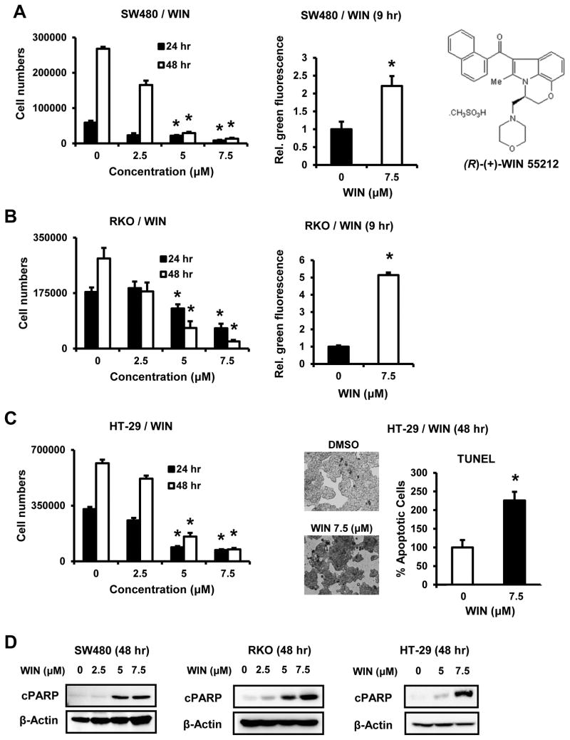
Treatment of SW480, RKO and HT-29 colon cancer cells with 2.5, 5.0 and 7.5 μM WIN for 24 or 48 hr significantly inhibited cell proliferation at the two higher concentrations (Fig. 1A – 1C), whereas minimal growth inhibition was observed for non-transformed CCD-18Co colon cells (Suppl. Fig. 1A). In addition, WIN also induced Annexin V staining in SW480 and RKO cells within 9 hr after treatment, indicating rapid induction of early apoptosis, whereas Annexin V was not induced in HT-29 cells at the early time point (data not shown). In contrast, TUNEL staining was increased in HT-29 cells treated with WIN for 48 hr, indicating differences between the timing of WIN-induced apoptosis in SW480/RKO vs. HT-29 cells. The red staining in RKO cells treated with 7.5 μM WIN was associated with dead cells and necrosis (Fig. 1B). Figure 1D shows that treatment with WIN for 48 hr also induced PARP cleavage in all three colon cancer cell lines as previously reported for WIN in SW480 cells (13).
Figure 1.
WIN inhibits colon cancer cell growth, induces apoptosis and PARP cleavage. SW480 (A), RKO (B) and HT-29 (C) cells were treated with DMSO and 2.5–7.5 μM WIN for 24 and 48 hr and counted, or treated with DMSO and 7.5 μM of WIN for 9 hr (A and B) and 48 hr (C) and effects on Annexin V (SW480 and RKO) and TUNEL (HT-29) staining was determined. Results are expressed as means ± SE for at least three separate determinations, and significant (p < 0.05) growth inhibition or induction of apoptosis by by WIN is indicated (*). (D) Cells were treated with DMSO and 2.5–7.5 μM of WIN for 48 hr and whole cell lysates were examined for expression of cleaved PARP by western blots. β-Actin served as a loading control for all western blots.
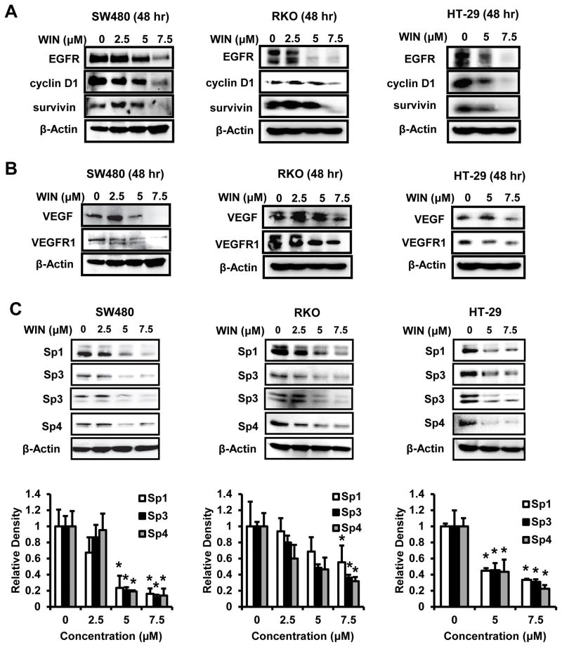
WIN-induced growth inhibition and apoptosis was also accompanied by decreased expression of growth promoting (EGFR, cyclin D1) and survival (survivin) genes products in SW480, RKO and HT29 cells (Fig. 2A). Treatment of SW480, RKO and HT29 cells with 2.5 – 7.5 μM WIN decreased levels of VEGF and VEGFR1 protein in the 3 cell lines, although the effects in HT-29 cells were less than observed in the other two cell lines (Fig. 2B). Treatment of SW480, RKO and HT-29 cells with 2.5, 5.0or 7.5 μM WIN for 48 hr decreased expression of Sp1, Sp3 and Sp4 proteins as indicated by quantitation of the band intensities (relative to β-actin) (Fig. 2C). Both the high and low molecular weight forms of Sp3 were observed as previously reported in colon cancer cell lines (37–39). This is the first example of a synthetic cannabinoid decreasing expression of Sp proteins and Sp-regulated gene products in cancer cells; however, we have recently reported that betulinic acid (BA), a triterpenoid compound that also decreases Sp1, Sp3 and Sp4 protein levels in multiple cancer cell lines, exhibits CB receptor agonist activity (35).
Figure 2.
WIN downregulates Sp-regulated proliferative, survival (A) and angiogenic (B) gene products and Sp proteins (C) in SW480, RKO and HT-29 cells. Cells were treated with 2.5–7.5 μM of WIN for 48 hr, whole cell lysates were analyzed by western blots, and expression of Sp1, Sp3 and Sp4 protein was quantitated (C) relative to βactin (levels in the DMSO group were set at 1.0). Results are expressed as means ± SE (three replicate experiments), and significantly (p < 0.05) decreased proteins are indicated (*). Data in Figures 2A and 2B were from the same experiment.
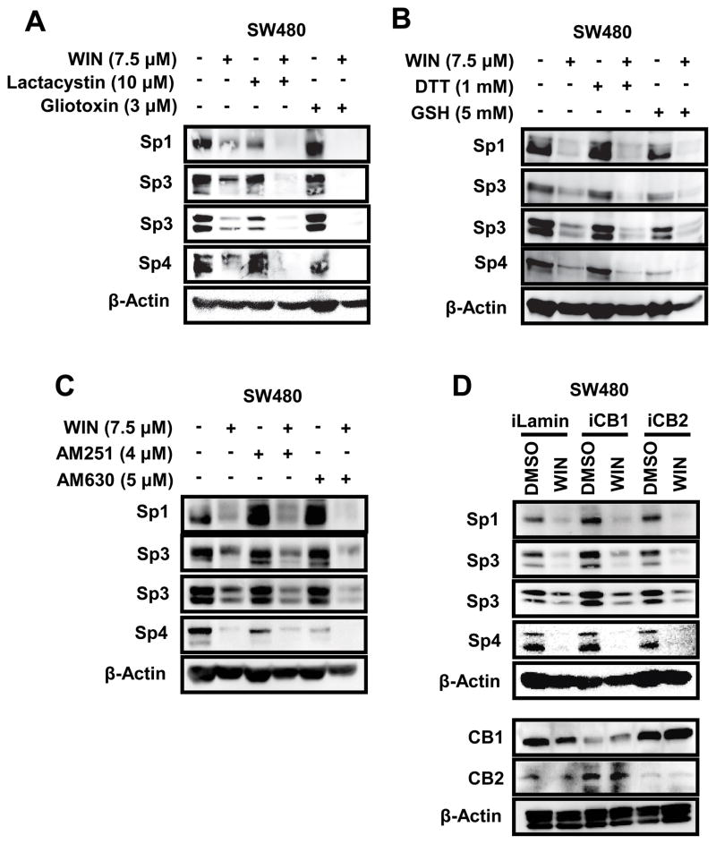
Drug-induced downregulation of Sp transcription factors has been linked to activation of proteasomes or induction of ROS (31, 36–39). Using SW480 cells as a model, we show that WIN-induced downregulation of Sp1, Sp3 and Sp4 was not reversed in cells cotreated with WIN plus the proteasome inhibitors lactacystin or gliotoxin (Fig. 3A). Moreover, treatment of SW480 cells with WIN in combination with the antioxidants DTT or GSH also did not inhibit WIN-induced downregulation of Sp1, Sp3 or Sp4 proteins (Fig. 3B), whereas BA and GT-094 (a nitro-NSAID)-induced downregulation of Sp1, Sp3 and Sp4 in SW480 cells was significantly inhibited after cotreatment with the antioxidants (38, 39). The CB1 and CB2 receptor antagonists AM251 and AM560 had minimal effects on Sp protein expression and, in combination with WIN, did not inhibit WIN-induced downregulation of Sp1, Sp3 or Sp4 proteins (Fig. 3C). Moreover, similar results were observed in SW480 cells after knockdown of the CB1 (iCB1) or the CB2 (iCB2) receptor by RNA interference (Fig. 3D), demonstrating that the effects of WIN on downregulation of Sp transcription factors was CB receptor-independent.
Figure 3.
Effects of proteasome inhibitors, antioxidants, cannabinoid receptor antagonists, and cannabinoid receptor knockdown on WIN-mediated downregulation of Sp proteins. SW480 cells were pre-treated with lactacystin (10 μM) or gliotoxin (3 μM) (A), DTT (1 mM) or GSH (5 mM) (B), AM251 (4 μM) and AM630 (5 μM) (C) for 45 min followed by treatment with 7.5 μM of WIN for 48 hr, and whole cell lysates were analyzed by western blots. (D) Cells were transfected with siRNAs against CB1 and CB2 receptors, followed by treatment with WIN for 48 hr, and whole cell lysates were analyzed by western blots. iLamin was used as a control oligonucleotide.
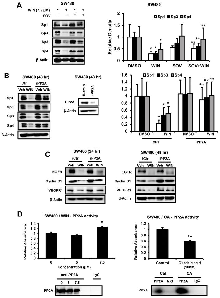
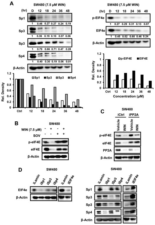
WIN induced several phosphatase mRNAs in SW480 cells and the phosphatase inhibitor sodium orthovanadate (SOV) partially blocked WIN-induced PARP cleavage (13) and, therefore, we investigated the effects of SOV on WIN-induced downregulation of Sp1, Sp3 and Sp4 proteins using SW480 cells as a model. Figure 4A shows that WIN-induced downregulation of Sp1, Sp3 and Sp4 was reversed after cotreatment with SOV and these results are consistent with the inhibition of WIN-induced PARP cleavage by SOV (13) since knockdown of Sp1 (by RNAi) also enhances PARP cleavage (31). A recent study showed that α-tocopherol succinate-induced downregulation of Sp1 in prostate cancer cells was PP2A-dependent (40) and western blot analysis of whole cell lysates from SW480 cells transfected with a non-specific oligonucleotide (iCtrl) or a specific oligonucleotide that targets PP2A (iPP2A) and treated with 7.5 μM WIN or DMSO showed that WIN-mediated downregulation of Sp1, Sp3 and Sp4 proteins was significantly attenuated in cells transfected with iPP2A (Fig. 4B). Similar results were also observed in RKO cells with some differences in the relative effectiveness of PP2A knockdown (Suppl. Fig. 1B). PP2A knockdown also attenuated WIN-induced downregulation of cyclin D1 and VEGFR1 but had minimal effect on EGFR protein levels (Fig. 4C), suggesting that decreased expression of EGFR in SW480 cells treated with WIN was phosphatase- and Sp-independent.
Figure 4.
Effects of phosphatase inhibition on WIN-dependent downregulation of Sp proteins and Sp-regulated gene products. (A) Cells were pre-treated with 0.35 mM SOV for 45 min followed by treatment with 7.5 μM of WIN for 48 hr, and whole cell lysates were analyzed by western blots. Cells were transfected with siRNA against phosphatase PP2A (iPP2A), followed by treatment with WIN for 48 hr, and whole cell lysates were examined for expression of Sp1, Sp3, Sp4 and PP2A proteins (B) and Sp-regulated gene products (C) by western blot analysis. Quantitation of changes in Sp1, Sp3 and Sp4 (A and B) relative to β-actin were determined from three replicate experiments; significant (p < 0.05) decreases (*) or attenuation of the response (**) are indicated. (D) Cells were treated with DMSO, 5 and 7.5 μM of WIN, and PP2A activity was determined in the absence and presence of the phosphatase inhibitor okadaic acid. Results are expressed as means ± SE for at least three separate determinations, and significant (p < 0.05) induction (*) or inhibition (**) by okadaic acid is indicated. Lysates used for phosphatase assay were analyzed by western blots to determine expression of PP2A protein.
We also used an in vitro assay to confirm that WIN induced PP2A activity since previous studies did not observe induction of PP2A mRNA levels in SW480 cells treated with WIN (13). PP2A activity in SW480 was measured using the PP2A Immunoprecipitation Phosphatase Assay Kit from Millipore. Treatment of SW480 cells with 7.5 μM WIN induced an approximate 30% increase in PP2A activity and this was comparable to the induction of PP2A phosphatase activity by metformin (41). Figure 4D shows that 10 nM okadaic acid significantly inhibited PP2A activity in SW480 cells (positive control). Cell lysates used for the phosphatase activity assays were then subjected to western blot analysis and probed with PP2A antibody to confirm the presence of PP2A in the cell lysates (Fig. 4D).
PP2A decreases expression of the phosphorylated form of the pro-oncogenic cap protein eIF4E (42) (p-eIF4E) and therefore the time-dependent effects of 7.5 μM WIN on expression of Sp1, Sp3, Sp4, p-eIF4E and eIF4E proteins were investigated (Fig. 5A). Sp1, Sp4 and Sp3 (high molecular weight forms) expression was significantly decreased after 12 hr, whereas the lower molecular weight forms of Sp3 were more slowly decreased over the 48 hr period. p-eIF4E and eIF4E proteins were significantly decreased within 12 hr after treatment with WIN and continued to decrease (over 48 hr); the decrease of eIF4E (total protein) was more gradual than observed for the phospho-protein. Treatment of SW480 cells with WIN alone or in combination with SOV showed that the phosphatase inhibitor blocked WIN-induced effects on p-eIF4E and eIF4E, but not PP2A (Fig. 5B), demonstrating that WIN-induced downregulation of p-eIF4E, eIF4E, Sp1, Sp3 and Sp4 was phosphatase dependent. The role of PP2A in this process was investigated by RNAi, and WIN-mediated repression of p-eIF4E and eIF4E (total) proteins was significantly inhibited by knockdown of PP2A (Fig. 5C), demonstrating a role for PP2A in mediating downregulation of eIF4E and peIF4E and this parallels results observed for phosphatase-dependent downregulation of Sp transcription factors (Fig. 4), suggesting possible cross-regulation of eIF4E and Sp transcription factors. WIN also decreased PP2A protein levels after treatment for 48 hr compared to 24 hr (Fig. 4D) and this is currently being investigated. Knockdown of Sp1 (iSp1), Sp3 (iSp3) and Sp4 (iSp4) in SW480 cells showed that iSp1 and, to a lesser extent, iSp3 also decreased eIF4E protein expression (Fig. 5D), whereas Sp4 knockdown had minimal effects, suggesting that in SW480 cells eIF4E expression is primarily regulated by Sp1. The knockdown of the Sp transcription factors was specific for Sp4 and Sp3; however, iSp1 also decreased expression of Sp4 as previously reported, indicating that Sp4 is an Sp1-regulated gene in SW480 cells (43). In contrast, knockdown of eIF4E by RNAi decreased expression of the targeted protein and slightly increased expression of Sp1, Sp3 or Sp4 proteins, suggesting that cross-regulation between Sp transcription factors and eIF4E is unidirectional. A comparable experiment was carried out in RKO cells; iSp1, iSp3 and iSp4 decreased eIF4E protein expression and the combination of all 3 oligonucleotides caused a marked decrease of eIF4E protein (Suppl. Fig. 1C), indicating that eIF4E is an Sp-regulated gene in colon cancer cells.
Figure 5.
Effects of WIN and Sp protein knockdown on total and phospho-eIF4E in SW480 cells. (A) Cells were treated with DMSO or 7.5 μM WIN for 12, 18, 24, 36 and 48 hr, and whole cell lysates were analyzed by western blots, and levels of expression relative to DMSO (set at 1.0) are indicated. Cells were pretreated with SOV for 45 min (B) or transfected with siRNA against PP2A (C), followed by treatment with WIN for 48 hr, and whole cell lysates were analyzed by western blots. (D) SW480 cells were transfected with various siRNA and whole cell lysates were analyzed for expression of Sp proteins and eIF4E by western blots. iLamin was used as a control oligonucleotide.
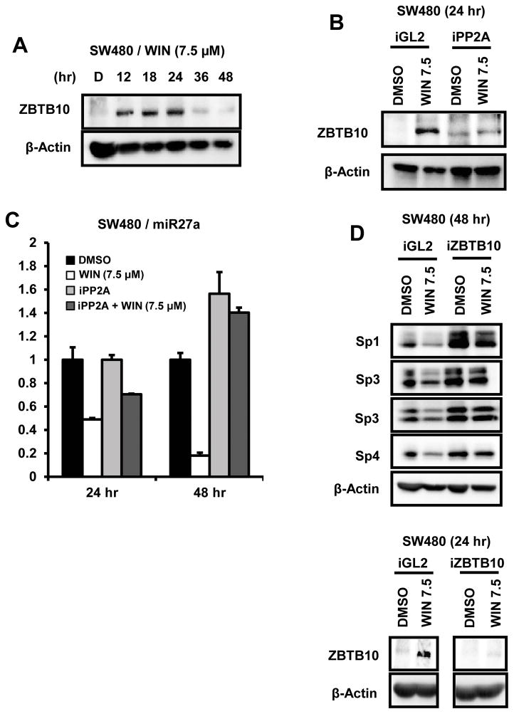
Drug-induced proteasome-independent downregulation of Sp1, Sp3 and Sp4 has been related to transcriptional repression due to downregulation of miR-27a and miR-20a/miR-17-5p which regulate the “Sp repressors” ZBTB10 and ZBTB4 (34, 44). Treatment of SW480 cell with 7.5 μM WIN induced ZBTB10 protein within 12 hr and induction was high after 24 hr but markedly decreased after 36 and 48 hr (Fig. 6A). In contrast, induction of ZBTB4 protein was not observed (data not shown). SW480 cells were treated with 7.5 μM WIN and also transfected with iGL2 (control) or iPP2A oligonucleotides, and the induction of ZBTB10 in the control cells was significantly decreased after knockdown of PP2A (Fig. 6B). In a parallel experiment, WIN also decreased expression of miR-27a after treatment for 24 or 48 hr and, after knockdown of PP2A by RNAi, downregulation of miR-27a by WIN was attenuated (Fig. 6C). The role of WIN-induced ZBTB10 on suppression of Sp proteins was confirmed in SW480 cells treated with WIN and transfected with iGL2 (control) or iZBTB10 oligonucleotides in an RNAi assay; knockdown of ZBTB10 inhibited downregulation of Sp1, Sp3 and Sp4 by WIN (Fig. 6D). We observed that WIN also induced ZBTB10 in RKO cells and this response was attenuated in cells transfected with iPP2A (RNAi), and knockdown of ZBTB10 by RNA also inhibited WIN-mediated suppression of Sp1, Sp3 and Sp4 proteins in RKO cells (Suppl. Figs. 1D and 1E). These results demonstrate that WIN-mediated downregulation of Sp proteins in SW480 cells is due to activation of PP2A and PP2A-dependent disruption of the miR-27a:ZBTB10 which results in induction of ZBTB10 and ZBTB10-dependent repression of Sp proteins and Sp-regulated genes. The miR-27a promoter contains binding sites for both c-Myc and NFκB (45) which are Sp-regulated genes in some cell lines (30, 31, 33); however, knockdown of Myc or p65 by RNA interference did not decrease miR-27a expression (Suppl. Fig. 2) and, currently, we are investigating the mechanism of phosphatase-induced downregulation of miR-27a.
Figure 6.
WIN modulates miR27a and ZBTB10 expression. (A) SW480 cells were treated with DMSO or 7.5 μM of WIN for 12, 18, 24, 36 and 48 hr, and whole cell lysates were analyzed for ZBTB10 protein by western blots. SW480 cells were transfected with siRNA against PP2A, followed by treatment with 7.5 μM WIN for 24 and 48 hr. Whole cell lysates were analyzed by western blot analysis for ZBTB10 expression (B) and miR27a levels (C) were determined by qPCR as described in the Materials and Methods. (D) SW480 cells were transfected with siRNA against ZBTB10 followed by treatment with 7.5 μM WIN for 24 and 48 hr, and whole cell lysates were analyzed by western blots. Results for RNA expression are expressed as means ± SE for at least three separate determinations, and significant (p < 0.05) inhibition (*) by WIN is indicated and reversal of these effects by iPP2A (**) are indicated.
DISCUSSION
Sp1, Sp3 and Sp4 proteins are highly expressed in multiple cancer cell lines and tumors, whereas levels of these transcription factors are low to non-detectable in non-tumor tissue (29–33) and these observations are consistent with reports that Sp1 expression in rodent and human tissues decreases with age (46, 47). The pro-oncogenic activity of Sp proteins is primarily due to Sp-regulated genes which include several that play pivotal roles in cancer cell proliferation [cyclin D1, c-Met, epidermal growth factor (EGFR)], survival (survivin, bcl-2), angiogenesis [vascular endothelial growth factor (VEGF) and its receptors (VEGFR1 and VEGFR2)], and inflammation (NFκB, p65). Thus, Sp transcription factors are an excellent example of non-oncogene addiction by cancer cells (48) and therefore an important target for mechanism-based anticancer drugs. Reports from different laboratories have identified diverse agents that decrease expression of Sp transcription factors and these include various phytochemical anticancer compounds including BA and their synthetic analogs, NSAIDs, bortezimob, α-tocopherol succinate, arsenic trioxide and other ROS inducers (29–40, 49).
BA decreases expression of Sp1, Sp3 and Sp4 in prostate, colon, bladder and breast cancer cell lines (32, 35, 37, 38). Results illustrated in Figures 1 and 2 confirm that the cannabinoid WIN also inhibited growth and induced apoptosis in SW480, RKO and HT-29 colon cancer cells and downregulated Sp1, Sp3, Sp4 and Sp-regulated genes. Since knockdown of Sp1 or multiple Sp transcription factors alone in cancer cells inhibits growth and induces apoptosis (29–32), then the anticancer activity of WIN is due, in part, to downregulation of Sp proteins. However, in contrast to BA and other agents that downregulate Sp transcription factors, WIN-induced Sp downregulation was proteasome- and ROS-independent and knockdown or inhibition of CB1 or CB2 receptors did not affect this response in SW480 cells (Fig. 3). The receptor-independent effects of WIN were consistent with previous studies showing that the anticancer activity of WIN was both receptor-dependent and -independent (11–13).
We recently reported that WIN-induced apoptosis in SW480 and LNCaP cells is inhibited by the phosphatase inhibitor SOV but not by CB receptor knockdown and WIN also induces multiple phosphatase mRNAs (13). These results, coupled with a report that PP2A plays a role in α-tocopherol succinate-induced downregulation of Sp1 in prostate cancer cells (40) prompted us to investigate the effects of SOV and PP2A knockdown on WIN-induced repression of Sp proteins. Both SOV and PP2A knockdown attenuated WIN-mediated downregulation of Sp1, Sp3, Sp4 and Sp-regulated gene products (Fig. 4), confirming a role for PP2A and possibly other identified phosphatases in mediating the anticancer activities of WIN. An important proteasome-independent pathway for drug-induced downregulation of Sp1, Sp3 and Sp4 involves induction of the transcriptional repressors ZBTB10 and ZBTB4 which exhibit low expression in cancer cell lines due to their regulation (repression) by miR-27a and miR-20a/miR-17-5p, respectively (38, 39, 44, 49). WIN did not induce ZBTB4 in SW480 cells; however, a time-dependent induction of ZBTB10 protein was observed (Fig. 6A) and this response was abrogated after knockdown of PP2A (Fig. 6B), and these results were paralleled by PP2A-dependent repression of miR-27a (Fig. 6C). Moreover, since knockdown of ZBTB10 by RNAi blocks WIN-mediated downregulation of Sp1, Sp3 and Sp4 proteins (Fig. 6D) and since miR-27a antogomirs and ZBTB10 expression decrease Sp protein expression in colon cancer cells (49), it is clear that disruption of miR-27a:ZBTB10 is critical for the observed responses, and results of c-Myc and p65(NFκB) silencing did not decrease miR-27a expression (Suppl. Fig. 2). The mechanism of PP2A-mediated effects on miR-27a:ZBTB10 are unknown and are currently being investigated.
The induction of PP2A activity by WIN suggested that WIN may also regulate phosphorylation of the important cap protein eIF4E since it has been reported that PP2A dephosphorylates p-eIF4E which has been characterized as a pro-oncogenic phospho-protein (42). WIN clearly decreased expression of p-eIF4E; however, this was unexpectedly accompanied by downregulation of eIF4E (total protein). Since eIF4E and Sp transcription factors regulate expression of several genes in common (e.g. cyclin D1 and VEGF), we used RNAi to show for the first time that eIF4E is an Sp-regulated gene in colon cancer cells (Fig. 5D). This observation extends the number of pro-oncogenic factors that are regulated by Sp1, Sp3 and Sp4 in cancer cells emphasizing the potential clinical applications of drugs that target Sp proteins. Previous studies show that other cannabinoids such as cannabidiol, other synthetic cannabinoids, and endocannabinoids inhibit colon cancer cell and tumor growth (22–26), and in vivo studies show that loss of the CB1 receptor enhances intestinal adenoma growth in APCmin/+ mice (23). The effects of these compounds on Sp transcription factors and eIF4E expression and the role of the CB receptors in mediating these responses are currently being investigated.
Supplementary Material
Acknowledgments
Funding: This research was supported by the National Institutes of Health (R01-CA136571) (S. Safe) and Texas AgriLife (S. Safe).
Footnotes
Disclosures/Conflicts of Interest: None
References
- 1.Bernadette H. Cannabinoid therapeutics: high hopes for the future. Drug Discov Today. 2005;10:459–62. doi: 10.1016/S1359-6446(05)03417-3. [DOI] [PubMed] [Google Scholar]
- 2.Pacher P, Batkai S, Kunos G. The endocannabinoid system as an emerging target of pharmacotherapy. Pharmacol Rev. 2006;58:389–462. doi: 10.1124/pr.58.3.2. [DOI] [PMC free article] [PubMed] [Google Scholar]
- 3.Pertwee RG. The pharmacology of cannabinoid receptors and their ligands: an overview. Int J Obes (Lond) 2006;30 (Suppl 1):S13–8. doi: 10.1038/sj.ijo.0803272. [DOI] [PubMed] [Google Scholar]
- 4.Maccarrone M, Lorenzon T, Bari M, Melino G, Finazzi-Agro A. Anandamide induces apoptosis in human cells via vanilloid receptors. Evidence for a protective role of cannabinoid receptors. J Biol Chem. 2000;275:31938–45. doi: 10.1074/jbc.M005722200. [DOI] [PubMed] [Google Scholar]
- 5.Ryberg E, Larsson N, Sjogren S, Hjorth S, Hermansson NO, Leonova J, et al. The orphan receptor GPR55 is a novel cannabinoid receptor. Br J Pharmacol. 2007;152:1092–101. doi: 10.1038/sj.bjp.0707460. [DOI] [PMC free article] [PubMed] [Google Scholar]
- 6.Guzman M, Duarte MJ, Blazquez C, Ravina J, Rosa MC, Galve-Roperh I, et al. A pilot clinical study of Delta9-tetrahydrocannabinol in patients with recurrent glioblastoma multiforme. Br J Cancer. 2006;95:197–203. doi: 10.1038/sj.bjc.6603236. [DOI] [PMC free article] [PubMed] [Google Scholar]
- 7.Guzman M. Cannabinoids: potential anticancer agents. Nat Rev Cancer. 2003;3:745–55. doi: 10.1038/nrc1188. [DOI] [PubMed] [Google Scholar]
- 8.Hall W, Christie M, Currow D. Cannabinoids and cancer: causation, remediation, and palliation. Lancet Oncol. 2005;6:35–42. doi: 10.1016/S1470-2045(04)01711-5. [DOI] [PubMed] [Google Scholar]
- 9.Sarfaraz S, Adhami VM, Syed DN, Afaq F, Mukhtar H. Cannabinoids for cancer treatment: progress and promise. Cancer Res. 2008;68:339–42. doi: 10.1158/0008-5472.CAN-07-2785. [DOI] [PubMed] [Google Scholar]
- 10.Flygare J, Sander B. The endocannabinoid system in cancer-potential therapeutic target? Semin Cancer Biol. 2008;18:176–89. doi: 10.1016/j.semcancer.2007.12.008. [DOI] [PubMed] [Google Scholar]
- 11.Xian XS, Park H, Cho YK, Lee IS, Kim SW, Choi MG, et al. Effect of a synthetic cannabinoid agonist on the proliferation and invasion of gastric cancer cells. J Cell Biochem. 2010;110:321–32. doi: 10.1002/jcb.22540. [DOI] [PubMed] [Google Scholar]
- 12.Gustafsson K, Christensson B, Sander B, Flygare J. Cannabinoid receptor-mediated apoptosis induced by R(+)-methanandamide and Win55,212-2 is associated with ceramide accumulation and p38 activation in mantle cell lymphoma. Mol Pharmacol. 2006;70:1612–20. doi: 10.1124/mol.106.025981. [DOI] [PubMed] [Google Scholar]
- 13.Sreevalsan S, Joseph S, Jutooru I, Chadalapaka G, Safe SH. Induction of apoptosis by cannabinoids in prostate and colon cancer cells is phosphatase dependent. Anticancer Res. 2011;31:3799–807. [PMC free article] [PubMed] [Google Scholar]
- 14.Greenhough A, Patsos HA, Williams AC, Paraskeva C. The cannabinoid delta(9)-tetrahydrocannabinol inhibits RAS-MAPK and PI3K-AKT survival signalling and induces BAD-mediated apoptosis in colorectal cancer cells. Int J Cancer. 2007;121:2172–80. doi: 10.1002/ijc.22917. [DOI] [PubMed] [Google Scholar]
- 15.Sarfaraz S, Afaq F, Adhami VM, Mukhtar H. Cannabinoid receptor as a novel target for the treatment of prostate cancer. Cancer Res. 2005;65:1635–41. doi: 10.1158/0008-5472.CAN-04-3410. [DOI] [PubMed] [Google Scholar]
- 16.Sarfaraz S, Afaq F, Adhami VM, Malik A, Mukhtar H. Cannabinoid receptor agonist-induced apoptosis of human prostate cancer cells LNCaP proceeds through sustained activation of ERK1/2 leading to G1 cell cycle arrest. J Biol Chem. 2006;281:39480–91. doi: 10.1074/jbc.M603495200. [DOI] [PubMed] [Google Scholar]
- 17.Carracedo A, Gironella M, Lorente M, Garcia S, Guzman M, Velasco G, et al. Cannabinoids induce apoptosis of pancreatic tumor cells via endoplasmic reticulum stress-related genes. Cancer Res. 2006;66:6748–55. doi: 10.1158/0008-5472.CAN-06-0169. [DOI] [PubMed] [Google Scholar]
- 18.Carracedo A, Lorente M, Egia A, Blazquez C, Garcia S, Giroux V, et al. The stress-regulated protein p8 mediates cannabinoid-induced apoptosis of tumor cells. Cancer Cell. 2006;9:301–12. doi: 10.1016/j.ccr.2006.03.005. [DOI] [PubMed] [Google Scholar]
- 19.Blazquez C, Gonzalez-Feria L, Alvarez L, Haro A, Casanova ML, Guzman M. Cannabinoids inhibit the vascular endothelial growth factor pathway in gliomas. Cancer Res. 2004;64:5617–23. doi: 10.1158/0008-5472.CAN-03-3927. [DOI] [PubMed] [Google Scholar]
- 20.Portella G, Laezza C, Laccetti P, De Petrocellis L, Di Marzo V, Bifulco M. Inhibitory effects of cannabinoid CB1 receptor stimulation on tumor growth and metastatic spreading: actions on signals involved in angiogenesis and metastasis. FASEB J. 2003;17:1771–3. doi: 10.1096/fj.02-1129fje. [DOI] [PubMed] [Google Scholar]
- 21.Mimeault M, Pommery N, Wattez N, Bailly C, Henichart JP. Anti-proliferative and apoptotic effects of anandamide in human prostatic cancer cell lines: implication of epidermal growth factor receptor down-regulation and ceramide production. Prostate. 2003;56:1–12. doi: 10.1002/pros.10190. [DOI] [PubMed] [Google Scholar]
- 22.Cianchi F, Papucci L, Schiavone N, Lulli M, Magnelli L, Vinci MC, et al. Cannabinoid receptor activation induces apoptosis through tumor necrosis factor alpha-mediated ceramide de novo synthesis in colon cancer cells. Clin Cancer Res. 2008;14:7691–700. doi: 10.1158/1078-0432.CCR-08-0799. [DOI] [PubMed] [Google Scholar]
- 23.Wang D, Wang H, Ning W, Backlund MG, Dey SK, DuBois RN. Loss of cannabinoid receptor 1 accelerates intestinal tumor growth. Cancer Res. 2008;68:6468–76. doi: 10.1158/0008-5472.CAN-08-0896. [DOI] [PMC free article] [PubMed] [Google Scholar]
- 24.Aviello G, Romano B, Borrelli F, Capasso R, Gallo L, Piscitelli F, et al. Chemopreventive effect of the non-psychotropic phytocannabinoid cannabidiol on experimental colon cancer. J Mol Med (Berl) 2012;90:925–34. doi: 10.1007/s00109-011-0856-x. [DOI] [PubMed] [Google Scholar]
- 25.Izzo AA, Aviello G, Petrosino S, Orlando P, Marsicano G, Lutz B, et al. Increased endocannabinoid levels reduce the development of precancerous lesions in the mouse colon. J Mol Med (Berl) 2008;86:89–98. doi: 10.1007/s00109-007-0248-4. [DOI] [PMC free article] [PubMed] [Google Scholar]
- 26.Kargl J, Haybaeck J, Stancic A, Andersen L, Marsche G, Heinemann A, et al. O-1602, an atypical cannabinoid, inhibits tumor growth in colitis-associated colon cancer through multiple mechanisms. J Mol Med (Berl) 2013;91:449–58. doi: 10.1007/s00109-012-0957-1. [DOI] [PMC free article] [PubMed] [Google Scholar]
- 27.Abdelrahim M, Smith R, 3rd, Burghardt R, Safe S. Role of Sp proteins in regulation of vascular endothelial growth factor expression and proliferation of pancreatic cancer cells. Cancer Res. 2004;64:6740–9. doi: 10.1158/0008-5472.CAN-04-0713. [DOI] [PubMed] [Google Scholar]
- 28.Higgins KJ, Abdelrahim M, Liu S, Yoon K, Safe S. Regulation of vascular endothelial growth factor receptor-2 expression in pancreatic cancer cells by Sp proteins. Biochem Biophys Res Commun. 2006;345:292–301. doi: 10.1016/j.bbrc.2006.04.111. [DOI] [PubMed] [Google Scholar]
- 29.Chadalapaka G, Jutooru I, Chintharlapalli S, Papineni S, Smith R, III, Li X, et al. Curcumin decreases specificity protein expression in bladder cancer cells. Cancer Res. 2008;68:5345–54. doi: 10.1158/0008-5472.CAN-07-6805. [DOI] [PMC free article] [PubMed] [Google Scholar]
- 30.Jutooru I, Chadalapaka G, Lei P, Safe S. Inhibition of NFkappaB and pancreatic cancer cell and tumor growth by curcumin is dependent on specificity protein down-regulation. J Biol Chem. 2010;285:25332–44. doi: 10.1074/jbc.M109.095240. [DOI] [PMC free article] [PubMed] [Google Scholar]
- 31.Jutooru I, Chadalapaka G, Abdelrahim M, Basha MR, Samudio I, Konopleva M, et al. Methyl 2-cyano-3,12-dioxooleana-1,9-dien-28-oate decreases specificity protein transcription factors and inhibits pancreatic tumor growth: role of microRNA-27a. Mol Pharmacol. 2010;78:226–36. doi: 10.1124/mol.110.064451. [DOI] [PMC free article] [PubMed] [Google Scholar]
- 32.Chadalapaka G, Jutooru I, Burghardt R, Safe S. Drugs that target specificity proteins downregulate epidermal growth factor receptor in bladder cancer cells. Mol Cancer Res. 2010;8:739–50. doi: 10.1158/1541-7786.MCR-09-0493. [DOI] [PMC free article] [PubMed] [Google Scholar]
- 33.Chintharlapalli S, Papineni S, Lee SO, Lei P, Jin UH, Sherman SI, et al. Inhibition of pituitary tumor-transforming gene-1 in thyroid cancer cells by drugs that decrease specificity proteins. Mol Carcinog. 2011;50:655–67. doi: 10.1002/mc.20738. [DOI] [PMC free article] [PubMed] [Google Scholar]
- 34.Mertens-Talcott SU, Chintharlapalli S, Li X, Safe S. The oncogenic microRNA-27a targets genes that regulate specificity protein transcription factors and the G2-M checkpoint in MDA-MB-231 breast cancer cells. Cancer Res. 2007;67:11001–11. doi: 10.1158/0008-5472.CAN-07-2416. [DOI] [PubMed] [Google Scholar]
- 35.Liu X, Jutooru I, Lei P, Kim K, Lee SO, Brents LK, et al. Betulinic acid targets YY1 and ErbB2 through cannabinoid receptor-dependent disruption of microRNA-27a:ZBTB10 in breast cancer. Mol Cancer Ther. 2012;11:1421–31. doi: 10.1158/1535-7163.MCT-12-0026. [DOI] [PMC free article] [PubMed] [Google Scholar]
- 36.Abdelrahim M, Baker CH, Abbruzzese JL, Safe S. Tolfenamic acid and pancreatic cancer growth, angiogenesis, and Sp protein degradation. J Natl Cancer Inst. 2006;98:855–68. doi: 10.1093/jnci/djj232. [DOI] [PubMed] [Google Scholar]
- 37.Chintharlapalli S, Papineni S, Ramaiah SK, Safe S. Betulinic acid inhibits prostate cancer growth through inhibition of specificity protein transcription factors. Cancer Res. 2007;67:2816–23. doi: 10.1158/0008-5472.CAN-06-3735. [DOI] [PubMed] [Google Scholar]
- 38.Chintharlapalli S, Papineni S, Lei P, Pathi S, Safe S. Betulinic acid inhibits colon cancer cell and tumor growth and induces proteasome-dependent and -independent downregulation of specificity proteins (Sp) transcription factors. BMC Cancer. 2011;11:371. doi: 10.1186/1471-2407-11-371. [DOI] [PMC free article] [PubMed] [Google Scholar]
- 39.Pathi SS, Jutooru I, Chadalapaka G, Sreevalsan S, Anand S, Thatcher GR, et al. GT-094, a NO-NSAID, inhibits colon cancer cell growth by activation of a reactive oxygen species-microRNA-27a: ZBTB10-specificity protein pathway. Mol Cancer Res. 2011;9:195–202. doi: 10.1158/1541-7786.MCR-10-0363. [DOI] [PMC free article] [PubMed] [Google Scholar]
- 40.Huang PH, Wang D, Chuang HC, Wei S, Kulp SK, Chen CS. alpha-Tocopheryl succinate and derivatives mediate the transcriptional repression of androgen receptor in prostate cancer cells by targeting the PP2A-JNK-Sp1-signaling axis. Carcinogenesis. 2009;30:1125–31. doi: 10.1093/carcin/bgp112. [DOI] [PMC free article] [PubMed] [Google Scholar]
- 41.Kickstein E, Krauss S, Thornhill P, Rutschow D, Zeller R, Sharkey J, et al. Biguanide metformin acts on tau phosphorylation via mTOR/protein phosphatase 2A (PP2A) signaling. Proc Natl Acad Sci U S A. 2010;107:21830–5. doi: 10.1073/pnas.0912793107. [DOI] [PMC free article] [PubMed] [Google Scholar]
- 42.Li Y, Yue P, Deng X, Ueda T, Fukunaga R, Khuri FR, et al. Protein phosphatase 2A negatively regulates eukaryotic initiation factor 4E phosphorylation and eIF4F assembly through direct dephosphorylation of Mnk and eIF4E. Neoplasia. 2010;12:848–55. doi: 10.1593/neo.10704. [DOI] [PMC free article] [PubMed] [Google Scholar]
- 43.Pathi S, Jutooru I, Chadalapaka G, Nair V, Lee SO, Safe S. Aspirin inhibits colon cancer cell and tumor growth and downregulates specificity protein (Sp) transcription factors. PLoS One. 2012;7:e48208. doi: 10.1371/journal.pone.0048208. [DOI] [PMC free article] [PubMed] [Google Scholar]
- 44.Kim K, Chadalapaka G, Lee SO, Yamada D, Sastre-Garau X, Defossez PA, et al. Identification of oncogenic microRNA-17-92/ZBTB4/specificity protein axis in breast cancer. Oncogene. 2011 doi: 10.1038/onc.2011.296. [DOI] [PMC free article] [PubMed] [Google Scholar]
- 45.Lee Y, Kim M, Han J, Yeom KH, Lee S, Baek SH, et al. MicroRNA genes are transcribed by RNA polymerase II. EMBO J. 2004;23:4051–60. doi: 10.1038/sj.emboj.7600385. [DOI] [PMC free article] [PubMed] [Google Scholar]
- 46.Adrian GS, Seto E, Fischbach KS, Rivera EV, Adrian EK, Herbert DC, et al. YY1 and Sp1 transcription factors bind the human transferrin gene in an age-related manner. J Gerontol A Biol Sci Med Sci. 1996;51:B66–75. doi: 10.1093/gerona/51a.1.b66. [DOI] [PubMed] [Google Scholar]
- 47.Oh JE, Han JA, Hwang ES. Downregulation of transcription factor, Sp1, during cellular senescence. Biochem Biophys Res Commun. 2007;353:86–91. doi: 10.1016/j.bbrc.2006.11.118. [DOI] [PubMed] [Google Scholar]
- 48.Luo J, Solimini NL, Elledge SJ. Principles of cancer therapy: oncogene and non-oncogene addiction. Cell. 2009;136:823–37. doi: 10.1016/j.cell.2009.02.024. [DOI] [PMC free article] [PubMed] [Google Scholar]
- 49.Chintharlapalli S, Papineni S, Abdelrahim M, Abudayyeh A, Jutooru I, Chadalapaka G, et al. Oncogenic microRNA-27a is a target for anticancer agent methyl 2-cyano-3,11-dioxo-18beta-olean-1,12-dien-30-oate in colon cancer cells. Int J Cancer. 2009;125:1965–74. doi: 10.1002/ijc.24530. [DOI] [PMC free article] [PubMed] [Google Scholar]
Associated Data
This section collects any data citations, data availability statements, or supplementary materials included in this article.


