

Abstract
A method for measuring sulcal and gyral patterns, using data derived from magnetic resonance (MR) scanning, is described. This method can be applied through two newly developed computer programs, BRAINPLOT and BRAINMAP. These programs provide quantitative measures of brain surface pattern. The method has been validated with postmortem brains, phantoms, and human MR data. The method is robust to detecting differences in brain surface anatomy between atrophic and nonatrophic brains. It appears to offer an efficient, fully automated, and accurate method for analyzing the large amounts of information generated through in vivo neuroimaging techniques.
Full text
PDF
Images in this article
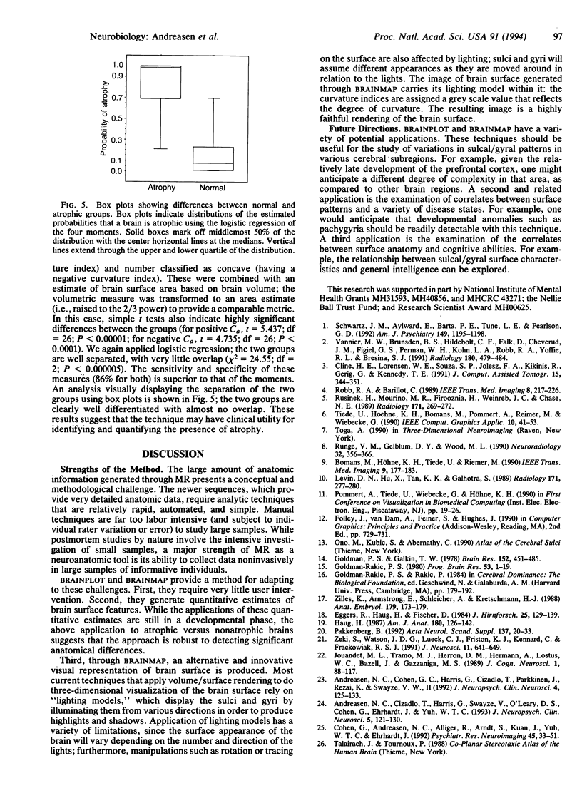
Selected References
These references are in PubMed. This may not be the complete list of references from this article.
- Andreasen N. C., Cizadlo T., Harris G., Swayze V., 2nd, O'Leary D. S., Cohen G., Ehrhardt J., Yuh W. T. Voxel processing techniques for the antemortem study of neuroanatomy and neuropathology using magnetic resonance imaging. J Neuropsychiatry Clin Neurosci. 1993 Spring;5(2):121–130. doi: 10.1176/jnp.5.2.121. [DOI] [PubMed] [Google Scholar]
- Andreasen N. C., Cohen G., Harris G., Cizadlo T., Parkkinen J., Rezai K., Swayze V. W., 2nd Image processing for the study of brain structure and function: problems and programs. J Neuropsychiatry Clin Neurosci. 1992 Spring;4(2):125–133. doi: 10.1176/jnp.4.2.125. [DOI] [PubMed] [Google Scholar]
- Cline H. E., Lorensen W. E., Souza S. P., Jolesz F. A., Kikinis R., Gerig G., Kennedy T. E. 3D surface rendered MR images of the brain and its vasculature. J Comput Assist Tomogr. 1991 Mar-Apr;15(2):344–351. doi: 10.1097/00004728-199103000-00035. [DOI] [PubMed] [Google Scholar]
- Cohen G., Andreasen N. C., Alliger R., Arndt S., Kuan J., Yuh W. T., Ehrhardt J. Segmentation techniques for the classification of brain tissue using magnetic resonance imaging. Psychiatry Res. 1992 May;45(1):33–51. doi: 10.1016/0925-4927(92)90012-s. [DOI] [PubMed] [Google Scholar]
- Eggers R., Haug H., Fischer D. Preliminary report on macroscopic age changes in the human prosencephalon. A stereologic investigation. J Hirnforsch. 1984;25(2):129–139. [PubMed] [Google Scholar]
- Goldman-Rakic P. S. Morphological consequences of prenatal injury to the primate brain. Prog Brain Res. 1980;53:1–19. [PubMed] [Google Scholar]
- Goldman P. S., Galkin T. W. Prenatal removal of frontal association cortex in the fetal rhesus monkey: anatomical and functional consequences in postnatal life. Brain Res. 1978 Sep 8;152(3):451–485. doi: 10.1016/0006-8993(78)91103-4. [DOI] [PubMed] [Google Scholar]
- Haug H. Brain sizes, surfaces, and neuronal sizes of the cortex cerebri: a stereological investigation of man and his variability and a comparison with some mammals (primates, whales, marsupials, insectivores, and one elephant). Am J Anat. 1987 Oct;180(2):126–142. doi: 10.1002/aja.1001800203. [DOI] [PubMed] [Google Scholar]
- Levin D. N., Hu X. P., Tan K. K., Galhotra S. Surface of the brain: three-dimensional MR images created with volume rendering. Radiology. 1989 Apr;171(1):277–280. doi: 10.1148/radiology.171.1.2928539. [DOI] [PubMed] [Google Scholar]
- Pakkenberg B. Stereological quantitation of human brains from normal and schizophrenic individuals. Acta Neurol Scand Suppl. 1992;137:20–33. doi: 10.1111/j.1600-0404.1992.tb05034.x. [DOI] [PubMed] [Google Scholar]
- Runge V. M., Gelblum D. Y., Wood M. L. 3-D imaging of the CNS. Neuroradiology. 1990;32(5):356–366. doi: 10.1007/BF00588469. [DOI] [PubMed] [Google Scholar]
- Rusinek H., Mourino M. R., Firooznia H., Weinreb J. C., Chase N. E. Volumetric rendering of MR images. Radiology. 1989 Apr;171(1):269–272. doi: 10.1148/radiology.171.1.2928536. [DOI] [PubMed] [Google Scholar]
- Schwartz J. M., Aylward E., Barta P. E., Tune L. E., Pearlson G. D. Sylvian fissure size in schizophrenia measured with the magnetic resonance imaging rating protocol of the Consortium to Establish a Registry for Alzheimer's Disease. Am J Psychiatry. 1992 Sep;149(9):1195–1198. doi: 10.1176/ajp.149.9.1195. [DOI] [PubMed] [Google Scholar]
- Vannier M. W., Brunsden B. S., Hildebolt C. F., Falk D., Cheverud J. M., Figiel G. S., Perman W. H., Kohn L. A., Robb R. A., Yoffie R. L. Brain surface cortical sulcal lengths: quantification with three-dimensional MR imaging. Radiology. 1991 Aug;180(2):479–484. doi: 10.1148/radiology.180.2.2068316. [DOI] [PubMed] [Google Scholar]
- Zeki S., Watson J. D., Lueck C. J., Friston K. J., Kennard C., Frackowiak R. S. A direct demonstration of functional specialization in human visual cortex. J Neurosci. 1991 Mar;11(3):641–649. doi: 10.1523/JNEUROSCI.11-03-00641.1991. [DOI] [PMC free article] [PubMed] [Google Scholar]
- Zilles K., Armstrong E., Schleicher A., Kretschmann H. J. The human pattern of gyrification in the cerebral cortex. Anat Embryol (Berl) 1988;179(2):173–179. doi: 10.1007/BF00304699. [DOI] [PubMed] [Google Scholar]