Dear Editor,
Burned-out tumors of the testis are extremely rare form of testicular malignancies, and they could disappear or regress spontaneously and completely. These tumors could be presented by metastatic form in retroperitoneum, lungs, retroperitoneal lymph nodes, and liver. In recent studies, differential diagnosis, clinic features, treatment modalities and follow-up outcome of burned-out tumor from primary retroperitoneal germ cell tumor are discussed. If patient with retroperitoneal lymph node involvement and histology of “testicular tumor”, burned-out tumor of the testis was suspected. Although screening of scrotal contents may be useful to identify intratesticular abnormalities in these cases, radical orchidectomy should be performed if burned-out tumor were suspected.
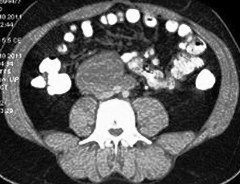
We present a case of a 39-year-old male complained of intermittent and increasing abdominal pain over the previous 2 months. The patient had no relevant past medical history. Abdominal examination revealed no palpable and tender mass. Scrotal examination revealed a normal left testis, but the right testis was atrophic, and no discrete mass was detected. Laboratory investigations were within normal ranges, with α-fetoprotein (AFP) 2.83 IU l−1 (0–9 IU l−1), β-human chorionic gonadotropin (HCG) hormone <0.1 IU l−1 (<0.1 IU l−1), and lactate dehydrogenase 183 IU l−1 (100–190 IU l−1). Scrotal ultrasonography revealed a normal left testis and atrophic right testicle with heterogeneous architecture and scattered micro-calcifications which may result with postorchitis sequel. No mass was visualized. Abdominal computed tomography (CT) showed multiple retroperitoneal masses with hypodens and soft tissue components extending from below the bilateral renal arteries down to the bifurcation of the aorta. The biggest one of the lymph nodes was 6 cm × 4 cm × 3 cm in diameter (Figure 1).
Figure 1.

The big retroperitoneal lymph node among multiple lymph nodes on para-aortic area.
A right radical orchiectomy was performed. The testicular size was 6 cm × 3 cm, 5 cm × 2.5 cm. Histopathology showed subtotal atrophy of the testis due to collected with stromal degenerative collagenous deposits in testicular parenchyma. There was no evidence of germ cell tumor, and immunostains including placental alkaline phosphatase (PLAP), Masson-trichroma, Kongo-red, and Periodic acid Schiff were negative. The patient subsequently underwent a CT-guided retroperitoneal lymph node biopsy which revealed no evidence of malignancy, but detected 4–5 mm tumor necrosis foci in some areas. The patient followed by three cycles of chemotherapy with bleomycin, etoposide, and cisplatin protocol. Follow-up CT after chemotherapy revealed that the para-aortic lymphadenopathies had decreased in size to 3.4 cm × 2 cm × 1.6 cm. The patient then underwent a retroperitoneal lymph node dissection. Pathology result reported that the lymph nodes metastases of testicular germ cell tumor. Immunohistochemistry was positive for PLAP (focal), PANCK, HCG (focal), and CD117, and negative for AFP, HMB45, CD30, and CD45.
Extragonadal germ cell tumors are rare and 5%–10% of all germ cell tumors, and they are predominantly affect young population.1 Extragonadal germ cell tumors are usually detected in the retroperitoneal, cervical, supraclavicular, and axillary lymph nodes and occasionally in the lung and liver. New epidemiologic study designed from Network of German Cancer Registries (GEKID) gave clue that extragonadal germ cell tumors usually appear not only mediastinal and retroperitoneal, but also in brain.2 Patients with retroperitoneal germ cell tumors usually detected lately after tumors getting larger to be a symptomatic. The symptoms are presented with a palpable mass, weight loss, constipation, back pain, dyspnea, leg edema, fever, and urinary retention. “Burned-out” phenomenon of testicular tumor is the spontaneous regression of a testicular germ cell tumor with or without metastasis. In our case report, we did not found the primary testicular tumor with histological characteristics of a germ cell tumor because of it was regressed after the development of metastasis in the retroperitoneum.
There are two ways to explain “burned-out” phenomenon. The first one is spontaneous regression of the primary germ cell tumor after metastasis of the germ cell tumor. Possible mechanisms are an immune response or ischemia caused by the neoplasm disseminate due to its high metabolic rate. The second way is the de-novo development of a primary germ cell tumor in extragonadal tissues.3 Histological features of testicular specimen are helpful in establishing a diagnosis of a regressed testicular germ cell tumor include, apart from the scar formation, intratubular calcifications, lymphoplasmacytic infiltrate, hemosiderin-containing macrophages, and testicular atrophy.4 Due to burned-out tumors may cause some confusion in the diagnosis, careful examination of the testis is essential for identifying the primary lesion site; if suspect abnormalities were detected with clinical or sonographic methods, orchiectomy was considered.5
As we know that the prognosis is poor due to late diagnosis in cases of patients with extragonadal germ cell tumor with histological patterns of nonseminomatous extragonadal germ cell tumor than seminomatous tumor in either the mediastinum or retroperitoneum. Unfortunately, the majority of patients with “burned-out” extragonadal tumors are germ cell tumors (80%) and thereby have a poor prognosis.6
When we look at the treatment modalities; primary extragonadal germ cell tumors are more aggressive than primary testicular cancer and are more resistant to chemotherapeutic agents and nonseminomatous extragonadal germ cell tumors are frequently chemoresistant and 5-year survival rates of 65%. The initial treatment modality of burned-out tumor is standard cisplatinum-based chemotherapy, after radical orchiectomy.7 In our case, patient was received three cycles chemotherapy after orchidectomy and retroperitoneal lymph node dissection was done when lymph nodes size was decreased due to chemotherapy response. Patient's follow-up is doing with serum tumor markers levels and thoraco-abdominal computerized tomography and normal during 1 year.8
As a result, in patients who present with a retroperitoneal mass, diagnosis of metastatic progression of a germ cell neoplasia should be considered. It is mandatory to suspect with a testicular lesion to use scrotal screening and physical examination of the scrotum and bilateral testis in the work-up of male patients presenting with a retroperitoneal mass. Retroperitoneal metastatic disease due to “burned-out” phenomenon testicular tumor has the same treatment modalities after diagnosis as a primary testicular malignancy; therefore, it is important that it is early recognized.
AUTHOR CONTRIBUTIONS
SB, OC, and HT designed of the study; OC and YOI carried out the study; OC, TS, and YOI drafted the manuscript. All authors read and approved the final manuscript.
COMPETING INTERESTS
The authors declare no competing interests.
REFERENCES
- 1.Schmoll HJ. Extragonadal germ cell tumors. Ann Oncol. 2002;13(Suppl 4):265–72. doi: 10.1093/annonc/mdf669. [DOI] [PubMed] [Google Scholar]
- 2.Rusner C, Trabert B, Katalinic A, Kieschke J, Emrich K, et al. Incidence patterns and trends of malignant gonadal and extragonadal germ cell tumors in Germany, 1998-2008. Cancer Epidemiol. 2013;37:370–3. doi: 10.1016/j.canep.2013.04.003. [DOI] [PMC free article] [PubMed] [Google Scholar]
- 3.Hainsworth JD, Greco FA. Extragonadal germ cell tumors and unrecognized germ cell tumors. Semin Oncol. 1992;19:119–27. [PubMed] [Google Scholar]
- 4.Balalaa N, Selman M, Hassen W. Burned-out testicular tumor: a case report. Case Rep Oncol. 2011;4:12–5. doi: 10.1159/000324041. [DOI] [PMC free article] [PubMed] [Google Scholar]
- 5.Kontos S, Doumanis G, Karagianni M, Politis V, Simaioforidis V, et al. Burned-out testicular tumor with retroperitoneal lymph node metastasis: a case report. J Med Case Rep. 2009;3:8705. doi: 10.4076/1752-1947-3-8705. [DOI] [PMC free article] [PubMed] [Google Scholar]
- 6.Geldart TR, Simmonds PD, Mead GM. Orchidectomy after chemotherapy for patients with metastatic testicular germ cell cancer. BJU Int. 2002;90:451–5. doi: 10.1046/j.1464-410x.2002.02916.x. [DOI] [PubMed] [Google Scholar]
- 7.Israel A, Bosl GJ, Golbey RB, Whitmore W, Jr, Martini N. The results of chemotherapy for extragonadal germ-cell tumors in the cisplatin era: the Memorial Sloan-Kettering Cancer Center experience (1975 to 1982) J Clin Oncol. 1985;3:1073–8. doi: 10.1200/JCO.1985.3.8.1073. [DOI] [PubMed] [Google Scholar]
- 8.Albers P, Albrecht W, Algaba F, Bokemeyer C, Cohn-Cedermark G, et al. EAU guidelines on testicular cancer: 2011 update. European Association of Urology. Actas Urol Esp. 2012;36:127–45. doi: 10.1016/j.acuro.2011.06.017. [DOI] [PubMed] [Google Scholar]


