


Abstract
Background
Central retinal vein occlusion (CRVO) is a relatively common retinal vascular disorder in which macular oedema may develop, with a consequent reduction in visual acuity. Until recently there has been no treatment of proven benefit, but growing evidence supports the use of anti‐vascular endothelial growth factor (anti‐VEGF) agents.
Objectives
To investigate the effectiveness and safety of anti‐VEGF therapies for the treatment of macular oedema secondary to CRVO.
Search methods
We searched CENTRAL (which contains the Cochrane Central Register of Controlled Trials (CENTRAL) and the Cochrane Eyes and Vision Group Trials Register) (The Cochrane Library 2013, Issue 10), Ovid MEDLINE (January 1950 to October 2013), EMBASE (January 1980 to October 2013), Latin American and Caribbean Health Sciences Literature Database (LILACS) (January 1982 to October 2013), Cumulative Index to Nursing and Allied Health Literature (CINAHL) (January 1937 to October 2013), OpenGrey, OpenSIGLE (January 1950 to October 2013), the metaRegister of Controlled Trials (mRCT) (www.controlled‐trials.com), ClinicalTrials.gov (www.clinicaltrials.gov), the WHO International Clinical Trials Registry Platform (ICTRP) (www.who.int/ictrp/search/en) and Web of Science Conference Proceedings Citation Index‐Science (CPCI‐S). There were no language or date restrictions in the electronic search for trials. The electronic databases and clinical trials registers were last searched on 29th October 2013.
Selection criteria
We considered randomised controlled trials (RCTs) that compared intravitreal anti‐VEGF agents of any dose or duration to sham injection or no treatment. We focused on studies that included individuals of any age or gender and a minimum of six months follow‐up.
Data collection and analysis
Two review authors independently assessed trial quality and extracted data. The primary outcome was the proportion of participants with a gain in best‐corrected visual acuity (BCVA) from baseline of greater than or equal to 15 letters (3 lines) on the Early Treatment of Diabetic Retinopathy Study (ETDRS) chart. Secondary outcomes included the proportion of participants with a loss of 15 letters or more of BCVA, the mean change from baseline BCVA, the mean change in central retinal thickness (CRT), the number and type of complications or adverse outcomes, and the number of additional interventions administered. Where available, we also presented quality of life and economic data.
Main results
We found six RCTs that met the inclusion criteria after independent and duplicate review of the search results. These RCTs included 937 participants and compared outcomes at six months to sham injection for four anti‐VEGF agents: aflibercept (VEGF Trap‐Eye, Eylea), bevacizumab (Avastin), pegaptanib sodium (Macugen) and ranibizumab (Lucentis). Three trials were conducted in Norway, Sweden and the USA, and three trials were multicentre, one including centres in the USA, Canada, India, Israel, Argentina and Columbia, a second including centres in the USA, Australia, France, Germany, Israel, and Spain, and a third including centres in Austria, France, Germany, Hungary, Italy, Latvia, Australia, Japan, Singapore and South Korea. We performed meta‐analysis on three key visual outcomes, using data from up to six trials. High‐quality evidence from six trials revealed that participants receiving intravitreal anti‐VEGF treatment were 2.71 times more likely to gain at least 15 letters of visual acuity at six months compared to participants treated with sham injections (risk ratio (RR) 2.71; 95% confidence intervals (CI) 2.10 to 3.49). High‐quality evidence from five trials suggested anti‐VEGF treatment was associated with an 80% lower risk of losing at least 15 letters of visual acuity at six months compared to sham injection (RR 0.20; 95% CI 0.12 to 0.34). Moderate‐quality evidence from three trials (481 participants) revealed that the mean reduction from baseline to six months in central retinal thickness was 267.4 µm (95% CI 211.4 µm to 323.4 µm) greater in participants treated with anti‐VEGF than in participants treated with sham. The meta‐analyses demonstrate that treatment with anti‐VEGF is associated with a clinically meaningful gain in vision at six months. One trial demonstrated sustained benefit at 12 months compared to sham. No significant ocular or systemic safety concerns were identified in this time period.
Authors' conclusions
Compared to no treatment, repeated intravitreal injection of anti‐VEGF agents in eyes with CRVO macular oedema improved visual outcomes at six months. All agents were relatively well tolerated with a low incidence of adverse effects in the short term. Future trials should address the relative efficacy and safety of the anti‐VEGF agents and other treatments, including intravitreal corticosteroids, for longer‐term outcomes.
Plain language summary
Anti‐vascular endothelial growth factor for macular oedema secondary to central retinal vein occlusion
Review question We reviewed the evidence about the effect of anti‐vascular endothelial growth factor (anti‐VEGF) agents in people with macular oedema secondary to central retinal vein occlusion (CRVO).
Background CRVO affects approximately one person per 1000 at any one time, and is associated with increasing age, high blood pressure, diabetes, glaucoma and various disorders of the blood. It frequently causes sudden, painless vision loss in one eye, although sometimes the vision loss may be minimal. If the vein blockage leads to inadequate oxygen delivery to the sensitive retinal tissue, the CRVO is the 'non‐perfused' or 'ischaemic' subtype. More commonly, blood flow and oxygen delivery are restored following the vein blockage and the CRVO is the 'perfused' or 'non‐ischaemic' subtype, and has a better visual outcome. Various other complications may develop over hours, days, weeks or months. These include macular oedema, in which fluid collects within the retina and causes reduction in vision. Until relatively recently there has been no evidence‐based treatment for this condition. Anti‐VEGF agents have been used successfully to treat patients with other retinal vascular disorders, including several conditions associated with macular oedema.
Study characteristics This systematic review identified six trials which included 937 participants with macular oedema secondary to CRVO (as of 29 October 2013). The trials compared sham injections with one of four types of anti‐VEGF agents: aflibercept (VEGF Trap‐Eye, Eylea), bevacizumab (Avastin), pegaptanib sodium (Macugen) and ranibizumab (Lucentis). All trials treated participants for at least six months. Three trials were multicentre, international trials and three were conducted in Norway, Sweden or the USA.
Key results Overall, treatment with anti‐VEGF agents increased the chance of a significant gain in vision (at least 3 lines on the vision chart) at six months by more than two and a half times, compared to no treatment. Furthermore, the risk of losing significant vision (at least 3 lines on the vision chart) was reduced by 80% in those receiving anti‐VEGF therapy compared to those receiving no treatment. No significant safety concerns were identified at six or 12 months, but the available studies do not allow a conclusion about their long‐term effectiveness and safety to be drawn. Nevertheless, the availability of anti‐VEGF treatment for CRVO macular oedema represents an important advance in the clinical management options for this sight‐threatening disease.
Quality of the evidence The six trials included in this review were high quality and consistently demonstrated visual benefit from anti‐VEGF injections.
Summary of findings
for the main comparison.
| Anti‐VEGF compared with sham injection for CRVO macular oedema | ||||||
|
Patient or population: participants with CRVO macular oedema of duration less than 9 months (mean < 3 months) and no prior treatment Settings: presenting baseline visual acuity ranging from 6 (< 6/120) letters to 73 letters (˜6/12), both non‐ischaemic and ischaemic eyes (% ischaemic at baseline of included trials ranged from 0% to 16%) Intervention: intravitreal injection with ranibizumab, bevacizumab, aflibercept or pegaptanib sodium Comparison: sham injection | ||||||
| Outcomes | Illustrative comparative risks* (95% CI) | Relative effect (95% CI) | No of participants (studies) | Quality of the evidence (GRADE) | Comments | |
| Assumed risk | Corresponding risk | |||||
| Sham injection | Anti‐VEGF injection | |||||
| BCVA gain of 15 letters or more (follow‐up: 6 months) |
182 per 1000 | 493 per 1000 (382 to 635) | RR 2.71 (2.10 to 3.49) | 937 (6 studies) | ⊕⊕⊕⊕ high | Includes all 4 anti‐VEGF agents |
| BCVA loss of 15 letters or more (follow‐up: 6 months) |
219 per 1000 | 44 per 1000 (26 to 74) | RR 0.20 (0.12 to 0.34) | 766 (5 studies) | ⊕⊕⊕⊕ high | Includes all 4 anti‐VEGF agents |
| Mean change in BCVA from baseline (follow‐up: 6 months) |
The mean change across control groups ranged from loss of 4 letters to gain of 3 letters | The mean gain across treatment groups ranged from +7.1 letters to + 18.0 letters | MD 15.23 letters (11.57 to 18.89) | 937 (6 studies) | ⊕⊕⊕⊝ moderate | Standard deviation or 95% CI not reported in 2 studies (aflibercept and pegaptanib sodium) |
| Mean change from baseline in central retinal thickness (follow‐up: 6 months) |
The mean reduction from baseline in CRT across control groups ranged from ‐102 to ‐169 microns (6 studies, 937 participants) | The mean reduction from baseline in CRT in the intervention groups was ‐267 microns greater (‐211 to ‐323 microns) | MD ‐267.4 µm (211.4 to 323.4) | 481 (3 studies) | ⊕⊕⊕⊝ moderate | Standard deviation or 95% CI not reported in 3 studies (aflibercept and pegaptanib sodium) |
| Complication: iris or retinal neovascularisation (follow‐up: 6 months) |
75 per 1000 | 14 per 1000 (7 to 27) |
RR 0.18 (0.09 to 0.36) | 936 (6 studies) |
⊕⊕⊕⊕ high | Includes all 4 anti‐VEGF agents |
| Ocular adverse events: endophthalmitis at 6 months | No cases in 347 participants treated with sham injection | 1 case in 590 participants treated with intravitreal injection of anti‐VEGF | ‐ | 937 (6 studies) |
⊕⊕⊕⊕ high | Includes all 4 anti‐VEGF agents |
| Mean change from baseline in quality of life score (NEI VFQ‐25 instrument) | The mean gain in quality of life score from baseline across control groups ranged from +0.8 to +3.5 points | The mean gain in quality of life score from baseline across control groups ranged from +6.2 to +7.5 points | ‐ | 743 (3 studies) |
⊕⊕⊕⊝ moderate | Standard deviation or 95% CI not reported in 2 studies (aflibercept) > 4‐point increase is considered a clinically relevant improvement |
| *The basis for the assumed risk (e.g. the median control group risk across studies) is provided in footnotes. The corresponding risk (and its 95% confidence interval) is based on the assumed risk in the comparison group and the relative effect of the intervention (and its 95% CI). Anti‐VEGF: anti‐vascular endothelial growth factor; BCVA: best‐corrected visual acuity; CI: confidence interval;CRT: central retinal thickness; CRVO: central retinal vein occlusion; MD: mean difference; NEI‐VFQ 25: National Eye Institute Visual Functioning Questionnaire 25 question instrument; RR: risk ratio | ||||||
| GRADE Working Group grades of evidence High quality: Further research is very unlikely to change our confidence in the estimate of effect. Moderate quality: Further research is likely to have an important impact on our confidence in the estimate of effect and may change the estimate. Low quality: Further research is very likely to have an important impact on our confidence in the estimate of effect and is likely to change the estimate. Very low quality: We are very uncertain about the estimate. | ||||||
*Assumed risk was taken from the mean baseline risk from studies in the meta‐analysis and was equal to the total number of events in the control groups divided by the total number of participants in the control groups.
Judgement of high quality: most evidence comes from RCTs at low risk of bias, with no unexplained heterogeneity and consistent results, low probability of publication bias, a large magnitude of effect or an apparent dose‐response gradient.
Judgement of moderate quality: most of the evidence comes from RCTs with some limitations. For example, limitations include an unclear risk of bias in one or several domains or few participants and wide confidence intervals suggesting imprecision of evidence.
Background
Description of the condition
Central retinal vein occlusion (CRVO) is a common retinal vascular disorder in which macular oedema, including the cystoid type, may develop with a consequent reduction in visual acuity (Guex‐Crosier 1999; Hayreh 1983). Cystoid macular oedema is characterised by the collection of fluid in intercellular spaces within the outer plexiform layer of the retina and results from the breakdown of the capillary endothelium blood‐retinal barrier and leakage of fluid from the vasculature (Guex‐Crosier 1999; Vinores 1999). This process is promoted by vascular endothelial growth factor (VEGF) (Vinores 1999), and the aqueous concentration of VEGF has been found to be significantly elevated in patients with CRVO macular oedema compared to controls (Funk 2009). Furthermore, there is a significant inverse association between duration since onset of CRVO macular oedema and the aqueous VEGF concentration (Funk 2009). Macular oedema can be visualised with slit‐lamp biomicroscopy and its thickness and pattern of distribution can be investigated and quantified using optical coherence tomography (OCT) (Catier 2005; Hee 1995).
Central retinal vein occlusion is classified into two subtypes, 'non‐ischaemic' (also called 'perfused') and 'ischaemic' (also called 'non‐perfused'), although it is unlikely that the disease process is binary. No single test reliably differentiates the two subtypes with 100% sensitivity and specificity during the early acute phase of CRVO (Hayreh 1990a). Fundus fluorescein angiography (FFA) is frequently used to define ischaemic CRVO (Clarkson 1994; CVOS Group 1997). However, standard FFA may be reliable in fewer than 60% of cases (Hayreh 1990a), on account of masking from extensive retinal haemorrhages, which frequently persist in the first six months, poor images resulting from media opacities (vitreous haemorrhage or lens opacities, which are frequent in the affected age group), and inadequate assessment of the peripheral retina (Hayreh 1990a). However, ultra wide‐field FFA is increasingly used in clinical practice to provide up to 200º imaging of the retina using a non‐contact lens‐based system (Prasad 2010).
Epidemiology
The burden of CRVO macular oedema in the population and the incidence of macular oedema following CRVO are unknown (McIntosh 2010), and there is still a paucity of data on risk factors for developing macular oedema following CRVO (Hayreh 1994).
The age‐ and sex‐standardised prevalence of CRVO has been estimated at 0.80 per 1000 persons (95% confidence interval (CI) 0.61 to 0.99), giving an estimated global burden (for the 2008 population) of 2.5 million affected adults (Rogers 2010). In this pooled analysis, including 11 population‐based studies on 49,869 participants in the United States, Asia, Europe and Australia, the prevalence of CRVO was found to increase with age, with a crude prevalence ranging from 0.27 per 1000 (95% CI 0 to 0.65) in those aged 40 to 49 years, to 5.44 per 1000 (95% CI 2.78 to 8.10) in those aged 80 years and over (Rogers 2010). Of these participants, 43.7% were male, 48.4% were white, 27.1% were Asian, 17.2% were Hispanic and 7.2% were black, and the prevalence did not differ by race or by gender. In another study of 1302 people diagnosed with CRVO in the USA, black people had a 58% increased risk of CRVO compared to white people (hazard ratio (HR) 1.58; 95% CI 1.25 to 1.99) and women had a 25% decreased risk compared to men (HR 0.75; 95% CI 0.66 to 0.85), after adjustment for known confounders (Stem 2013). Numerous other factors increase the hazard of CRVO, including a hypercoagulable state (HR 2.92; 95% CI 1.52 to 2.42), a diagnosis of stroke (HR 1.44; 95% CI 1.23 to 1.68), end‐organ damage from hypertension (HR 1.92; 95% CI 1.52 to 2.42) and end‐organ damage from diabetes mellitus (HR 1.53; 95% CI 1.28 to 1.84), relative to people without these conditions (Stem 2013). After developing a CRVO there is a 1% chance per year of a retinal vein occlusion in the fellow eye (CVOS Group 1997). CRVO is associated with an increase in mortality on account of its statistical association with comorbid diabetes or cardiovascular disease (Bertelsen 2013).
Natural history and prognosis
There is a paucity of population‐level data on the prevalence and incidence of ischaemic versus non‐ischaemic CRVO macular oedema, but the distinction is important because their presentation and natural history differ (Hayreh 2011). The ischaemic subtype of CRVO accounts for approximately 20% of acute presentations (Hayreh 1983; Hayreh 1994) and is associated with worse baseline acuity and a poor visual prognosis, even after the resolution of macular oedema (CVOS Group 1997; Hayreh 2011). In a natural history cohort study (n = 697 eyes with CRVO), baseline visual acuity was 6/30 (20/100) or better in 78% of patients with non‐ischaemic CRVO and in only 1% with ischaemic CRVO, and visual field defects were mild in 91% and 8%, respectively (Hayreh 2011). Final visual acuity, after resolution of macular oedema, was 6/30 (20/100) or better in 83% with non‐ischaemic CRVO and 12% with ischaemic CRVO (Hayreh 2011). In non‐ischaemic CRVO, development of foveal pigmentary degeneration, epiretinal membrane or both has been identified as the main cause of poor final acuity (Hayreh 2011). Visual morbidity, including blindness, may also result from persistent macular oedema, ocular neovascularisation or secondary glaucoma (Campochiaro 2008). The risk of developing these complications also differs between the subtypes, with 16.6% of eyes with more than 5.5 disc areas of non‐perfusion on FFA at baseline, versus 4.0% of eyes with less than 5.5 disc areas of non‐perfusion, developing neovascular glaucoma at 36 months (Chan 2011). The non‐ischaemic type may convert to the ischaemic type in an estimated one‐third of cases within three years, and conversion is most frequent in the initial four months (CVOS Group 1997).
Presenting visual acuity is also a key predictor of visual outcome at three years, considering ischaemic and non‐ischaemic subtypes together (CVOS Group 1997). Out of 714 eyes with CRVO, 65% of patients with presenting visual acuity better than or equal to 6/12 (20/40) were found to maintain this level of vision; those with presenting visual acuity 6/12 to 6/60 (20/40 to 20/200) have a variable prognosis with 19% improving to better than 6/15 (20/50), 44% showing no improvement and 37% worsening to less than 6/60 (20/200); whilst 80% of those with vision less than 6/60 (20/200) at presentation have no visual improvement. In another natural history study, amongst non‐ischaemic eyes (n = 588) with an initial visual acuity of approximately 6/24 (20/70) or worse, 59% improved, 27% showed no change and 14% deteriorated on resolution of macular oedema, whereas amongst those with ischaemic CRVO (n = 109), 41% improved, 41% did not change and 18% deteriorated (Hayreh 2011). An intact external limiting membrane in the outer retina at baseline, which can be visualised with spectral‐domain OCT, has also been found to predict better visual outcomes after treatment (Wolf‐Schnurrbusch 2011).
History of the management of CRVO macular oedema
Historically, there was no evidence‐based treatment for CRVO macular oedema. Various medical and surgical interventions had been tried, including argon laser panretinal photocoagulation (Hayreh 1990b), macular grid photocoagulation (CVOS Group 1995), tissue plasminogen activator (Everett 2006), oral pentoxifylline (Park 2007), hyperbaric oxygen therapy (Wright 2007) and pars plana vitrectomy with internal limiting membrane peeling (DeCroos 2009; Park 2010; Raszewska‐Steglinska 2009), but these were not effective. Evidence for the effect of optic neurotomy has been mixed (Arevalo 2008; Hasselbach 2007; Opremcak 2006; Zambarakji 2005), with one recent randomised controlled trial (RCT) suggesting possible benefit (Aggermann 2012). A RCT comparing laser‐induced chorioretinal venous anastomosis to sham treatment in adults with non‐ischaemic CRVO macular oedema of three to 12 months' duration demonstrated moderate efficacy, especially in the 76% who developed a functioning anastomosis following laser treatment, and the visual acuity advantage compared to control was maintained at 18 months (McAllister 2010). However, 18% in the treatment arm developed neovascularisation at the treatment site and 9% required vitrectomy surgery for macular traction or non‐resolving vitreous haemorrhage (McAllister 2010).
More significantly, two RCTs on corticosteroids for CRVO macular oedema demonstrated short‐term effectiveness, but concerns have been raised about the incidence of complications (Gewaily 2009). Firstly, the 'Standard Care vs. Corticosteroid for Retinal Vein Occlusion' trial treated participants with non‐ischaemic CRVO macular oedema with either 1 mg or 4 mg intravitreal triamcinolone (IVTA) injections repeated every four months, or observation alone (SCORE 2009). Both treatment doses were associated with a five times increase in the odds of achieving a 15 letter gain in visual acuity at 12 months (P = 0.001). However, 35% of patients treated with 4 mg, and 20% of patients treated with 1 mg, required intraocular pressure lowering treatment by 12 months compared to 8% in the observation group (SCORE 2009). Similarly, at 12 months there was new lens opacity, or progression of existing lens opacity, in 33% of the 4 mg group and 26% of the 1 mg group, compared to 18% of the observation group (SCORE 2009). Secondly, the Ozurdex GENEVA trial compared a single dexamethasone implant (Ozurdex, Allergan, Inc., Irvine, CA) at a dose of 0.7 mg or 0.35 mg, to a sham implant, in adults with non‐ischaemic branch retinal vein occlusion (BRVO) or CRVO macular oedema of six weeks to nine months duration, and baseline acuity of 6/15 (68 letters, 20/50) to 6/60 (34 letters, 20/20) (Ozurdex GENEVA 2010). The percentage of eyes with CRVO macular oedema achieving a 15 letter improvement in visual acuity was significantly higher in both Ozurdex groups at day 30 and day 60 than in the sham group (P < 0.001), with the maximal effect at day 60. However, by days 90 and 180 there was no significant difference between groups (Ozurdex GENEVA 2010). The incidence of ocular adverse events was not reported for CRVO macular oedema separately, but did not differ significantly between the 0.35 mg and 0.7 mg dose groups. In both groups there was a higher incidence of ocular hypertension at day 60 (P < 0.002), and by day 180 approximately 24% of patients with dexamethasone implants required intraocular pressure (IOP)‐lowering medication, and five patients required a surgical or laser procedure to reduce IOP (Ozurdex GENEVA 2010). There was also a significant increase in anterior chamber activity in the 0.35 mg (P = 0.007) and 0.7 mg (P = 0.03) treatment groups compared to sham (P = 0.03), but no significant increase in the risk of incident cataract at 180 days (Ozurdex GENEVA 2010). In a subsequent six‐month, open‐label extension, all patients were eligible for a single 0.7 mg Ozurdex implant, regardless of initial treatment assignment, if BCVA was < 84 letters (6/6, 20/20) or CRT was > 250 μm (Ozurdex GENEVA 2011). Amongst patients who received two treatments with 0.7 mg implants for CRVO, six months apart (n = 114), mean BCVA improvements were similar after the first and second injections, with a peak improvement of approximately 8 to 10 letters at 60 days. Patients randomised to receive sham injection in the first six months who subsequently received 0.7 mg dexamethasone (n = 117) had an average gain of approximately 6 to 7 letters at 60 days. There was no apparent sustained visual acuity gain at 12 months in either group. Summed safety data for CRVO and BRVO patients who received a 0.7 mg implant at day 180 (n = 997) were reported. Over 12 months, cataract progression occurred in 29.8% of phakic eyes that received two 0.7 mg implants, versus 5.7% of sham‐treated phakic eyes. In the former treatment group (n = 341), a > 10 mmHg IOP rise was observed in 12.6% after the first treatment and 15.4% after the second treatment, and in the majority this was transient or controlled with topical medication. However, a laser or surgical procedure to reduce IOP was required for 14 treated eyes compared to no eyes in the untreated group (Ozurdex GENEVA 2011). There are currently no RCTs exploring the efficacy or safety of more frequent treatment with dexamethasone implants, or with follow‐up beyond 12 months. Despite this, the UK National Institute of Clinical Excellence (NICE) have recently approved Ozurdex implants as a treatment option for CRVO macular oedema, with a suggested repeat interval of six months, to a total of six implants per patient.
Description of the intervention
Monoclonal antibodies against VEGF were first developed as an intravenous treatment for metastatic colorectal cancer (Homsi 2007; Los 2007). The first drug licensed for this purpose was bevacizumab (Avastin®), which received Food and Drug Administration (FDA) approval in 2004 (Genentech/Roche 2009b). Bevacizumab is a 149kDa recombinant humanised monoclonal whole immunoglobulin G1 antibody that binds to VEGF and blocks the binding of VEGF to receptors (Flt‐1 and KDR) on endothelial cells (Genentech 2009). It is not licensed for intraocular use, although there has been widespread off‐licence intravitreal use in the USA and Europe. Pegaptanib sodium (Macugen®) is a 50kDa aptamer; a pegylated modified oligonucleotide, which adopts a three‐dimensional configuration in vivo that allows it to bind to extracellular VEGF‐165 and antagonise its biological effects (Eyetech 2008; Gragoudas 2004). It was approved by the FDA in 2004, and the European Medicines Agency in 2006, for use in neovascular age‐related macular degeneration (wet AMD) (Eyetech 2008). Ranibizumab (Lucentis®) is a 48kDa recombinant humanised monoclonal immunoglobulin G1 antibody fragment (kappa isotype) that binds to the receptors of all biologically active isoforms of VEGF‐A and blocks the binding of VEGF‐A to VEGFR1 and VEGFR2 receptors on endothelial cells (Genentech 2008). Ranibizumab has a binding affinity for VEGF approximately 100 times greater than bevacizumab (Ferrara 2006). It was approved for the treatment of wet AMD by the FDA in 2006 and by NICE in 2008 (Genentech/Roche 2009a); for the treatment of retinal vein occlusion by the FDA in June 2010; and for the treatment of diabetic macular oedema by the FDA in August 2012, and by NICE in October 2012. Aflibercept (Eylea®, vascular endothelial growth factor Trap‐Eye; Regeneron Pharmaceuticals, Tarrytown, NY) is a 115kDa decoy receptor fusion protein comprising the second domain of human VEGF receptor 1 and the third domain of VEGF receptor 2 fused to the constant Fc domain of human immunoglobulin G1 (Economides 2003). It has a greater binding affinity for VEGF than bevacizumab and ranibizumab (Stewart 2012), and mathematical modelling indicates that it may require less frequent dosing than shorter‐acting anti‐VEGF agents (Stewart 2008). It was approved by the FDA for the treatment of wet AMD in November 2011 and for CRVO macular oedema in September 2012.
The pharmacokinetics of 1.25 mg bevacizumab and 0.5 mg ranibizumab intravitreal injections have been investigated in an experimental rabbit model (Bakri 2007a; Bakri 2007b). The vitreous concentration of both drugs declined in a monoexponential function, with a half‐life of 4.32 days for bevacizumab, and 2.88 days for ranibizumab. At 30 days both drugs persisted in the vitreous, at a concentration of > 0.1 μg/ml for ranibizumab versus > 10 μg/ml for bevacizumab. No ranibizumab was detected in the fellow eye or serum, whilst a peak serum concentration of bevacizumab of 3.3 μg/ml was reached at eight days, with a half‐life of 6.86 days, and very low concentrations (ng/ml) were detected in the fellow eye throughout the 29‐day study. The aqueous half‐life of a single 1.5 mg intravitreal injection of bevacizumab has also been studied in humans with various causes of macular oedema and has been found to be approximately 9.8 days (Krohne 2008). Patients with CRVO demonstrate moderate variability in the aqueous concentration of ranibizumab measured one month after a first intravitreal injection of 0.3 mg or 0.5 mg, but measurements one month following subsequent injections are highly correlated for a given patient (Campochiaro 2009).
The anti‐VEGF agents have demonstrated promise in treating CRVO macular oedema in many retrospective and prospective case series, and in a number of RCTs in recent years.
How the intervention might work
Vascular endothelial growth factor is a cytokine produced by cells in response to hypoxia that promotes vascular leakage by binding to receptors on endothelial cells. It has been observed that transgenic mice over‐expressing VEGF in the photoreceptors exhibited blood‐retina barrier failure (Vinores 1999). Another study observed that injecting VEGF intravitreally induces a time and dose‐dependent breakdown of the blood‐aqueous and blood‐retinal barriers in a rabbit model, with maximal vascular leakage occurring 48 hours after injection (Edelman 2005). Animal and human studies have identified that the expression of VEGF mRNA is significantly upregulated in regions of ischaemic retina of various causes, including CRVO (Pe'er 1995; Pe'er 1998; Shima 1996). Serum amyloid A, a major acute phase protein, and the cytokine IL‐6, which is derived from activated T lymphocytes and induces expression of VEGF and vascular permeability, have been found to be significantly elevated in the aqueous humour of eyes with CRVO macular oedema compared to control eyes (Feng 2013). In a rat model of CRVO, injection of bevacizumab fully prevented the upregulation of VEGF‐A after one day and the upregulation of pigment‐epithelium‐derived factor after three days, which is known to influence the development of vascular oedema (Drechsler 2012). Bevacizumab also decreased the upregulation of the proinflammatory cytokine interleukin (IL)‐1B which otherwise developed one day after a CRVO (Drechsler 2012). Furthermore, the concentration of VEGF in human aqueous demonstrates close temporal correlation with the course of neovascularisation and permeability in CRVO, and injecting anti‐VEGF antibodies inhibits VEGF‐driven neovascularisation both in vitro and in vivo (Adamis 1996; Aiello 1995; Boyd 2002). Significant correlations have been identified between the aqueous VEGF concentration in patients with CRVO macular oedema and different components of the full‐field electroretinography (ERG) including the b/a ratio of the single flash ERG, implicit times of the cone a‐wave, cone b‐wave and 30 Hz flicker ERG leading to the suggestion that full‐field ERGs could be used to detect patients at high risk of developing neovascularisation (Yasuda 2011).
Whilst VEGF‐A has been identified as an important anti‐angiogenic target in retinal diseases, it has more recently also been recognised to play an important role in neuroprotection in the retina (Nishijima 2007). In a model of ischaemia‐reperfusion injury VEGF‐A exposure resulted in a dose‐dependent reduction in retinal neuron apoptosis. Furthermore, ischaemic preconditioning, which increases VEGF‐A levels, was found to reduce the number of apoptotic retinal cells after injury, suggesting its role in the adaptive response to retinal ischaemia, and this protective effect was reversed with VEGF‐A inhibition (Nishijima 2007). Chronic VEGF‐A inhibition in adult animals was also found to result in a significant loss of retinal ganglion cells, and the requirement for VEGF‐A in the maintenance of normal vasculature has now been recognised (Nishijima 2007). Interestingly, pegaptanib sodium, which does not bind to VEGF‐120, did not reduce retinal ganglion cell viability in this animal model (Nishijima 2007). In a small RCT of 19 participants with neovascular glaucoma secondary to ischaemic CRVO and poor baseline visual acuity, six months after randomisation to a single bevacizumab injection with panretinal photocoagulation (PRP) (n = 10) versus PRP alone (n = 9), neovascularisation had resolved in the bevacizumab group, but the a‐ and b‐wave amplitudes of the combined rod‐cone response and the b‐wave amplitudes of the 30 Hz flicker response were markedly reduced, suggesting a potential adverse effect of anti‐VEGF treatment on photoreceptor function (Wittstrom 2012). These basic science and clinical studies illustrate that it should not be assumed that different anti‐VEGF agents will have the same biological and clinical effects, or that all effects of VEGF blockade are beneficial.
Why it is important to do this review
The visual prognosis in CRVO macular oedema is poor in a substantial proportion of patients, especially those with the ischaemic subtype, and until recently there was no treatment of proven benefit (Everett 2006; Hayreh 2003; Prisco 2002). Ranibizumab and aflibercept have now been approved in the US for the treatment of CRVO macular oedema, and clinicians internationally are increasingly using various anti‐VEGF agents both on‐ and off‐label for the treatment of CRVO macular oedema, based on emerging clinical experience and short‐term trial evidence. This systematic review was therefore designed to investigate the effectiveness and safety of anti‐VEGF agents for the treatment of CRVO macular oedema.
Objectives
To investigate the effectiveness and safety of anti‐VEGF therapies for the treatment of macular oedema secondary to CRVO.
Methods
Criteria for considering studies for this review
Types of studies
We included RCTs with a minimum of six months follow‐up.
Types of participants
We included trials involving participants of all ages who had unilateral or bilateral macular oedema secondary to CRVO.
Types of interventions
We included trials in which anti‐VEGF treatment was compared to placebo or no treatment, and trials that investigated dosage and duration of treatment. We excluded studies in which anti‐VEGF agents were only compared to, or used in combination with, other agents.
Types of outcome measures
Primary outcomes
The primary outcome for this review was the proportion of participants with an improvement from baseline in best‐corrected visual acuity (BCVA) of greater than or equal to 15 letters (3 lines) on the Early Treatment in Diabetic Retinopathy Study (ETDRS) Chart at four metres, after six months of follow‐up, and any additional follow‐up times. A gain of 15 letters represents a doubling of the visual angle, and whilst this binary cut‐off considerably exceeds the amount of change required to have a high degree of certainty that the observed change is real, even in the presence of poor vision, it has been the standard primary outcome measure for evaluating the efficacy of treatments for retinal diseases for more than a decade (Beck 2007).
Secondary outcomes
We included the following secondary outcomes, at six months and any additional follow‐up times:
The proportion of participants with a loss of 15 letters or more (ETDRS) compared to baseline.
Mean visual acuity change.
Objective assessment of macular oedema regression measured by mean change in central retinal thickness (CRT) on ocular coherence tomography (OCT).
The number and type of complications relating to CRVO.
The number of anti‐VEGF or sham injections administered.
The number and type of additional interventions administered.
Adverse outcomes
We documented any ocular or systemic adverse outcomes reported in the trials, which were potentially related to the intervention or to intravitreal injection. We specifically aimed to report the proportion of participants experiencing potentially serious systemic or ocular adverse events including, but not limited to, retinal tears, retinal detachment, ocular inflammation, endophthalmitis, thromboembolic events, ocular hypertension, glaucoma (excluding neovascular) and cataract.
Economic data
We reported any cost‐benefit data included in the primary studies.
Quality of life data
We reported any data relating to impact on health‐ or vision‐related quality of life or daily functioning included in the primary studies.
Search methods for identification of studies
Electronic searches
We searched CENTRAL (which contains the Cochrane Central Register of Controlled Trials (CENTRAL) and the Cochrane Eyes and Vision Group Trials Register) (The Cochrane Library 2013, Issue 10), Ovid MEDLINE (January 1950 to October 2013), EMBASE (January 1980 to October 2013), Latin American and Caribbean Health Sciences Literature Database (LILACS) (January 1982 to October 2013), Cumulative Index to Nursing and Allied Health Literature (CINAHL) (January 1937 to October 2013), OpenGrey, OpenSIGLE (January 1950 to October 2013), the metaRegister of Controlled Trials (mRCT) (www.controlled‐trials.com), ClinicalTrials.gov (www.clinicaltrials.gov), the WHO International Clinical Trials Registry Platform (ICTRP) (www.who.int/ictrp/search/en) and Web of Science Conference Proceedings Citation Index‐Science (CPCI‐S). There were no language or date restrictions in the electronic search for trials. The electronic databases and clinical trials registers were last searched on 29th October 2013.
See: Appendices for details of search strategies for CENTRAL (Appendix 1), MEDLINE (Appendix 2), EMBASE (Appendix 3), LILACS (Appendix 4), CINAHL (Appendix 5), OpenSIGLE, mRCT (Appendix 6) and ClinicalTrials.gov (Appendix 8).
Searching other resources
We manually searched references of included studies and used the Science Citation Index to identify additional studies citing trials.
Data collection and analysis
Selection of studies
Two review authors independently screened the titles and abstracts resulting from the electronic and manual searches. We classified abstracts as relevant, potentially relevant or not relevant for this review. We obtained full‐text copies of articles for those abstracts that were designated relevant or potentially relevant. Two review authors independently assessed each article and determined whether to definitely include, definitely exclude or record each trial as unclear. We documented agreement between review authors and resolved discrepancies by consensus. For any studies classified as unclear we contacted the authors in an attempt to include or exclude the study from the review. We reported any studies that were definitely excluded.
Data extraction and management
We extracted the following participant and trial characteristics and reported them in a table format.
Methodology (group size, randomisation and masking).
Participant characteristics (gender, age, type of CRVO and diagnostic criteria used, baseline visual acuity, OCT‐determined thickness of macular oedema).
Intervention (agent, dose, timing of first dose in relation to diagnosis, delivery route, frequency and treatment length).
Primary and secondary outcomes (proportion with 15 letter gain in visual acuity at six months, proportion with 15 letter loss in visual acuity at six months, mean difference in visual acuity at six months compared to baseline, central retinal thickness, adverse events and outcomes at longer follow‐up intervals).
Additional data (economic, quality of life and visual functioning data).
Treatment compliance and losses to follow‐up.
Two review authors independently extracted the data using a form developed by the Cochrane Eyes and Vision Group. We contacted trial authors for more information when data were missing or difficult to interpret. We resolved any discrepancies between the two review authors by discussion and consensus. One review author entered the data into Review Manager 5 (RevMan 2011) and the second author checked the entered data for any errors or inconsistencies.
Assessment of risk of bias in included studies
Two review authors assessed the methodological quality of the selected trials according to the methods set out in Chapter 8 of the Cochrane Handbook for Systematic Reviews of Interventions (Higgins 2011). We considered the following domains:
random sequence generation (selection bias);
allocation concealment (selection bias);
masking of participants and personnel (performance bias);
masking of outcome assessment (detection bias);
incomplete outcome data (attrition bias);
selective reporting (reporting bias); and
other sources of bias.
We documented relevant information on each domain in a 'Risk of bias' table for each study. Each assessor assigned a judgement of 'high risk', 'low risk' or 'unclear risk' relating to whether the study was adequate with regard to the risk of bias for each domain's entry. We contacted the authors of trials for additional information on domains judged to be 'unclear'. When authors did not respond within four weeks, we assigned a judgement on the domain based on the available information. We documented agreement between review authors and resolved discrepancies by consensus.
Measures of treatment effect
We reported dichotomous variables as risk ratios (RRs) with 95% confidence intervals (CIs), unless the outcome of interest occurred at very low frequency (< 1%), in which case we used the Peto odds ratio. We reported continuous variables as mean differences between treatment groups with 95% CIs. We did not check for skewness of data as both continuous outcomes of interest (mean change in visual acuity and mean change in central retinal thickness) were measured as mean changes from baseline.
Unit of analysis issues
The unit of analysis was the eye for data on visual acuity and macular oedema measurements. The unit of analysis was the individual for ocular adverse events, demographic characteristics, economic data and quality of life data. In all trials, only one eye from each patient was enrolled, and we reviewed the method for selecting the study eye to assess for potential selection bias.
Dealing with missing data
We attempted to contact authors for missing data. When authors did not respond within four weeks, we imputed data where possible using available information such as P values or confidence intervals (CIs).
Assessment of heterogeneity
We assessed clinical diversity (variability in the participants, interventions and outcomes studied), methodological diversity (variability in study design and risk of bias) and statistical heterogeneity (variability in the intervention effects being evaluated) by examining study characteristics and forest plots of the results. We used the I2 statistic to quantify inconsistency across studies and the Chi2 test to assess statistical heterogeneity for meta‐analysis. We interpreted an I2 value of 50% or more to be substantial, as this suggests that more than 50% of the variability in effect estimates was due to heterogeneity rather than sampling error (chance). We considered P < 0.10 to represent significant statistical heterogeneity for the Chi2 test.
Assessment of reporting biases
We accessed the primary and secondary outcomes registered on clinicaltrials.gov for each trial to look for possible selective outcome reporting. We did not examine funnel plots for publication bias as fewer than 10 studies were included in the review. Where summary estimates of treatment effect across multiple studies (i.e. more than 10) are included in the future, we will examine funnel plots from each meta‐analysis to assess publication bias.
Data synthesis
Where data from three or more trials were available, we considered performing meta‐analysis using a random‐effects model. We considered a fixed‐effect model if synthesising data from fewer than three trials. If significant heterogeneity was found, we reported results in tabular form, rather than performing meta‐analysis. The dichotomous outcome variables were the proportion of patients with at least a 15 letter gain or loss in visual acuity. Continuous outcome variables included the mean changes from baseline in visual acuity and central retinal thickness.
Additional dichotomous outcomes were the proportion of patients experiencing each ocular or systemic adverse event, and the proportion requiring additional treatments (e.g. panretinal photocoagulation), at six months and other follow‐up times. We reported the total number of events at six months, in the combined treatment groups and combined control groups. Since the sample size was tailored to the primary outcome, these secondary outcomes may well lack power to detect important differences. We used the Peto odds ratio method to combine data on a given outcome across multiple studies at event rates below 1%, providing there was no substantial imbalance between the treatment and control group sizes.
Subgroup analysis and investigation of heterogeneity
We planned to conduct subgroup analyses and investigate possible sources of heterogeneity based on type of anti‐VEGF agent, clinical subtype (ischaemic an non‐ischaemic), duration since onset and baseline BCVA. Data were not sufficient to conduct subgroup analyses for this review (only six studies were included and outcome‐specific data were not always available for each study); however, we documented when individual trials noted subgroup differences. If sufficient and comparable data are reported in future updates to this review, we will conduct subgroup analyses based on the criteria listed above.
Sensitivity analysis
We considered performing sensitivity analyses to examine how strongly related our review results were to decisions and assumptions that were made during the review. If there were a sufficient number of studies to obtain an informative result, we planned to investigate the impact of studies with lower methodological quality (e.g. domains judged to be inadequate with regard to risk of bias, marked 'high risk' or 'unclear' in the 'Risk of bias' table). We also planned to perform sensitivity analyses if there were any unpublished data or if studies differed with regard to their funding source (e.g. industry‐funded studies).
Summary of findings
We produced a 'Summary of findings' tables of the primary and secondary outcomes included in our review for the comparison of anti‐VEGF therapy versus sham injection. We judged the quality of evidence by consensus and assessed each outcome as follows.
High quality: most evidence comes from RCTs at low risk of bias, consistent results with no unexplained heterogeneity, low probability of publication bias, and a large magnitude of effect or an apparent dose‐response gradient.
Moderate quality: most evidence comes from RCTs with some limitations. For example, limitations may include an unclear risk of bias in one or several domains, few participants and wide confidence intervals suggesting imprecision of evidence.
Low quality: most evidence comes from studies with limitations. For example, limitations may include an unclear or high risk of bias in one or several domains, few studies reporting this outcome, few participants and wide confidence intervals suggesting imprecision of evidence.
Very low quality: most evidence comes from studies with major limitations. For example, limitations may include high risk of bias in one or several domains, few studies reporting this outcome and great uncertainty about the estimate.
Results
Description of studies
Results of the search
The electronic search on 10 August 2010 yielded a total of 123 non‐duplicate titles with accompanying abstracts. We screened full‐text articles corresponding to 34 possibly relevant titles and two definitely relevant titles (CRUISE 2010; Wroblewski 2009), which identified two RCTs comparing an anti‐VEGF treatment to sham injection. There were no RCTs comparing anti‐VEGF agents to observation only. We excluded one trial because it compared treatment with bevacizumab to combined treatment with bevacizumab and timolol‐dorzolamide, with no group that did not receive anti‐VEGF treatment (Byeon 2009). Two studies are awaiting classification as they are potentially relevant to this review, yet no results have been made available (EBOVER; Habibabadi 2008).
An updated electronic search on 29 October 2013 yielded 217 additional non‐duplicate titles with accompanying abstracts. We screened full‐text articles corresponding to 22 possibly relevant titles, of which 18 were pertinent to this review. From these 18 reports we identified four new RCTs comparing an anti‐VEGF treatment to sham injection (Copernicus 2012; Epstein 2012; GALILEO 2013; ROCC 2010). We excluded four reports from three randomised studies which did not include a control or observation group (Campochiaro 2008; Ding 2011; Wang 2011) (See Figure 1). There were no RCTs comparing anti‐VEGF agents to observation.
1.
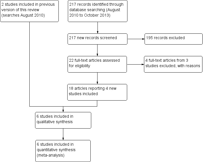
Results from searching for studies for inclusion in the review (as of 29 October 2013).
Included studies
We found six trials that met our inclusion criteria (Copernicus 2012; CRUISE 2010; Epstein 2012; GALILEO 2013; ROCC 2010; Wroblewski 2009). These investigated four anti‐VEGF agents in comparison to sham injection: VEGF Trap Eye (Aflibercept), bevacizumab (Avastin), ranibizumab (Lucentis) and Pegaptanib sodium (Macugen). The study design, treatments, doses and duration of treatment are summarised in the following table. The table Characteristics of included studies details a full summary of each of these trials.
Table: Summary of included studies
| Study | Study design | Treatment group 1 | Treatment group 2 | Control group | Number of injections | Treatment period |
| Copernicus 2012 | Phase III dmRCT | VEGF Trap‐Eye 2.0 mg (n = 114) | ‐ | Sham injection (n = 74) | 6 | 6 months |
| CRUISE 2010 | Phase III dmRCT | Ranibizumab 0.3 mg (n = 132) | Ranibizumab 0.5 mg (n = 130) | Sham injection (n = 130) | 6 | 6 months |
| Epstein 2012 | Phase III dmRCT | Bevacizumab 1.25 mg (n = 30) | ‐ | Sham injection (n = 30) | 4 | 24 weeks |
| GALILEO 2013 | Phase III dmRCT | VEGF Trap‐Eye 2.0 mg (n = 106) | ‐ | Sham injection (n = 71) | 6 + PRN | 52 weeks |
| ROCC 2010 | Phase III dmRCT | Ranibizumab 0.5 mg (n = 16) | ‐ | Sham injection (n = 16) | 3 + 3 (PRN) | 6 months |
| Wroblewski 2009 | Phase II dmRCT | Pegaptanib sodium 0.3 mg (n = 33) | Pegaptanib sodium 1.0 mg (n = 33) | Sham injection (n = 32) | 5 | 30 weeks |
PRN = pro re nata (as the circumstances require) dmRCT = double‐masked randomised controlled trial
Extension studies
In an open‐label, six‐month extension of the bevacizumab trial (Epstein 2012), all participants (n = 60, 100%) in both the sham control and bevacizumab 1.25 mg groups were treated with bevacizumab 1.25 mg every six weeks, to 12 months.
In an open‐label, six‐month observation period (i.e. months 7 to 12) participants in the CRUISE 2010 trial were reviewed monthly and offered ranibizumab if they met retreatment criteria including central retinal thickness > 250 or BCVA < 6/12. Those in the original treatment groups received either 0.3 mg or 0.5 mg ranibizumab, according to their original randomisation, whilst participants in the original sham control group received 0.5 mg ranibizumab. Of 392 participants randomised at baseline, 363 (92.6%) completed the study to six months, and 349 (89.0%) completed the open‐label extension study to 12 months. A further 12‐month, open‐label extension (i.e. months 13 onwards), the HORIZON trial, explored safety and efficacy in 304 (77.5%) of those originally recruited to the CRUISE trial. Participants were seen at least every three months and offered 0.5 mg ranibizumab if they met retreatment criteria. During the study, ranibizumab was approved by the FDA for the treatment of retinal vein occlusion and according to the protocol, all participants were discontinued from the study by 30 days after the approval date. The duration of follow‐up in the HORIZON trial was therefore variable, with a mean of 14 months (standard deviation (SD) 4.7, range 1 to 24 months), and missing outcome data at 24 months were considerable, with the consequent potential for significant attrition bias in the available results.
In an open‐label, six‐month extension of the Copernicus 2012 trial (i.e. months 7 to 12), all participants were offered 2.0 mg aflibercept (VEGF Trap‐Eye) monthly as needed, according to retreatment criteria. One hundred and sixty‐four (86.8%) of the participants randomised at baseline completed 52 weeks.
Baseline characteristics
The six trials included patients with broadly similar baseline characteristics, which are summarised in the table below. There was some difference in the proportion of recruited patients who were ischaemic at baseline, with negligible ischaemia at baseline in patients recruited to CRUISE 2010 and Wroblewski 2009. The mean age and percentage of male participants was similar across the six trials. The mean time between occlusive event and study entry was also broadly similar across the groups. The baseline mean BCVA measured in ETDRS letters was broadly similar between studies, ranging from a mean of 52 letters in GALILEO 2013 (Snellen approximately 6/30 or 20/100) to 43 letters (Snellen approximately 6/42 or 20/138) in the smaller of the ranibizumab trials (ROCC 2010). The percentage of patients with a poor presenting BCVA of less than 35 letters (Snellen approximately 6/60 or 20/200) ranged from 17% (GALILEO 2013) to 32% (Epstein 2012). The mean baseline central retinal thickness (CRT) was broadly similar across the six trials, ranging between 619 μm (Wroblewski 2009) and 721 μm (Epstein 2012).
Table: Summary of baseline characteristics
| Study | Duration of CRVO‐MO | % Ischaemic | Mean age (years) | % Male | Duration | Baseline mean VA (letters) | % BCVA less than 35 letters (6/60) | Mean CRT (μm) |
| Copernicus 2012 | < 9 months | 15.5% (29/187) ischaemic 16.6% (31/187) undetermined |
66 | 57 | Mean 2.40 months (SD 2.80) | 50.0 (SD 14.1) | 24.6% | 665.8 (SD 239.8) |
| CRUISE 2010 | < 3 months | 0.5% (2/392) | 68 | 57 | Mean 3.3 months Median 2 months (range 0 to 27) |
48.1 (14.6) | 30% | 680 (242) to 689 (253) |
| Epstein 2012 | < 6 months | 11.7% (7/60)* | 71 | 60 | Mean 8.8 weeks (SD 5.7) | 44.1 (SD 15.5) letters | 32% | 721 (SD 269) |
| GALILEO 2013 | < 9 months | 8.2% (14/171) ischaemic 8.2% (14/171) undetermined |
62 | 56 | Mean 82 days (SD 85) | 52.2 (SD 15.7) | 17% | 665.5 (SD 231.0) |
| ROCC 2010 | < 6 months | 15.6% (5/32) | 72 | 55 | Mean 78 days (range 10 to 163) | 43 (SD 22) | NR | 625 (SD 159) |
| Wroblewski 2009 | < 6 months | 0% (0/98) | 63 | 53 | Mean 80 days | 48.5 | 22% | 619 to 675 |
* Personal communication SD = standard deviation VA = visual acuity, measured in ETDRS letters CRT = central retinal thickness
Excluded studies
We excluded four randomised studies including an anti‐VEGF intervention group because they did not include a sham control or observation arm. One non‐masked trial randomised 20 patients with CRVO macular oedema to receive 0.3 mg (n = 10) or 0.5 mg (n = 10) ranibizumab given monthly for three months (Campochiaro 2008). In a subsequent extension trial to 24 months, patients were reviewed every two months and treated with the same dose of ranibizumab as their initial treatment assignment, if retreatment criteria were met (Campochiaro 2010). A second trial compared treatment with bevacizumab to combined treatment with bevacizumab and timolol‐dorzolamide, without comparison to a sham injection or observation group. This trial combined patients with both BRVO and CRVO, and the small sample size precluded analysis of CRVO macular oedema specifically (Byeon 2009). A third open‐label study randomised patients with CRVO macular oedema to receive intravitreal injection of either 4 mg preservative‐free triamcinolone acetonide (n = 16) or 1.25 mg bevacizumab (n = 16) at baseline, with subsequent 'as required' injections from three months (Ding 2011). A fourth open‐label study randomised patients with CRVO macular oedema to a single injection of either 1.25 mg bevacizumab or to combination therapy with 1.25 mg bevacizumab plus 2.0 mg triamcinolone acetonide, with follow‐up over 12 weeks (Wang 2011). (See the Characteristics of excluded studies table for further details).
We did not conduct a separate electronic search for study designs less rigorous than the randomised controlled trial. We identified numerous interventional case series and case reports and summarised them in tables in the first publication of this review (Braithwaite 2010). These studies did not meet the inclusion criteria for the systematic review, and these tables were not updated or included in the current review.
Risk of bias in included studies
The six included studies had a low risk of bias in a majority of domains (see Figure 2 and Figure 3). In all studies one eye was enrolled in the study per participant. In CRUISE 2010 the authors specified that where both eyes met the inclusion criteria, the eye with the worse BCVA at screening was selected. In Copernicus 2012 and GALILEO 2013 the patient was excluded if both eyes had a retinal vein occlusion at baseline. In ROCC 2010 patients were only recruited to the study if they had unilateral CRVO macular oedema. Whilst one eye per patient was included in the remaining two trials, Wroblewski 2009 and Epstein 2012, no statement regarding the method of selection of the study eye was given to cover the uncommon event that both eyes met the eligibility criteria.
2.
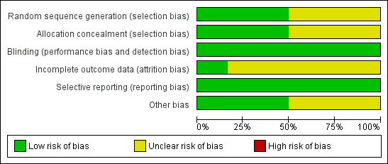
'Risk of bias' graph: review authors' judgements about each risk of bias item presented as percentages across all included studies.
3.
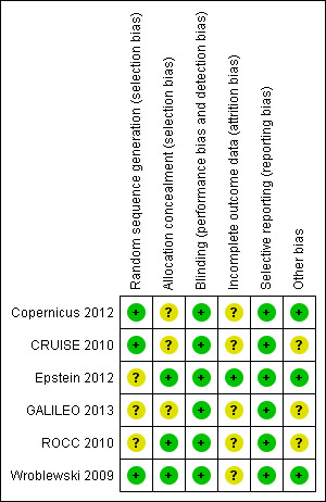
'Risk of bias' summary: review authors' judgements about each risk of bias item for each included study.
Allocation
We judged there to be a low risk of bias in random sequence generation in three studies (Copernicus 2012CRUISE 2010; Wroblewski 2009). These studies all reported a centralised randomisation method. We considered the risk of bias 'unclear' in the other three studies (Epstein 2012;GALILEO 2013; ROCC 2010). We gave a judgement of unclear risk of bias where the study investigators did not explicitly report the process of random sequence generation, and either did not respond to a request for further information, or did not provide sufficient additional explanation via personal communication.
We judged there to be a low risk of bias in allocation concealment in three studies (Epstein 2012; ROCC 2010; Wroblewski 2009). We considered the risk of bias to be low in studies that explicitly reported the method of protection of the allocation sequence between the time of randomisation and of treatment assignment to injection or sham injection. For example, if the medication kits were identical in appearance and identified by randomisation number only, and if the allocation was conveyed to the injecting ophthalmologist by the study co‐ordinator, or another third party, in a way that did not inform the injecting ophthalmologist of the allocation until the time of the injection procedure, then we considered the risk of bias low. When sealed, opaque envelopes drawn by staff not involved in patient treatment or follow‐up were used to conceal allocation until the first day of injection, we judged the risk of bias to be low, even if the envelopes were not sequentially numbered based on the randomisation code, although we note that methods utilising envelopes may be subject to manipulation. We felt the risk of bias in this domain was unclear in the remaining three studies (Copernicus 2012; CRUISE 2010; GALILEO 2013), in which the method of concealment of the allocations was not described explicitly.
Blinding
We considered all six studies to have a low risk of bias in this domain. In these studies, the participants, examining ophthalmologists and other clinical examiners were masked to the treatment allocation. In addition, in the aflibercept trial (Copernicus 2012), pegaptanib sodium trial (Wroblewski 2009) and the CRUISE 2010 trial, grading of OCT and FFA images was done by an independent grading centre. The ophthalmologists performing the intravitreal injections, who performed either a sham injection or a drug injection, were not involved in reviewing the participants at their follow‐up assessments. In all studies an attempt was made to mask patients randomised to the control group by treating them similarly to those in the treatment groups, except that the hub of a syringe was placed against the injection site and the plunger depressed to mimic an injection, without globe penetration (i.e. sham injection). It is unclear whether sham injection effectively masks patients to their treatment allocation, given that patients sometimes report 'feeling' the needle despite topical anaesthesia, and that the injection can cause a subconjunctival haemorrhage which would not necessarily be expected with a sham injection. However, a majority of the outcomes in these studies are relatively objective measures, so that even if a patient suspected their assignment this would be unlikely to bias the outcome data.
Incomplete outcome data
We considered there to be a low risk of attrition bias in one study reporting no loss to follow‐up (Epstein 2012). We also would have considered the risk of bias to be low if the losses to follow‐up were small and balanced between the groups, with analysis by intention‐to‐treat, or if imputation methods that provide valid type 1 error rates under explicitly stated assumptions were used to take moderate missing data into account.
Where we considered the losses to be slightly more considerable as a proportion of the total sample (an issue with small studies), or where we felt that losses to follow‐up were unbalanced between groups, but where the investigators reported reasons for losses, we considered the risk of bias 'unclear' (Copernicus 2012; CRUISE 2010; GALILEO 2013; ROCC 2010; Wroblewski 2009). Unbalanced loss of patients with potentially more severe and visually significant disease from the sham group in these trials might have introduced bias, reducing any apparent benefit associated with anti‐VEGF therapy. We also considered the risk of bias 'unclear' where investigators did not account for missing data, performing only a 'per protocol' analysis (ROCC 2010), restricted to those participants who fulfilled the criteria for eligibility, received all prescribed interventions and attended all outcome assessments. Non‐random loss of participants is better handled through analysis by the intention‐to‐treat approach, in which data are analysed according to randomisation group, regardless of whether the participants received or adhered to their allocated intervention, as this provides fair comparisons among the groups.
Where investigators accounted for losses to follow‐up or unbalanced missing data using the last‐observation‐carried‐forward (LOCF) method, without performing sensitivity analyses to assess the impact of assumptions about the method of accounting for missing data on trial outcomes, we also considered the risk 'unclear' (Copernicus 2012; CRUISE 2010; GALILEO 2013). The LOCF method limits the number of patients eliminated from the analysis. However, it assumes that patients do not change from their last follow‐up (i.e. they could improve or get worse and in either case this would not be captured). Treating missing data as if there has been no change from a previous visit generally yields a conservative estimate of treatment effect. If patients who are given treatment drop out because they get worse or experience harmful side effects then the LOCF methods may over‐report efficacy or under‐report harmful safety problems.
Selective reporting
We accessed ClinicalTrials.gov to review the prespecified primary and secondary outcomes for the trials, where these were available. We considered there to be a low risk of selective reporting bias in all six studies (Copernicus 2012; CRUISE 2010; GALILEO 2013; ROCC 2010; Wroblewski 2009), in which the main prespecified primary and secondary outcomes were reported. In Epstein 2012, an additional secondary outcome of mean change in BCVA was added in the published report. In Copernicus 2012, two additional secondary outcomes were reported: the proportion of eyes progressing to ocular neovascularisation and the change in total score on the National Eye Institute 25‐item Visual Function Questionnaire (NEI VFQ‐25) from baseline. In ROCC 2010, the investigators prespecified that they would include mean change from baseline in the NEI VFQ‐25 near activities subscale as a secondary outcome, but did not include this in their published report. However, as the subscale on this instrument is not a validated outcome measure, we felt that failure to report these results had no impact on the key outcome measures of interest. The ClinicalTrials.gov entry for Wroblewski 2009 did not include prespecified outcome measures. However, all outcomes that would be expected were included, and both positive and negative results were reported without apparent bias, so although we were not able to exclude the possibility of selective reporting, we considered the study 'low risk' in this domain. Four studies did not report measures of variance for continuous variables including mean change in best‐corrected visual acuity from baseline and mean change in central retinal thickness from baseline (Copernicus 2012; Epstein 2012;GALILEO 2013; Wroblewski 2009); unpublished data were provided for the bevacizumab trial (personal communication).
Other potential sources of bias
We considered the risk of bias to be low in three studies where no other threats to validity were identified, or where we considered potential sources of bias to be very small (Copernicus 2012; Epstein 2012; Wroblewski 2009). We considered the risk of bias to be 'unclear' in three studies where protocol violations were reported, resulting from recruitment of small numbers of patients who either did not meet the prespecified inclusion criteria, or who did not receive all planned treatment (CRUISE 2010; GALILEO 2013; ROCC 2010).
Effects of interventions
See: Table 1
Primary outcome
Gain of at least 15 letters best‐corrected visual acuity at six months
Five studies reported this outcome (Copernicus 2012; CRUISE 2010; Epstein 2012; GALILEO 2013; Wroblewski 2009), and unpublished data were provided by study investigators for the sixth study (ROCC 2010). The table below summarises and compares the data across the six randomised controlled trials (RCTs) at six months. The proportion gaining 15 letters or more of visual acuity at six months ranged from 12.3% to 28.1% in the sham groups, and from 36.4% to 60.2% in the treatment groups. Data from six trials were included in the meta‐analysis. Meta‐analysis indicated that patients receiving intravitreal anti‐vascular endothelial growth factor (anti‐VEGF) treatment were 2.71 times more likely to gain 15 letters or more of visual acuity at six months compared to patients treated with sham injections, and the 95% confidence interval (CI) suggested a statistically significant effect (RR 2.71; 95% CI 2.10 to 3.49) (see Analysis 1.1; Figure 4). There was no significant statistical heterogeneity (I2 = 10%), and also no significant difference between the different anti‐VEGF subgroups (I2 = 11.7%). This demonstrates a clinically significant gain in visual acuity at six months associated with anti‐VEGF therapy.
1.1. Analysis.
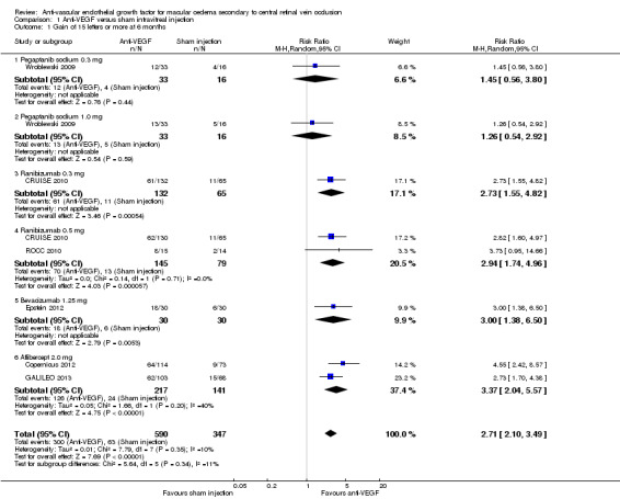
Comparison 1 Anti‐VEGF versus sham intravitreal injection, Outcome 1 Gain of 15 letters or more at 6 months.
4.
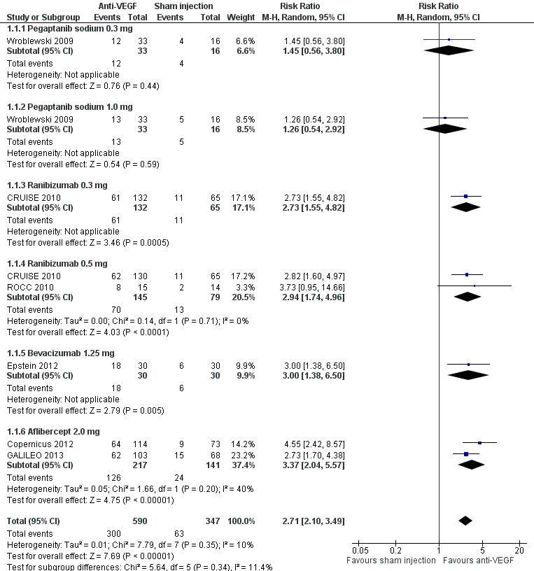
Forest plot of comparison: Anti‐VEGF versus sham intravitreal injection, outcome: 1.1 Gain of 15 letters or more at 6 months.
| Study | Treatment group 1 | % > 15 letters gain at 6 months (n) | Treatment group 2 | % > 15 letters gain at 6 months (n) | Control group | % > 15 letters gain at 6 months (n) | P value |
| Copernicus 2012 | VEGF Trap‐Eye 2.0 mg (n = 114) | 56.1 (64/114) | ‐ | ‐ | Sham injection | 12.3 (9/73) | < 0.001 |
| CRUISE 2010 | Ranibizumab 0.3 mg (n = 132) | 46.2 (61/132) | Ranibizumab 0.5 mg (n = 130) | 47.7 (62/130) | Sham injection | 16.9 (22/130) | < 0.0001 |
| Epstein 2012 | Bevacizumab 1.25 mg (n = 30) | 60.0 (18/30) | ‐ | ‐ | Sham injection | 20.0 (6/30) | 0.003 |
| GALILEO 2013 | VEGF Trap‐Eye 2.0 mg (n = 106) | 60.2 (62/103) | ‐ | ‐ | Sham injection | 22.1 (15/68) | < 0.0001 |
| ROCC 2010 | Ranibizumab 0.5 mg (n = 16) | 53.3 (8/15) | ‐ | ‐ | Sham injection | 14.3 (2/14) | ‐ |
| Wroblewski 2009 | Pegaptanib sodium 0.3 mg (n = 33) | 36.4 (12/33) | Pegaptanib sodium 1.0 mg (n = 33) | 39.4 (13/33) | Sham injection | 28.1 (9/32) | 0.35 |
Only one RCT reported 12‐month outcomes for 15‐letter gain in visual acuity, comparing treatment with 2 mg intravitreal aflibercept versus sham control, using PRN (as needed) dosing between months 6 and 12 (GALILEO 2013). Meta‐analysis was therefore not performed. The proportion receiving aflibercept who gained 15 letters or more was largely maintained between months 6 and 12, at 60.2%. A mean 2.5 injections (standard deviation (SD) 1.7) were required during the PRN phase. The proportion gaining 15 letters of visual acuity improved in those randomised to sham injection who received PRN aflibercept between 6 and 12 months, from 22.1% to 32.4% (GALILEO 2013), but remained significantly worse than those randomised to aflibercept (P = 0.0004).
In the three open‐label extension studies the gain in visual acuity seen at six months in those treated with anti‐VEGF agents was also largely maintained at 12 months (Copernicus 2012; CRUISE 2010; Epstein 2012). Fifteen letters or more of visual acuity was gained at 12 months by 55.3% (63/114) of those treated with aflibercept monthly for six months then PRN with monthly reassessment for a further six months (Copernicus 2012); by 60% (18/30) of those treated with bevacizumab every six weeks for 12 months (Epstein 2012); and by 47.0% (62/132) and 50.8% (66/130) of those treated with 0.3 mg and 0.5 mg ranibizumab, respectively, every month for six months then PRN with monthly reassessment for a further six months (CRUISE 2010). Patients randomised to sham injection in these three trials crossed over to treatment with anti‐VEGF from months 7 to 12, and in all sham/anti‐VEGF groups the proportion gaining 15 letters or more at 12 months was higher than it had been at six months prior to treatment with anti‐VEGF. Specifically, 15 letters or more gain in BCVA was reported in 30.1% (22/73) receiving delayed 2.0 mg aflibercept PRN for six months (Copernicus 2012), 33.1% (43/130) receiving delayed 0.5 mg ranibizumab PRN for six months (CRUISE 2010) and 33.3% (10/30) receiving delayed 1.25 mg bevacizumab every six weeks for six months (Epstein 2012). However, in all three trials the sham/anti‐VEGF cross‐over groups remained significantly worse with respect to this primary outcome than the groups initially randomised to anti‐VEGF treatment.
Subgroup comparison of patients with baseline ischaemic central retinal vein occlusion (CRVO) macular oedema versus non‐ischaemic CRVO macular oedema, defined as over 10 disc areas of retinal non‐perfusion on a seven standard field fluorescein angiogram, was reported in one trial (Copernicus 2012). The proportion of eyes with baseline ischaemia who gained 15 letters at six months was 51.4% versus 4.3% in those treated with aflibercept and sham respectively (Copernicus 2012). The proportion of eyes without ischaemia at baseline who gained 15 letters at six months was 58.4% versus 16.0% in those treated with aflibercept and sham, respectively. At 52 weeks, the proportion gaining 15 letters or more of visual acuity following anti‐VEGF treatment was largely maintained in both ischaemic and non‐ischaemic treatment groups, at 48.6% and 58.4%, respectively. Following cross‐over PRN treatment of the sham group from months 7 to 12, the proportion gaining 15 letters or more improved in both ischaemic and non‐ischaemic sham randomised patients, to 30.4% and 30.0%. This suggests a beneficial effect of anti‐VEGF treatment in both patients with ischaemic CRVO and with non‐ischaemic CRVO, and a lesser benefit to both ischaemic and non‐ischaemic subgroups if treatment is delayed by six months.
Subgroup comparison of patients randomised to receive treatment within or beyond two months of diagnosis was performed in two trials (Copernicus 2012; GALILEO 2013). The proportion of patients treated with aflibercept who gained 15 letters or more at six months was 64.1% versus 42.9% in those receiving first aflibercept treatment within or later than two months of diagnosis, respectively (Copernicus 2012). Similarly, in the GALILEO study, a 15 letter gain was seen in 70.9% versus 50.0% treated with aflibercept within or beyond two months of diagnosis, respectively (GALILEO 2013). This suggests a more beneficial effect of anti‐VEGF treatment when commenced early.
Subgroup comparison of patients with a baseline BCVA of 6/60 (20/200) or worse was performed in one trial (Copernicus 2012). The proportion gaining 15 letters or more at six months was 67.9% versus 16.7% in the aflibercept versus sham groups with a baseline visual acuity of 6/60 (20/200) or worse, and 52.3% versus 10.9% in the aflibercept versus sham treated groups with a baseline visual acuity better than 6/60. This suggests that anti‐VEGF treatment is beneficial even with a very poor presenting visual acuity.
Secondary outcomes
Loss of 15 letters or more best‐corrected visual acuity at six months
Four studies reported this outcome (Copernicus 2012; CRUISE 2010; Epstein 2012; Wroblewski 2009), and unpublished data were provided by study investigators for a fifth study (ROCC 2010). The table below summarises and compares the data across the five RCTs. The proportion losing 15 letters of visual acuity at six months ranged from 15.4% to 31.2% in the sham groups, and from 1.8% to 13.3% in the treatment groups. We included data from all five studies in the meta‐analysis. Meta‐analysis suggested that patients receiving intravitreal anti‐VEGF treatment had an 80% lower risk of losing 15 letters of visual acuity at six months compared to patients receiving a sham injection (RR 0.20; 95% CI 0.12 to 0.34) and the 95% CI suggested a statistically significant effect (see Analysis 1.2; Figure 5). There was no significant statistical heterogeneity (I2 = 0%) and also no significant difference between the different anti‐VEGF subgroups (I2 = 0%). This represents a clinically significant benefit of anti‐VEGF therapy at six months.
1.2. Analysis.
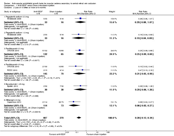
Comparison 1 Anti‐VEGF versus sham intravitreal injection, Outcome 2 Loss of 15 letters or more at 6 months.
5.
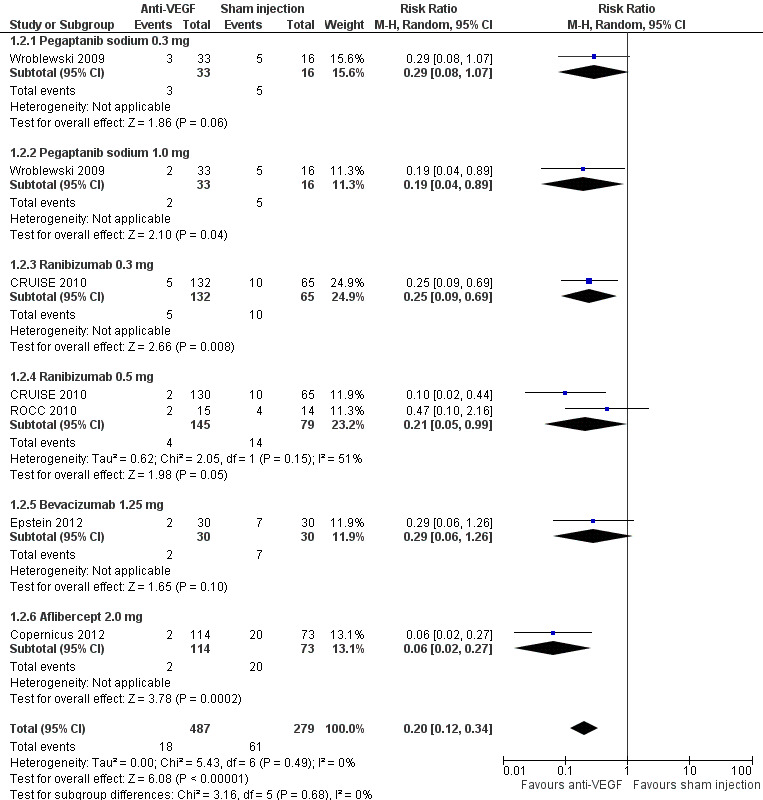
Forest plot of comparison: Anti‐VEGF versus sham intravitreal injection, outcome: 1.2 Loss of 15 letters or more at 6 months.
| Study | Treatment group 1 | % > 15 letters loss at 6 months | Treatment group 2 | % > 15 letters loss at 6 months | Control group | % > 15 letters loss at 6 months | P value |
| Copernicus 2012 | VEGF Trap‐Eye 2.0 mg (n = 114) | 1.8% (2/114) | ‐ | ‐ | Sham injection | 27.4% (20/73) | ‐ |
| CRUISE 2010 | Ranibizumab 0.3 mg (n = 132) | 3.8% (5/132) | Ranibizumab 0.5 mg (n = 130) | 1.5% (2/130) | Sham injection | 15.4% (20/130) | < 0.005 |
| Epstein 2012 | Bevacizumab 1.25 mg (n = 30) | 6.7% (2/30) | ‐ | ‐ | Sham injection | 23.3% (7/30) | 0.15 |
| GALILEO 2013 | VEGF Trap‐Eye 2.0 mg (n = 106) | Not reported | ‐ | ‐ | Sham injection | Not reported | ‐ |
| ROCC 2010 | Ranibizumab 0.5 mg (n = 16) | 13.3% (2/15) | ‐ | ‐ | Sham injection | 28.6% (4/14) | ‐ |
| Wroblewski 2009 | Pegaptanib sodium 0.3 mg (n = 33) | 9.1% (3/33) | Pegaptanib sodium 1.0 mg (n = 33) | 6.1% (2/33) | Sham injection | 31.2% (10/32) | 0.03 |
Only one trial reported 12‐month outcomes and so we did not perform meta‐analysis of this outcome at 12 months (GALILEO 2013). Loss of 15 letters or more visual acuity at 12 months developed in 1% (n = 1) of the treatment group versus 14.7% (n = 10) of the sham group (GALILEO 2013).
In the three open‐label extension trials the number of patients treated with anti‐VEGF therapy who lost 15 letters or more of visual acuity remained stable (CRUISE 2010; Epstein 2012), or worsened slightly (Copernicus 2012), between months 6 and 12. Specifically, 6.7% (2/30) treated with an additional six months of bevacizumab six‐weekly (Epstein 2012), 5.3% (6/114) treated with an additional six months of aflibercept PRN Copernicus 2012, 3.8% (5/132) treated with an extra six months of 0.3 mg ranibizumab PRN, and 2.3% (3/130) treated with an extra six months of 0.5 mg ranibizumab PRN (CRUISE 2010) had > 15 letters loss at 12 months. With six‐month delayed anti‐VEGF treatment, fewer patients in the sham cross‐over groups had > 15 L vision loss at 12 months compared to their six‐month outcomes. Specifically, 6.7% (2/30) (Epstein 2012), 10.0% (13/130) (CRUISE 2010) and 15.1% (11/73) (Copernicus 2012) of sham/anti‐VEGF cross‐over patients had more than 15 letters vision loss at 12 months, suggesting a beneficial effect of delayed anti‐VEGF therapy.
Mean change in best‐corrected visual acuity at six months
All six studies reported this outcome, but only three reported measures of dispersion (standard deviation or 95% CI) (Copernicus 2012; CRUISE 2010; ROCC 2010). Unpublished data from a fourth study were provided by the investigators (Epstein 2012). The table below summarises and compares the data across the six RCTs. The mean change in best‐corrected visual acuity letter score at six months ranged from a gain of 3.3 letters to a loss of 4.0 letters in the sham groups, and from a gain of 7.1 letters to a gain of 18.0 letters in the treatment groups. In all studies, the greatest gain in BCVA was seen within one to two months of treatment with the anti‐VEGF agents, with maintenance or more gradual improvement thereafter to six months. The mean difference (MD) between anti‐VEGF and sham was 15.23 letters (95% CI 11.57 to 18.89) at six months. Although the statistical heterogeneity was considerable (I2 = 63%), we combined data in meta‐analysis because the direction of effect was the same for all trials (Analysis 1.3).
1.3. Analysis.
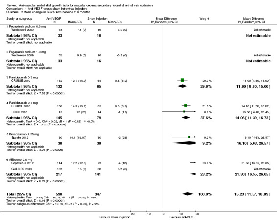
Comparison 1 Anti‐VEGF versus sham intravitreal injection, Outcome 3 Mean change in BCVA from baseline at 6 months.
| Study | Treatment group 1 | Mean change in BCVA (letters) | Treatment group 2 | Mean change in BCVA (letters) | Control group | Mean change in BCVA (letters) | P value |
| Copernicus 2012 | VEGF Trap‐Eye 2.0 mg (n = 114) | +17.3 | ‐ | ‐ | Sham injection | ‐4.0 | < 0.001 |
| CRUISE 2010 | Ranibizumab 0.3 mg (n = 132) | +12.7 | Ranibizumab 0.5 mg (n = 130) | +14.9 | Sham injection | +0.8 | < 0.0001 |
| Epstein 2012 | Bevacizumab 1.25 mg (n = 30) | +14.1 | ‐ | ‐ | Sham injection | ‐2.0 | ‐ |
| GALILEO 2013 | VEGF Trap‐Eye 2.0 mg (n = 106) | +18.0 | ‐ | ‐ | Sham injection | +3.3 | < 0.0001 |
| ROCC 2010 | Ranibizumab 0.5 mg (n = 16) | +12.0 | ‐ | ‐ | Sham injection | ‐1.0 | < 0.01 |
| Wroblewski 2009 | Pegaptanib sodium 0.3 mg (n = 33) | +7.1 | Pegaptanib sodium 1.0 mg (n = 33) | +9.9 L | Sham injection | ‐3.2 | 0.02 and 0.09 |
Only one trial reported change in mean BCVA at 12 months and so meta‐analysis was not performed (GALILEO 2013). The significant difference between treatment and sham groups seen at 24 weeks was maintained to 52 weeks, with a respective gain from baseline in BCVA of 16.9 letters and 3.8 letters (P < 0.0001).
In the three open‐label PRN extension studies the mean gain in BCVA from baseline seen at six months in those treated with anti‐VEGF agents was also largely maintained at 12 months (Copernicus 2012; CRUISE 2010; Epstein 2012). The mean gain in the treatment groups was 16.2 letters (Copernicus 2012), 16.1 letters (Epstein 2012) and 13.9 letters in those treated with both 0.3 mg and 0.5 mg ranibizumab (CRUISE 2010). Patients randomised to sham injection in these three trials crossed over to treatment with anti‐VEGF from months 7 to 12, and in all sham/anti‐VEGF groups the mean gain in BCVA at 12 months was higher than it had been at six months prior to treatment with anti‐VEGF. Specifically, a BCVA gain of 3.8 letters from baseline was reported in those receiving six‐month delayed 2.0 mg aflibercept PRN for six months (Copernicus 2012), a gain of 7.3 letters was reported in those receiving delayed 0.5 mg ranibizumab PRN for six months (CRUISE 2010), and a gain of 4.6 letters was reported in those receiving delayed 1.25 mg bevacizumab every six weeks for six months (Epstein 2012). However, in all three trials the sham/anti‐VEGF cross‐over groups remained significantly worse with respect to this outcome than the groups initially randomised to anti‐VEGF treatment, with P values for comparison between groups at 12 months of P < 0.001 (Copernicus 2012), P < 0.001 (CRUISE 2010) and P < 0.05 (Epstein 2012). Only one study, the HORIZON trial, considered longer‐term acuity outcomes, in 304 patients (87%) who completed the 12‐month CRUISE trial (CRUISE 2010). Patients originally randomised to sham (n = 98), 0.3 mg ranibizumab (n = 107) and 0.5 mg ranibizumab (n = 99) were eligible for PRN treatment with 0.5 mg ranibizumab during months 12 to 24, were typically reviewed every three months and received a mean of 2.9, 3.8 and 3.5 injections, respectively. At the end of 24 months of treatment in total, the mean BCVA gain from baseline was 7.6 letters, 8.2 letters and 12.0 letters in the sham/0.5 mg, 0.3 mg/0.5 mg and 0.5 mg groups, respectively.
Subgroup analysis in anti‐VEGF treatment groups, based on disease duration, was performed in three studies (Copernicus 2012; Epstein 2012; GALILEO 2013). Patients treated with bevacizumab within 90 days improved by 18.7 letters at six months, compared to 9.8 letters in patients with a longer disease duration (Epstein 2012). Patients treated with aflibercept within two months improved by 20.2 letters (versus a ‐5.5 loss in the sham group) at six months, compared to 13.4 letters (versus ‐0.5 letters loss in the sham group) in patients with a longer disease duration (Copernicus 2012). A similar difference was seen at 12 months in those treated with aflibercept within two months of diagnosis who gained 19.5 letters (versus 2.1 letters in the sham group), compared to a gain of 13.7 letters (versus 5.5 letters in the sham group) seen in those with a longer disease duration (GALILEO 2013).
Subgroup analysis according to baseline perfusion status was reported in two trials (Copernicus 2012; GALILEO 2013). Eyes with ischaemic CRVO macular oedema at baseline gained +17.8 letters by six months when treated with aflibercept, compared to ‐2.3 letters in the sham group (Copernicus 2012). Eyes with non‐ischaemic CRVO macular oedema at baseline gained +17.1 letters by six months when treated with aflibercept, compared to ‐4.8 letters in the sham group (Copernicus 2012). Similarly, the GALILEO 2013 trial demonstrated that patients stratified by baseline perfusion status and treated with aflibercept for six months plus six months PRN had a similar gain from baseline in BCVA at 12 months, of +16.8 (SD 14.7 letters) and 17.4 (SD 16.1 letters) in the non‐ischaemic and ischaemic groups, respectively (GALILEO 2013). In contrast, eyes with no baseline retinal ischaemia in the sham group gained a mean 6.8 letters (SD 17.5) compared to a loss of 8.0 letters (SD 15.8 letters) in those with baseline ischaemia (GALILEO 2013). Importantly, this suggests a benefit of anti‐VEGF treatment with aflibercept in eyes with both ischaemic and non‐ischaemic CRVO macular oedema, and possibly a relatively greater gain in those with ischaemic CRVO macular oedema at baseline, whose visual outcome without treatment is worse.
Subgroup analysis of patients according to baseline mean BCVA of better than or worse than 6/60 (20/200) was performed in two trials (Copernicus 2012; GALILEO 2013). In patients with a baseline BCVA of 6/60 or worse, the mean change in Early Treatment of Diabetic Retinopathy Study (ETDRS) letter score was +21.9 versus 0 in the anti‐VEGF versus sham groups; in patients with a baseline BCVA of better than 6/60 the mean change was +15.9 versus ‐5.4 letters in the anti‐VEGF and sham groups, respectively (Copernicus 2012). In the second trial, at 12 months, patients with a baseline BCVA of 6/60 or worse also had a greater BCVA gain than those with a baseline BCVA of better than 6/60 (9.4 versus 2.4 letters for sham and 21.1 versus 16.0 letters for aflibercept monthly for six months then PRN for six months, respectively) (GALILEO 2013). This suggests a benefit of anti‐VEGF treatment compared to sham, especially for patients with a worse presenting baseline BCVA.
Mean change in central retinal thickness (CRT) from baseline
All six studies measured this outcome but only two reported measures of dispersion (standard deviation or 95% CI) (CRUISE 2010; ROCC 2010); unpublished data were supplied for a third study (Epstein 2012, personal communication). The table below summarises and compares the data across the six RCTs. The mean change in CRT from baseline to six months ranged from a reduction of 102 µm to 169.3 µm in the sham groups, to a reduction of 243 µm to 457.2 µm in the treatment groups. Meta‐analysis of the data from three studies suggests that patients treated with anti‐VEGF agents have a mean reduction in CRT from baseline of 267.4 µm more than patients treated with sham, with 95% confidence that the true difference in the reduction lies between 211.4 µm and 323.4 µm. There was no significant statistical heterogeneity (I2 = 8%) and also no significant difference between the different anti‐VEGF subgroups (I2 = 0%) (see Analysis 1.4; Figure 6). This represents a clinically significant benefit of anti‐VEGF treatment over sham treatment at six months.
1.4. Analysis.
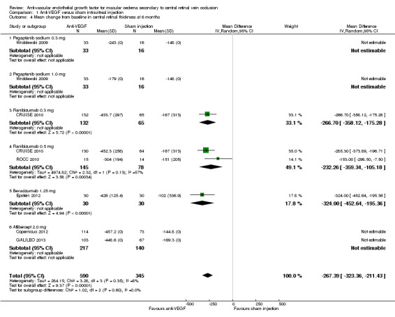
Comparison 1 Anti‐VEGF versus sham intravitreal injection, Outcome 4 Mean change from baseline in central retinal thickness at 6 months.
6.
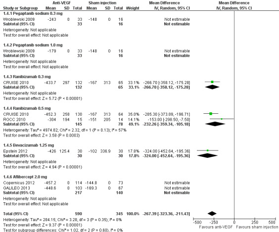
Forest plot of comparison: Anti‐VEGF versus sham intravitreal injection, outcome: 1.4 Mean change from baseline in central retinal thickness at 6 months.
| Study | Treatment group 1 | Mean change in CRT (µm) | Treatment group 2 | Mean change in CRT (µm) | Control group | Mean change in CRT |
| Copernicus 2012 | VEGF Trap‐Eye 2.0 mg (n = 114) | ‐457.2 | ‐ | ‐ | Sham injection | ‐144.8 |
| CRUISE 2010 | Ranibizumab 0.3 mg (n = 132) | ‐433.7 | Ranibizumab 0.5 mg (n = 130) | ‐452.3 | Sham injection | ‐167 |
| Epstein 2012 | Bevacizumab 1.25 mg (n = 30) | ‐426 | ‐ | ‐ | Sham injection | ‐102 |
| GALILEO 2013 | VEGF Trap‐Eye 2.0 mg (n = 106) | ‐448.6 | ‐ | ‐ | Sham injection | ‐169.3 |
| ROCC 2010 | Ranibizumab 0.5 mg (n = 16) | ‐304 | ‐ | ‐ | Sham injection | ‐151 |
| Wroblewski 2009 | Pegaptanib sodium 0.3 mg (n = 33) | ‐243 | Pegaptanib sodium 1.0 mg (n = 33) | ‐179 | Sham injection | ‐148 |
In all six trials, graphs of the mean change in CRT over time convincingly illustrated the rapid and beneficial effect of all the anti‐VEGF agents on resolution of macular oedema. The greatest reduction is seen within a month of the first injection in all trials, and the CRT reduction benefit is maintained throughout the treatment period. This is in marked contrast to the trajectory of change in CRT from baseline of the control patients, who demonstrate a more gentle, linear reduction in CRT over time.
Three open‐label extension trials investigated the impact of immediate versus six‐month delayed treatment with anti‐VEGF on central retinal thickness (CRT) at 12 months. In these trials, patients initially randomised to sham injection for six months crossed over to receive treatment with anti‐VEGF agents from months 7 to 12, either every six weeks (Epstein 2012), or monthly as required, and according to retreatment criteria (Copernicus 2012; CRUISE 2010). In all three trials, the mean reduction in CRT was largely maintained from months 6 to 12 in the anti‐VEGF treatment groups (Copernicus 2012; CRUISE 2010; Epstein 2012). No significant difference in CRT, or the proportion of patients with residual macular oedema, was found between the anti‐VEGF and sham/anti‐VEGF groups at 12 months (Copernicus 2012; CRUISE 2010; Epstein 2012). These trials suggest that patients not receiving anti‐VEGF therapy for an initial six months catch up, in terms of improvement in CRT, following six months of anti‐VEGF therapy (CRUISE 2010; Epstein 2012). It should be noted, however, that resolution towards normal CRT does not necessarily indicate restoration of normal structural integrity or neuroretinal function.
One trial reported subgroup analysis for 12‐month outcomes by baseline retinal perfusion status (GALILEO 2013). Patients treated with six months of monthly aflibercept followed by six months of PRN aflibercept, versus sham, had a greater reduction from baseline in mean CRT, regardless of baseline non‐ischaemia or ischaemia (412.4 (SD 238.1) versus 201.2 (SD 226.4) for the non‐ischaemic treatment and sham groups, and 494.6 (SD 318.4) versus 294.3 (SD 258.6) for the ischaemic treatment and sham groups) (GALILEO 2013).
Complications and ocular adverse events
Certain complications may develop in the natural history of untreated CRVO macular oedema, in particular neovascularisation of the iris and retina, associated neovascular glaucoma and vitreous haemorrhage. Objective differentiation of complications versus adverse events associated with intravitreal injection is not always possible, and so we considered these outcomes together without a priori assumption in this review, at six months of follow‐up. In addition, one study reported safety data at 12 months (GALILEO 2013). Three studies included open‐label extension to 12 months (Copernicus 2012; CRUISE 2010; Epstein 2012). One study included open‐label extension to 24 months (CRUISE 2010). However, since patients initially randomised to sham injection received PRN anti‐VEGF therapy beyond six months in these three trials, there was no untreated control group for inclusion in meta‐analysis to compare outcomes at 12 months. Where the outcome developed with greater frequency in the sham group, we suggest that anti‐VEGF reduced progression to this complication. Where the outcome occurred with greater frequency in the anti‐VEGF group, we suggest it to be an adverse event associated with intravitreal injection or with anti‐VEGF therapy. The six trials did not all report all potential adverse events or complications at six months. Where data on a specific outcome are incomplete, we do not know whether no outcomes occurred, whether outcomes that occurred were not documented or reported, or whether there was selective reporting bias (Analysis 1.5).
1.5. Analysis.
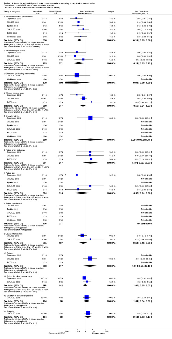
Comparison 1 Anti‐VEGF versus sham intravitreal injection, Outcome 5 Adverse events and complications at 6 months (ocular).
The development of iris or retinal neovascularisation was reported in all six studies. Across all studies, there were a total of 26/346 patients with this complication in the sham injection groups compared to 8/590 in the anti‐VEGF treatment groups at six months. The Peto odds ratio was 0.18 (95% CI 0.09 to 0.36), with no significant statistical heterogeneity (I2 = 0%). This suggests that treatment with anti‐VEGF therapy reduced the odds of progression to this recognised complication of CRVO to about 18% of what it would have been without treatment. Four studies specifically reported that many of these patients received panretinal photocoagulation treatment for this (Copernicus 2012; GALILEO 2013; ROCC 2010; Wroblewski 2009). Cases of neovascular glaucoma at six months were reported in three trials (Copernicus 2012; CRUISE 2010; GALILEO 2013) in a total of 1/479 patients treated with anti‐VEGF and 5/271 patients in the sham groups. The Peto odds ratio was 0.14 (95% CI 0.03 to 0.72), with no significant statistical heterogeneity (I2 = 0%), suggesting a beneficial effect of anti‐VEGF therapy in reducing progression to about 14% of what it would have been without anti‐VEGF treatment. Additional non‐neovascular glaucoma cases were reported in two trials, in 0/170 patients in the anti‐VEGF groups and 1/100 patients in the control groups (GALILEO 2013; Wroblewski 2009). Considering longer durations of follow‐up, new iris neovascularisation developing between 6 and 12 months did not occur in the sham or treatment groups of the aflibercept or bevacizumab trials (Copernicus 2012; Epstein 2012; GALILEO 2013), but did develop in an additional two people in the sham/0.5 mg ranibizumab PRN group, and in an additional four persons in the 0.5 mg/0.5 mg PRN treatment groups of the CRUISE 2010 trial.
Vitreous haemorrhage was specifically reported in three studies (Copernicus 2012; CRUISE 2010; ROCC 2010), in a total of 14/390 patients treated with anti‐VEGF and 13/217 patients in the sham groups. The Peto odds ratio was 0.55 (95% CI 0.24 to 1.23) suggesting that treatment with anti‐VEGF reduced the odds of vitreous haemorrhage to about 55% of what it would have been without treatment. However, there was significant statistical heterogeneity (I2 = 72%), perhaps reflecting that vitreous haemorrhage may result from intravitreal injection (i.e. an adverse event) or develop as a complication of CRVO and neovascularisation. Considering longer durations of follow‐up, vitreous haemorrhage developed in an additional one person in each of the treatment and sham/PRN treatment (Copernicus 2012) or sham (GALILEO 2013) groups in both the Copernicus 2012 and GALILEO 2013 trials between 6 and 12 months, and in two persons in the original 0.3 mg ranibizumab group, treated with PRN ranibizumab between months 6 and 12 (CRUISE 2010).
All six studies specifically reported on endophthalmitis, with 1/590 cases occurring in the anti‐VEGF groups (following intravital infection of aflibercept) (Copernicus 2012) and 0/347 occurring in the sham groups (Copernicus 2012; CRUISE 2010; Epstein 2012; GALILEO 2013; ROCC 2010; Wroblewski 2009). The Peto odds ratio was 5.20 (95% CI 0.09 to 287.41), reflecting that the odds of this outcome are best estimated at over five times higher in those treated with intravitreal injection or with anti‐VEGF therapy compared to no injection or anti‐VEGF treatment, although the CI reflects considerable uncertainty. This one case was culture‐positive for coagulase negative Staphylococcus and was considered to be associated with intravitreal injection. There were no additional cases of endophthalmitis at 12 months (Copernicus 2012; CRUISE 2010; Epstein 2012; GALILEO 2013). However, two cases of endophthalmitis were reported after 12 months in the HORIZON extension to the CRUISE trial, in patients in the 0.3 mg/0.5 mg group (CRUISE 2010).
Five trials reported no retinal detachments at six months (CRUISE 2010; Epstein 2012; GALILEO 2013; ROCC 2010; Wroblewski 2009). One retinal detachment developed between months 6 and 12 in the group treated with PRN aflibercept (GALILEO 2013), but there were no additional detachments in three open‐label extension trials (Copernicus 2012; CRUISE 2010; Epstein 2012).
Three studies reported retinal artery occlusion in 3/391 patients in the anti‐VEGF groups and 0/217 patients in the sham groups (Copernicus 2012; CRUISE 2010; ROCC 2010). The Peto odds ratio was 5.37 (95% CI 0.51 to 55.03), with no significant statistical heterogeneity (I2 = 0%), reflecting that the odds of this outcome are over five times higher in those treated with intravitreal injection or with anti‐VEGF therapy compared to no injection or anti‐VEGF treatment. No additional events were reported between 6 and 12 months (Copernicus 2012).
Cataract was reported in four out of 390 patients treated with anti‐VEGF at six months versus 0 out of 217 receiving sham (Copernicus 2012; CRUISE 2010; ROCC 2010). All patients developing cataract received ranibizumab. The Peto odds ratio was 4.51 (95% CI 0.56 to 36.48) reflecting that the odds of developing cataract may be more than four times higher in those treated with anti‐VEGF compared to sham. In the 12‐month open‐label extension trial, cataract was reported in a total of two patients treated with sham then PRN ranibizumab for six months, and an additional 10 patients in the treatment groups (CRUISE 2010). At 12 months one person in each group receiving PRN aflibercept between 6 and 12 months developed cataract (Copernicus 2012).
There was variable reporting of other more minor adverse events and complications in the studies, including subconjunctival haemorrhage, eye pain, elevation in intraocular pressure and ocular inflammation.
Non‐ocular adverse events potentially associated with anti‐VEGF therapy or intravitreal injection
Five trials reported two cases of myocardial infarction in 575 patients treated with anti‐VEGF and two cases out of 333 treated with sham (Copernicus 2012; CRUISE 2010; Epstein 2012; GALILEO 2013; Wroblewski 2009). The Peto odds ratio was 0.57 (95% CI 0.08 to 3.88), indicating that anti‐VEGF treatment may be associated with lower odds of myocardial infarction compared to sham (Analysis 1.6). Four trials reported one case of transient ischaemic attack (TIA) out of 575 patients, which occurred in the 0.5 mg treatment group of the CRUISE 2010 trial, compared to no cases in 333 control patients (CRUISE 2010; Epstein 2012; GALILEO 2013; Wroblewski 2009). The Peto odds ratio was 1.49 (95% CI 0.06 to 36.29) suggesting that anti‐VEGF treatment may be associated with higher odds of TIA, as compared to sham treatment. Hypertension was reported in 14 out of 479 patients receiving anti‐VEGF, compared to 4 out 271 receiving sham (Copernicus 2012; CRUISE 2010; GALILEO 2013). The Peto odds ratio was 1.27 (95% CI 0.13 to 12.29) suggesting that anti‐VEGF treatment may be associated with higher odds of hypertension compared to sham. Upper respiratory tract infections were reported more frequently in association with aflibercept treatment, in 14 out of 218 as compared to 6 out of 142 with sham treatment (Copernicus 2012; GALILEO 2013). The Peto odds ratio was 1.89 (95% CI 0.20 to 17.94) suggesting that aflibercept treatment may be associated with higher odds of nasopharyngitis than sham injection. No other significant systemic adverse events were reported in the six trials.
1.6. Analysis.
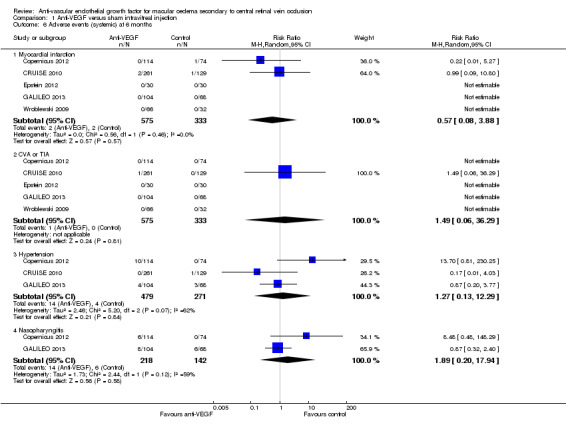
Comparison 1 Anti‐VEGF versus sham intravitreal injection, Outcome 6 Adverse events (systemic) at 6 months.
The number of anti‐VEGF or sham injections administered
The number of anti‐VEGF or sham injections was prespecified in the protocol of all trials for a fixed period, which ranged from three months (ROCC 2010) to six months (GALILEO 2013, Copernicus 2012, CRUISE 2010, Epstein 2012, Wroblewski 2009). Numerous trials included a pro re nata (PRN) treatment period ranging from a further three months (ROCC 2010) to a further six months (Copernicus 2012) or more (CRUISE 2010), in which the anti‐VEGF agent was given to all groups according to protocol‐specified retreatment criteria. One trial included a PRN treatment period of a further six months in which randomisation to sham or anti‐VEGF agent was maintained (GALILEO 2013). The following table summarises the mean injections given in each of the trials.
| Trial | Mean number of injections months 0 to 5 | Mean number of injections months 6 to 12 | Notes |
| Copernicus 2012 | 6 (sham or ranibizumab) | 3.9 (SE 0.3) in sham + PRN ranibizumab; 2.7 (SE 0.2) in ranibizumab + PRN ranibizumab | PRN ranibizumab 6 to 12 m |
| CRUISE 2010 | 6 (sham or 0.3 mg or 0.5 mg ranibizumab) | 3.8 in 0.3 mg group; 3.3 in 0.5 mg group; 3.7 in sham/0.5 mg group |
PRN ranibizumab 6 to 12m |
| Epstein 2012 | 5 (sham or bevacizumab) | 4 (bevacizumab for both groups) | Fixed injection schedule |
| GALILEO 2013 | 6 (sham or aflibercept) | From 0 to 52 weeks: 11.8 (SD 2.8) aflbercept; 10.5 (SD 4.2) sham | PRN ranibizumab or sham from 6 to 12 m |
| ROCC 2010 | 4.3 (SD 0.9) ranibizumab; 5.5 (SD 1.1) sham | ‐ | PRN ranibizumab or sham from 3 to 6 m |
| Wroblewski 2009 | 5 (sham or pegaptanib sodium) | ‐ | Fixed injection schedule |
The number and type of additional interventions administered
No additional interventions to treat CRVO‐MO were reported in any of the trials.
Economic data
No trials included economic data. Aside from these six RCTs, the remainder of the literature identified through this review's electronic search was based on open‐label, prospective studies, retrospective chart reviews, case reports and case series with relatively short follow‐up periods.
Quality of life
Three trials included the mean change in total score from baseline to six months on the National Eye Institute 25‐item Visual Function Questionnaire, NEI VFQ‐25 (Copernicus 2012; CRUISE 2010; GALILEO 2013); however, only one study reported data sufficient for analysis (Analysis 1.7). Specifically, in the CRUISE 2010 trial, patients in the 0.3 mg and 0.5 mg ranibizumab groups had a mean gain of 7.1 (95% CI 5.2 to 9.0) and 6.2 (95% 4.3 to 8.0) points, respectively, compared to 2.8 (95% CI 0.8 to 4.7) in the sham group (P < 0.05 for each ranibizumab group versus sham), even though the study eye was the worse seeing eye in most cases. Similarly, in the Copernicus 2012 and GALILEO 2013 trials, patients in the 2.0 mg aflibercept groups gained an average of 7.2 points and 7.5 points, respectively, compared to 0.8 points and 3.5 points, in the sham groups. At 52 weeks, further slight gains were reported from baseline in the treatment (7.8 points) and sham (4.5 points) groups in the GALILEO 2013 trial.
1.7. Analysis.
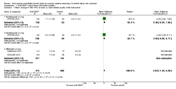
Comparison 1 Anti‐VEGF versus sham intravitreal injection, Outcome 7 Mean change in NEI VFQ 25 score (a vision‐related quality of life instrument).
Discussion
Summary of main results
All six trials demonstrated that repeated intravitreal anti‐vascular endothelial growth factor (anti‐VEGF) treatment was associated with (predominantly) significant improvements in the primary and secondary outcomes at six months compared to sham, and no significant safety concerns relating to the drug were identified in this time. Specifically, the proportion of treated patients gaining 15 letters or more of visual acuity ranged from 36.4% for 0.3 mg pegaptanib sodium (Wroblewski 2009), to 60.2% for 2.0 mg aflibercept (GALILEO 2013). The mean gain in visual acuity ranged from 7.1 letters in those treated with 0.3 mg pegaptanib sodium (Wroblewski 2009) to 18.0 letters in those treated with 2.0 mg aflibercept (GALILEO 2013). The proportion of treated patients losing 15 letters or more of visual acuity ranged from 1.5% (CRUISE 2010) to 13.3% (ROCC 2010), in patients receiving 0.5 mg ranibizumab. Participants receiving anti‐VEGF treatment had a greater reduction in mean central retinal thickness (CRT) from baseline than patients receiving sham, indicating enhanced resolution of macular oedema. The reduction ranged from ‐179 to ‐243 μm in patients receiving pegaptanib sodium (Wroblewski 2009), to between ‐426 and ‐457 μm in participants receiving ranibizumab, bevacizumab and aflibercept (Copernicus 2012; CRUISE 2010; Epstein 2012; GALILEO 2013). Participants in the treatment groups of the CRUISE 2010, Copernicus 2012 and GALILEO 2013 trials reported an improvement in quality of life, with a gain of approximately seven points on the National Eye Institute 25‐item Visual Function Questionnaire (NEI VFQ‐25) instrument at six months. The three open‐label extension trials demonstrated that in the anti‐VEGF treatment groups, the visual acuity and CRT gains seen at six months were maintained at 12 months, and no new safety concerns were identified in the treated groups (Copernicus 2012; CRUISE 2010; Epstein 2012).
In contrast, the outcomes in the sham groups were significantly worse and were similar across the six trials. Specifically, a gain of 15 or more letters at six months was reported in between 12% (Copernicus 2012) and 28% (Wroblewski 2009), whilst a loss of 15 letters or more was reported in between 15% (CRUISE 2010) and 31% (Wroblewski 2009). In general, after six months of sham injection there was a negligible mean change from baseline visual acuity, ranging from ‐4 letters (Copernicus 2012) to +3.3 letters (GALILEO 2013). There was some reduction in mean central retinal thickness from baseline, ranging from 102 μm (Epstein 2012) to 169 μm (GALILEO 2013). These outcomes are summarised in Table 1. Participants in the sham groups reported minimal functional gain in quality of life from baseline to six months on the NEI VFQ‐25 instrument, with an average change of 0.8 points (Copernicus 2012), 2.8 points (CRUISE 2010) and 3.5 points (GALILEO 2013).
Allowing for variability resulting from the relatively small sample sizes, the six randomised controlled trials (RCTs) included participants with broadly similar baseline characteristics in both the sham and treatment groups (Copernicus 2012; CRUISE 2010; Epstein 2012; GALILEO 2013; ROCC 2010; Wroblewski 2009). The mean duration from diagnosis to first treatment ranged from 61 days (Epstein 2012) to 80 days (Wroblewski 2009). The mean age ranged from 62 years (GALILEO 2013) to 72 years (ROCC 2010). The proportion of male patients ranged from 53% (Wroblewski 2009) to 60% (Epstein 2012). The mean baseline BCVA ranged from 52 letters (GALILEO 2013) to 43 letters (ROCC 2010), and the percentage of patients with a baseline visual acuity worse than 35 letters (6/60) ranged from 17% (GALILEO 2013) to 32% (Epstein 2012). The mean baseline central retinal thickness ranged from 619 µm (Wroblewski 2009) to 721 µm (Epstein 2012). Two trials included only patients with non‐ischaemic central retinal vein occlusion (CRVO) macular oedema (CRUISE 2010; Wroblewski 2009), and four trials included a mixed cohort of ischaemic and non‐ischaemic patients. The proportion of patients with baseline ischaemic CRVO macular oedema was 8.2% (plus 8.2% undetermined) in the GALILEO 2013 trial of aflibercept; 11.7% in the bevacizumab trial (Epstein 2012); 15.6% in the smaller ranibizumab trial (ROCC 2010) and 15.5% (plus 16.6% undetermined) in the other trial of aflibercept (Copernicus 2012).
Subgroup analyses to investigate the impact of treatment delay were performed in a few studies (Copernicus 2012; Epstein 2012; GALILEO 2013). This suggested that the greatest benefit of anti‐VEGF treatment occurs in patients with a shorter duration between diagnosis and treatment (analysis was based on before and after two months or 90 days). Three open‐label extension trials, in which patients randomised to sham injection for six months crossed over to receive PRN (as needed) anti‐VEGF between months 7 and 12, further corroborated this. Six‐month delayed anti‐VEGF therapy resulted in resolution of macular oedema, with no significant differences between groups at 12 months in mean CRT. However, whilst visual outcomes improved in groups treated with anti‐VEGF on a PRN basis after six months, the visual outcomes remained significantly worse at 12 months in these groups compared to the groups initially randomised to anti‐VEGF therapy (Copernicus 2012; CRUISE 2010; Epstein 2012). Only one open‐label extension trial, the HORIZON trial, considered longer‐term outcomes at 24 months (CRUISE 2010). It is difficult to determine whether the lack of sustained benefit of anti‐VEGF treatment at 24 months, with worsening of functional (but not anatomical) outcomes compared to 12 months, reflected reduced efficacy of anti‐VEGF over time, or whether it was related to the lower assessment and treatment frequency in months 12 to 24, or to the high probability of attrition bias in the outcome data resulting from early termination of the trial.
Subgroup analyses to investigate the impact of retinal perfusion status on visual outcomes were performed in a few studies (Copernicus 2012; GALILEO 2013). These demonstrated that without anti‐VEGF treatment, eyes with baseline ischaemic CRVO macular oedema have a worse visual prognosis than eyes with non‐ischaemic CRVO macular oedema, but that both groups experience similar gains in visual acuity and anatomical resolution of macular oedema with anti‐VEGF therapy. Subgroup analyses with stratification by baseline visual acuity (better or worse than 6/60) indicated that greater absolute gains in visual acuity are seen in patients with a baseline BCVA worse than 6/60 than with a baseline acuity better than 6/60, in both sham and treatment groups (Copernicus 2012; GALILEO 2013).
Anti‐VEGF therapy was associated with significant reduction in the odds of developing iris or retinal neovascularisation or neovascular glaucoma at six months compared to sham treatment (Copernicus 2012; CRUISE 2010; Epstein 2012; GALILEO 2013; ROCC 2010; Wroblewski 2009). Recognised complications of intravitreal injection therapy, including endophthalmitis, rhegmatogenous retinal detachment and retinal artery occlusion, were reported at very low frequency in some of the treatment groups. No significant association between anti‐VEGF therapy and serious systemic adverse events was identified at six months.
In summary, despite some variability in baseline characteristics between the trials, all four anti‐VEGF agents were demonstrated to enhance the anatomical resolution of macular oedema, to stabilise the visual acuity in some participants and to significantly improve the visual acuity in approximately half of those treated, with associated gains in quality of life in the trials which included this outcome, with no significant safety concerns up to 12 months. The 12‐month GALILEO 2013 trial and the three open‐label extension trials demonstrated a benefit to sham patients commenced on anti‐VEGF treatment following a delay of six months (Copernicus 2012; CRUISE 2010; Epstein 2012). Subgroup analyses indicated benefit of anti‐VEGF treatment to both ischaemic and non‐ischaemic CRVO macular oedema patients. The results of this systematic review and meta‐analysis demonstrate that treatment with anti‐VEGF agents confers significant clinical gains in all outcomes of interest compared to no treatment.
Overall completeness and applicability of evidence
One limitation of some of the trials was their relatively small sample sizes; these ranged from 14 participants per group (ROCC 2010) to 132 participants per group (CRUISE 2010). Whilst this may have reduced the power to identify a significant difference between the treatment and sham groups in the main and secondary outcomes of interest (Wroblewski 2009), the treatment effect associated with anti‐VEGF therapy in most studies was sufficiently large to yield significant differences from the control groups (Copernicus 2012; CRUISE 2010; Epstein 2012; GALILEO 2013; ROCC 2010). Two different treatment doses were investigated in comparison to sham in the CRUISE 2010 and Wroblewski 2009 trials, but neither trial had sufficient power to investigate outcome differences between these doses.
A second limitation in five out of six of the trials was the relatively short follow‐up period of approximately six months, which did not permit assessment of how long the apparent benefits of treatment with anti‐VEGF are sustained. One trial reported 12‐month outcomes compared to sham (GALILEO 2013), and three open‐label extension trials yielded safety data to 12 months (Copernicus 2012; Epstein 2012) and 24 months (CRUISE 2010), for the treatment and cross‐over groups. Ocular or systemic adverse events occurring at a longer latency from treatment may have been missed.
Whilst the treatment of CRVO macular oedema with these four anti‐VEGF agents appears very promising, the applicability of the available trial data to important subgroups of patients in clinical practice is not known. For example, since patients who had persisting CRVO macular oedema for more than one year were excluded from recruitment into the trials, the efficacy and safety of anti‐VEGF therapy in these patients is not known. Patients with previous retinal vein occlusion, or other co‐morbid eye disease including diabetic retinopathy and age‐related macular degeneration, were also excluded, even though age and diabetes are among the established risk factors for CRVO (Shahsuvaryan 2003). It is not uncommon for patients in clinical practice to have multiple ocular and systemic pathologies. Furthermore, the CVOS Study reported that 29% of patients with CRVO present with a visual acuity of 6/12 or better (CVOS Group 1997), but as these participants were mostly excluded it is not possible to determine whether treatment with anti‐VEGF might confer sufficient benefit in this group to outweigh the risks associated with intravitreal injection.
Quality of the evidence
The six included RCTs were small to moderate in size but were well designed with a low risk of bias in the majority of domains. They can therefore be considered to provide reasonably high‐quality evidence on 6‐ to 12‐month outcomes for the specific cohort of participants studied.
Data from numerous non‐randomised studies were reviewed in the previous publication of this review (Braithwaite 2010). A comprehensive search for such data was not performed, and these tables were not included or updated in the current review.
Agreements and disagreements with other studies or reviews
We are not aware of any other systematic reviews on the use of anti‐VEGF agents for the treatment of CRVO macular oedema. However, these agents have been administered intravitreally in numerous other ophthalmological contexts, and a good short‐term safety profile is emerging, with a low incidence of serious ocular and systemic adverse events. A systematic review including 278 studies on 9061 participants who received 49,584 intravitreal anti‐VEGF injections, specifically, ranibizumab (19,908 injections given over a mean of 16 months), bevacizumab (11,018 injections given over a mean of five months) and pegaptanib sodium (18,658 injections given over a mean of 11 months) reported a low incidence of all serious ocular adverse events with all three agents (van der Reis 2011). Specifically, there was a low cumulative incidence (per 100 injections) of endophthalmitis (0.04% to 0.11%), retinal detachment (0.01% to 0.08%), intraocular inflammation (0.25% to 1.06%), elevated intraocular pressure (IOP) (0.15% to 3.60%), intraocular haemorrhage (0.03% to 0.18%) and cataract progression (0.05% to 0.64%) (van der Reis 2011). Similarly, five cases of retinal detachment were reported over three years out of 35,942 anti‐VEGF intravitreal injections performed at six high‐volume centres in Germany (Meyer 2011). All retinal detachments occurred within two to six days of injection, and were more frequent in myopic patients (Meyer 2011). Other less serious side effects associated with intravitreal injections have included lid irritation, ocular discomfort and foreign body sensation, transient vision blurring, subconjunctival haemorrhage, mild anterior chamber inflammation and mild vitreitis, uveitis and raised IOP (Lynch 2007). Systemic adverse events including stroke, myocardial infarction and blood pressure elevation have been reported following intravenous administration of bevacizumab at doses more than 300 times higher than are used for ophthalmic indications (Lynch 2007). Wu et al reported systemic adverse events at one year in 1.5% of patients following intravitreal administration of 4303 bevacizumab injections into 1310 eyes. These adverse events included acute systolic blood pressure elevation (0.59%), cerebrovascular accident (0.5%), myocardial infarction (0.4%), iliac artery aneurysm (0.17%), toe amputation (0.17%) and death (0.4%) (Wu 2008). In the systematic review of three anti‐VEGF agents, systemic adverse events were similarly infrequent, with a low cumulative incidence (per 100 injections) of heart disease (0.05% to 0.34%), vascular disease (0.01% to 0.05%), hypertension (0.15% to 0.55%), cerebrovascular accident (CVA) or transient ischaemic attack (TIA) (0.01% to 0.07%) and thromboembolic events (0.07% to 0.19%) (van der Reis 2011). The available evidence for the short‐term safety profile of these agents is reassuring, but the follow‐up periods are still too short, and the total number of treated patients too small to detect serious adverse events that occur at very low incidence, or at long latency from the treatment period.
It will be important to determine the relative effectiveness and safety of anti‐VEGF agents versus other interventions for the treatment of CRVO macular oedema, but no head‐to‐head trial data are available yet. Comparison with RCT data on intravitreal corticosteroids will be particularly important, as both anti‐VEGF agents and steroid implants have now been approved for the treatment of CRVO macular oedema in various countries, and are being used extensively off‐label in others. Differences in baseline characteristics of participants recruited to the existing trials, comparing either anti‐VEGF agents or steroid treatments to sham, render outcome comparison challenging at present. For example, the average duration of disease was less than two months in 62% patients recruited to the Copernicus 2012 trial, and less than three months in 69% participants in the CRUISE 2010 trial, but in only 39% participants recruited to the SCORE 2009 trial and 17% recruited to the Ozurdex GENEVA 2010 trial. Numerous head‐to‐head RCTs comparing different anti‐VEGF agents and steroid treatments are currently underway and will begin to address this.
Authors' conclusions
Implications for practice.
The randomised controlled trial (RCT) evidence from six trials clearly demonstrates that repeated intravitreal injection therapy for central retinal vein occlusion (CRVO) macular oedema with the anti‐vascular endothelial growth factor (anti‐VEGF) agents ranibizumab, pegaptanib sodium, aflibercept and bevacizumab, improves visual and anatomical outcomes at six and 12 months, compared to sham treatment. Smaller subgroup analyses suggest that early initiation of treatment (within two months or 90 days of diagnosis) is probably more beneficial than delayed treatment. Clinical benefit has been demonstrated by a few trials in subgroups of patients with both ischaemic and non‐ischaemic CRVO macular oedema at baseline, and with baseline best‐corrected visual acuity (BCVA) both better than and worse than 6/60. The efficacy and safety of anti‐VEGF therapy, and of repeat intravitreal injections, over longer periods of follow‐up has yet to be determined.
The relative effectiveness and safety profile of anti‐VEGF agents versus steroid therapies for the treatment of different subgroups of CRVO macular oedema was not explored in this review and has yet to be determined. The impact of prior or combined treatment with intravitreal corticosteroid, or other treatments, was also not explored in this review.
In summary, anti‐VEGF therapy is a relatively safe and effective treatment for CRVO macular oedema in the short term, and this represents an important therapeutic advance for the treatment of this visually disabling disease. It is not yet possible to determine the potential economic impact of the use of anti‐VEGF agents in this clinical context, but we will be reassess this if data become available.
Implications for research.
That anti‐VEGF therapy confers clear clinical benefit for the treatment of CRVO macular oedema over no treatment (sham) in patients with certain baseline characteristics has been demonstrated, and marketing licenses have been granted to several agents accordingly. Future research should explore the relative efficacy and safety of different anti‐VEGF agents in head‐to‐head trials, and other treatments (including corticosteroid injections and implants) and combination therapies. Further research into the efficacy and safety of anti‐VEGF treatment for patients excluded from the trials reviewed here is needed. In particular, there is a clinical need to determine whether anti‐VEGF therapy is also beneficial to patients with a duration from diagnosis exceeding a year, to those with ocular comorbidity and to those with a baseline visual acuity of 6/12 or better. Trials including larger patient samples would be needed to ensure sufficient statistical power for important subgroup analyses to be performed. Longer‐term outcomes data on anti‐VEGF treatment groups would also be valuable, to investigate whether the apparently low incidence of ocular and systemic adverse events is maintained in the longer term, or if there are any ocular or systemic harms associated with chronic VEGF inhibition.
The burden placed on patients and healthcare systems of monthly reassessment and repeat injection is very considerable, and the relative efficacy of different treatment intervals and approaches to reassessment and retreatment needs to be investigated further. The length of treatment to achieve a stable visual outcome has also yet to be determined. Recent trials have demonstrated that patients with both ischaemic and non‐ischaemic CRVO macular oedema, with baseline BCVA worse than and better than 6/60, and with shorter and longer duration of disease at baseline all benefit, to a greater or lesser extent, from anti‐VEGF therapy. Comprehensive baseline assessment to permit stratification and subgroup analysis would facilitate exploration of the prognostic significance of these important baseline factors. The inclusion of patients with a baseline BCVA better than 6/12 in future trials would also provide valuable clinical management information for this important subset of patients to whom the existing trial data do not necessarily apply. Further epidemiological data from different populations on the risk factors for developing CRVO macular oedema would also be beneficial.
What's new
| Date | Event | Description |
|---|---|---|
| 14 January 2014 | New citation required and conclusions have changed | Issue 5, 2014: Four new RCTs identified, meta‐analysis performed and conclusions amended |
| 14 January 2014 | New search has been performed | Issue 5, 2014: Electronic searches updated |
Acknowledgements
We are especially grateful to Anupa Shah, Ann Ervin and Richard Wormald for their guidance on this review and to Iris Gordon for performing the electronic searches. We wish to thank Catey Bunce, Jenny Evans and Mariacristina Parravano for their peer review comments. We would also like to thank Barbara Hawkins, Michele Melia and Quan Dong Nguyen who made valuable comments on the protocol and first review manuscript.
Richard Wormald (Co‐ordinating Editor for Cochrane Eyes and Vision Group) acknowledges financial support for his CEVG research sessions¬from the Department of Health through the award made by the National Institute for Health Research to Moorfields Eye Hospital NHS Foundation Trust and UCL Institute of Ophthalmology for a Specialist Biomedical Research Centre for Ophthalmology. The views expressed in this publication are those of the authors and not necessarily those of the Department of Health.
Appendices
Appendix 1. CENTRAL search strategy
#1 MeSH descriptor Macular Edema, Cystoid #2 MeSH descriptor Edema #3 MeSH descriptor Macula Lutea #4 macula* near/3 oedema #5 macula* near/3 edema #6 CME or CMO #7 (#1 OR #2 OR #3 OR #4 OR #5 OR #6) #8 MeSH descriptor Retinal Vein Occlusion #9 MeSH descriptor Retinal Vein #10 retina* near/3 (vein* or occlu* or obstruct* or clos* or stricture* or steno* or block* or embolism*) #11 CRVO or CVO or RVO #12 (#8 OR #9 OR #10 OR #11) #13 MeSH descriptor Angiogenesis Inhibitors #14 MeSH descriptor Angiogenesis Inducing Agents #15 MeSH descriptor Endothelial Growth Factors #16 MeSH descriptor Vascular Endothelial Growth Factors #17 macugen* or pegaptanib* or lucentis* or rhufab* or ranibizumab* or bevacizumab* or avastin #18 anti near/2 VEGF* #19 endothelial near/2 growth near/2 factor* #20 (#13 OR #14 OR #15 OR #16 OR #17 OR #18 OR #19) #21 (#7 AND #12 AND #20)
Appendix 2. MEDLINE search strategy
1. randomized controlled trial.pt. 2. (randomized or randomised).ab,ti. 3. placebo.ab,ti. 4. dt.fs. 5. randomly.ab,ti. 6. trial.ab,ti. 7. groups.ab,ti. 8. or/1‐7 9. exp animals/ 10. exp humans/ 11. 9 not (9 and 10) 12. 8 not 11 13. exp macular edema cystoid/ 14. exp edema/ 15. exp macula lutea/ 16. (macula$ adj3 oedema).tw. 17. (macula$ adj3 edema).tw. 18. (CME or CMO).tw. 19. or/13‐18 20. exp retinal vein occlusion/ 21. exp retinal vein/ 22. ((vein$ or occlu$ or obstruct$ or clos$ or stricture$ or steno$ or block$ or embolism$) adj3 retina$).tw. 23. (CRVO or CVO or RVO).tw. 24. or/20‐23 25. exp angiogenesis inhibitors/ 26. exp angiogenesis inducing agents/ 27. exp endothelial growth factors/ 28. exp vascular endothelial growth factors/ 29. (macugen$ or pegaptanib$ or lucentis$ or rhufab$ or ranibizumab$ or bevacizumab$ or avastin).tw. 30. (anti adj2 VEGF$).tw. 31. (endothelial adj2 growth adj2 factor$).tw. 32. or/25‐31 33. 19 and 24 and 32
The search filter for trials at the beginning of the MEDLINE strategy is from the published paper by Glanville (Glanville 2006).
Appendix 3. EMBASE search strategy
1. exp randomized controlled trial/ 2. exp randomization/ 3. exp double blind procedure/ 4. exp single blind procedure/ 5. random$.tw. 6. or/1‐5 7. (animal or animal experiment).sh. 8. human.sh. 9. 7 and 8 10. 7 not 9 11. 6 not 10 12. exp clinical trial/ 13. (clin$ adj3 trial$).tw. 14. ((singl$ or doubl$ or trebl$ or tripl$) adj3 (blind$ or mask$)).tw. 15. exp placebo/ 16. placebo$.tw. 17. random$.tw. 18. exp experimental design/ 19. exp crossover procedure/ 20. exp control group/ 21. exp latin square design/ 22. or/12‐21 23. 22 not 10 24. 23 not 11 25. exp comparative study/ 26. exp evaluation/ 27. exp prospective study/ 28. (control$ or propspectiv$ or volunteer$).tw. 29. or/25‐28 30. 29 not 10 31. 30 not (11 or 23) 32. 11 or 24 or 31 33. exp retina macula cystoid edema/ 34. exp eye edema/ 35. exp retina macula lutea/ 36. (macula$ adj3 oedema).tw. 37. (macula$ adj3 edema).tw. 38. (CME or CMO).tw. 39. or/33‐38 40. exp retinal vein occlusion/ 41. exp retina vein/ 42. ((vein$ or occlu$ or obstruct$ or clos$ or stricture$ or steno$ or block$ or embolism$) adj3 retina$).tw. 43. (CRVO or CVO or RVO).tw. 44. or/40‐43 45. exp angiogenesis/ 46. exp angiogenesis inhibitors/ 47. exp angiogenic factor/ 48. exp endothelial cell growth factor/ 49. exp vasculotropin/ 50. (macugen$ or pegaptanib$ or lucentis$ or rhufab$ or ranibizumab$ or bevacizumab$ or avastin).tw. 51. (anti adj2 VEGF$).tw. 52. (endothelial adj2 growth adj2 factor$).tw. 53. or/45‐52 54. 39 and 44 and 53 55. 32 and 54
Appendix 4. LILACS search strategy
macula$ oedema or macula$ edema or CME or CMO AND vein$ or occlu$ or obstruct$ or CRVO or CVO or RVO AND angiogenesis or endothelial growth factor or macugen$ or pegaptanib$ or lucentis$ or rhufab$ or ranibizumab$ or bevacizumab$ or avastin
Appendix 5. CINAHL search strategy
macula$ oedema or macula$ edema or CME or CMO AND vein$ or occlu$ or obstruct$ or CRVO or CVO or RVO AND angiogenesis or endothelial growth factor or macugen$ or pegaptanib$ or lucentis or rhufab$ or ranibizumab$ or bevacizumab$ or avastin
Appendix 6. OpenSIGLE search strategy
macula* oedema or macula* edema or CME or CMO AND vein* or occlu* or obstruct* or CRVO or CVO or RVO AND angiogenesis or endothelial growth factor or macugen* or pegaptanib* or lucentis or rhufab* or ranibizumab* or bevacizumab* or avastin
Appendix 7. metaRegister of Controlled Trials search strategy
(angiogenesis or endothelial growth factor) AND macula oedema
Appendix 8. ClinicalTrials.gov search strategy
(Angiogenesis or Endothelial Growth Factor) AND (Macula Oedema)
Data and analyses
Comparison 1. Anti‐VEGF versus sham intravitreal injection.
| Outcome or subgroup title | No. of studies | No. of participants | Statistical method | Effect size |
|---|---|---|---|---|
| 1 Gain of 15 letters or more at 6 months | 6 | 937 | Risk Ratio (M‐H, Random, 95% CI) | 2.71 [2.10, 3.49] |
| 1.1 Pegaptanib sodium 0.3 mg | 1 | 49 | Risk Ratio (M‐H, Random, 95% CI) | 1.45 [0.56, 3.80] |
| 1.2 Pegaptanib sodium 1.0 mg | 1 | 49 | Risk Ratio (M‐H, Random, 95% CI) | 1.26 [0.54, 2.92] |
| 1.3 Ranibizumab 0.3 mg | 1 | 197 | Risk Ratio (M‐H, Random, 95% CI) | 2.73 [1.55, 4.82] |
| 1.4 Ranibizumab 0.5 mg | 2 | 224 | Risk Ratio (M‐H, Random, 95% CI) | 2.94 [1.74, 4.96] |
| 1.5 Bevacizumab 1.25 mg | 1 | 60 | Risk Ratio (M‐H, Random, 95% CI) | 3.0 [1.38, 6.50] |
| 1.6 Aflibercept 2.0 mg | 2 | 358 | Risk Ratio (M‐H, Random, 95% CI) | 3.37 [2.04, 5.57] |
| 2 Loss of 15 letters or more at 6 months | 5 | 766 | Risk Ratio (M‐H, Random, 95% CI) | 0.20 [0.12, 0.34] |
| 2.1 Pegaptanib sodium 0.3 mg | 1 | 49 | Risk Ratio (M‐H, Random, 95% CI) | 0.29 [0.08, 1.07] |
| 2.2 Pegaptanib sodium 1.0 mg | 1 | 49 | Risk Ratio (M‐H, Random, 95% CI) | 0.19 [0.04, 0.89] |
| 2.3 Ranibizumab 0.3 mg | 1 | 197 | Risk Ratio (M‐H, Random, 95% CI) | 0.25 [0.09, 0.69] |
| 2.4 Ranibizumab 0.5 mg | 2 | 224 | Risk Ratio (M‐H, Random, 95% CI) | 0.21 [0.05, 0.99] |
| 2.5 Bevacizumab 1.25 mg | 1 | 60 | Risk Ratio (M‐H, Random, 95% CI) | 0.29 [0.06, 1.26] |
| 2.6 Aflibercept 2.0 mg | 1 | 187 | Risk Ratio (M‐H, Random, 95% CI) | 0.06 [0.02, 0.27] |
| 3 Mean change in BCVA from baseline at 6 months | 6 | 937 | Mean Difference (IV, Random, 95% CI) | 15.23 [11.57, 18.89] |
| 3.1 Pegaptanib sodium 0.3 mg | 1 | 49 | Mean Difference (IV, Random, 95% CI) | 0.0 [0.0, 0.0] |
| 3.2 Pegaptanib sodium 1.0 mg | 1 | 49 | Mean Difference (IV, Random, 95% CI) | 0.0 [0.0, 0.0] |
| 3.3 Ranibizumab 0.3 mg | 1 | 197 | Mean Difference (IV, Random, 95% CI) | 11.90 [8.80, 15.00] |
| 3.4 Ranibizumab 0.5 mg | 2 | 224 | Mean Difference (IV, Random, 95% CI) | 14.06 [11.39, 16.73] |
| 3.5 Bevacizumab 1.25 mg | 1 | 60 | Mean Difference (IV, Random, 95% CI) | 16.1 [5.63, 26.57] |
| 3.6 Aflibercept 2.0 mg | 2 | 358 | Mean Difference (IV, Random, 95% CI) | 21.3 [16.55, 26.05] |
| 4 Mean change from baseline in central retinal thickness at 6 months | 6 | 935 | Mean Difference (IV, Random, 95% CI) | ‐267.39 [‐323.36, ‐211.43] |
| 4.1 Pegaptanib sodium 0.3 mg | 1 | 49 | Mean Difference (IV, Random, 95% CI) | 0.0 [0.0, 0.0] |
| 4.2 Pegaptanib sodium 1.0 mg | 1 | 49 | Mean Difference (IV, Random, 95% CI) | 0.0 [0.0, 0.0] |
| 4.3 Ranibizumab 0.3 mg | 1 | 197 | Mean Difference (IV, Random, 95% CI) | ‐266.7 [‐358.12, ‐175.28] |
| 4.4 Ranibizumab 0.5 mg | 2 | 223 | Mean Difference (IV, Random, 95% CI) | ‐232.26 [‐359.34, ‐105.18] |
| 4.5 Bevacizumab 1.25 mg | 1 | 60 | Mean Difference (IV, Random, 95% CI) | ‐324.0 [‐452.64, ‐195.36] |
| 4.6 Aflibercept 2.0 mg | 2 | 357 | Mean Difference (IV, Random, 95% CI) | 0.0 [0.0, 0.0] |
| 5 Adverse events and complications at 6 months (ocular) | 6 | Peto Odds Ratio (Peto, Fixed, 95% CI) | Subtotals only | |
| 5.1 Neovascularisation (iris or retina) | 6 | 936 | Peto Odds Ratio (Peto, Fixed, 95% CI) | 0.18 [0.09, 0.36] |
| 5.2 Neovascular glaucoma | 3 | 750 | Peto Odds Ratio (Peto, Fixed, 95% CI) | 0.14 [0.03, 0.72] |
| 5.3 Glaucoma (excluding neovascular) | 2 | 270 | Peto Odds Ratio (Peto, Fixed, 95% CI) | 0.08 [0.00, 4.39] |
| 5.4 Vitreous haemorrhage | 3 | 607 | Peto Odds Ratio (Peto, Fixed, 95% CI) | 0.55 [0.24, 1.23] |
| 5.5 Endophthalmitis | 6 | 937 | Peto Odds Ratio (Peto, Fixed, 95% CI) | 5.20 [0.09, 287.41] |
| 5.6 Retinal artery occlusion | 3 | 608 | Peto Odds Ratio (Peto, Fixed, 95% CI) | 5.37 [0.52, 55.03] |
| 5.7 Retinal tear | 5 | 839 | Peto Odds Ratio (Peto, Fixed, 95% CI) | 0.37 [0.04, 3.66] |
| 5.8 Retinal detachment | 5 | 747 | Peto Odds Ratio (Peto, Fixed, 95% CI) | 0.0 [0.0, 0.0] |
| 5.9 Ocular inflammation | 2 | 562 | Peto Odds Ratio (Peto, Fixed, 95% CI) | 0.58 [0.16, 2.06] |
| 5.10 Cataract | 3 | 607 | Peto Odds Ratio (Peto, Fixed, 95% CI) | 4.51 [0.56, 36.48] |
| 5.11 Subconjunctival haemorrhage | 2 | 360 | Peto Odds Ratio (Peto, Fixed, 95% CI) | 1.07 [0.55, 2.07] |
| 5.12 Elevation in intraocular pressure | 1 | 172 | Peto Odds Ratio (Peto, Fixed, 95% CI) | 1.64 [0.54, 5.01] |
| 5.13 Eye pain | 1 | 172 | Peto Odds Ratio (Peto, Fixed, 95% CI) | 2.44 [0.83, 7.17] |
| 6 Adverse events (systemic) at 6 months | 5 | Risk Ratio (M‐H, Random, 95% CI) | Subtotals only | |
| 6.1 Myocardial infarction | 5 | 908 | Risk Ratio (M‐H, Random, 95% CI) | 0.57 [0.08, 3.88] |
| 6.2 CVA or TIA | 5 | 908 | Risk Ratio (M‐H, Random, 95% CI) | 1.49 [0.06, 36.29] |
| 6.3 Hypertension | 3 | 750 | Risk Ratio (M‐H, Random, 95% CI) | 1.27 [0.13, 12.29] |
| 6.4 Nasopharyngitis | 2 | 360 | Risk Ratio (M‐H, Random, 95% CI) | 1.89 [0.20, 17.94] |
| 7 Mean change in NEI VFQ 25 score (a vision‐related quality of life instrument) | 3 | 743 | Mean Difference (IV, Fixed, 95% CI) | 3.84 [1.49, 6.20] |
| 7.1 Ranibizumab 0.3 mg | 1 | 193 | Mean Difference (IV, Fixed, 95% CI) | 4.3 [0.94, 7.66] |
| 7.2 Ranibizumab 0.5 mg | 1 | 192 | Mean Difference (IV, Fixed, 95% CI) | 3.4 [0.09, 6.71] |
| 7.3 Aflibercept 2.0 mg | 2 | 358 | Mean Difference (IV, Fixed, 95% CI) | 0.0 [0.0, 0.0] |
Characteristics of studies
Characteristics of included studies [ordered by study ID]
Copernicus 2012.
| Methods | Study design: double‐masked, randomised, sham injection‐controlled, phase III trial
Unit of randomisation and analysis: one study eye per participant Loss to follow‐up: 14 (18.9%) in control group, 5 (4.3%) in aflibercept (VEGF Trap‐Eye) 2.0 mg group Study duration: 6 months (2‐year follow‐up planned) |
|
| Participants | Countries: United States, Canada, India, Israel, Argentina and Columbia (70 sites) Enrolment: 189 eyes (74 control group, 115 in VEGF Trap‐Eye group) with CRVO‐MO of < 9 months duration. Non‐ischaemic* (67.9%), ischaemic (15.5%) and indeterminate (16.6%) included Baseline characteristics:
Inclusion criteria:
Exclusion criteria:
*Non‐ischaemic CRVO defined as < 10 disc areas of non‐perfusion on FFA |
|
| Interventions | Intervention #1 (n = 74): sham injection Intervention #2 (n = 114): aflibercept (VEGF Trap‐Eye) 2.0 mg Intravitreal injection (or sham injection) given every 4 weeks for 24 weeks, followed by an open‐label extension from weeks 24 to 52, in which all patients were offered 2 mg aflibercept as required, according to retreatment criteria Other treatments: All patients were eligible to receive panretinal photocoagulation at any time during the study at the discretion of the investigator if they progressed to neovascularisation of the anterior segment, disc or elsewhere |
|
| Outcomes | Primary outcome: proportion of eyes with a gain of 15 ETDRS letters or more in BCVA from baseline to week 24 Secondary outcomes: change from baseline to week 24 and 52 in the following variables: BCVA, CRT, proportion of eyes progressing to ocular neovascularisation, and NEI‐VFQ 25 total score Safety assessments included ocular adverse events (AEs) in the study and fellow eye, non‐ocular AEs, ocular and non‐ocular serious AE (SAEs), AEs of interest, laboratory assessments, vital signs and measurement of anti‐drug antibody in serum. Measurements: full ocular exam including visual acuity testing, slit‐lamp biomicroscopy, indirect ophthalmoscopy, intraocular pressure measurement (preinjection and 30 minutes after injection) and OCT: day 1, week 4 and every 4 weeks thereafter to week 24. Adverse events and concomitant medications were recorded, and vital signs were obtained at each visit Fundus photography and FFA: baseline, week 12 and week 24. NEI‐VFQ 25: baseline and week 24. Laboratory assessments, including anti‐VEGF Trap‐Eye antibodies: baseline and weeks 12 and 24 Unit of analysis: eye |
|
| Notes | Study dates: July 2009 to October 2010 (for 24‐week data) Funding source: Regeneron Pharmaceuticals, Inc, Tarrytown, New York, and Bayer Healthcare, Berlin, Germany Publication language: English |
|
| Risk of bias | ||
| Bias | Authors' judgement | Support for judgement |
| Random sequence generation (selection bias) | Low risk | Quote: "Randomization was stratified using a centralized interactive voice randomization system, by geographic region and by baseline BCVA score." 1 eye per patient was selected as the study eye; patients with bilateral retinal vein occlusion at baseline were excluded |
| Allocation concealment (selection bias) | Unclear risk | The method of allocation concealment was not described Comment: Probably done |
| Blinding (performance bias and detection bias) All outcomes | Low risk | Quote: "Sham injections were performed by pressing an empty, needleless syringe barrel to the conjunctival surface to simulate an injection"; "examiners masked to treatment arm… NEI‐VFQ25 administered by masked site personnel before intravitreal injection"; "OCT scans read at a masked independent central reading center…angiographic images ..transmitted to an independent reading centre for review by masked graders." |
| Incomplete outcome data (attrition bias) All outcomes | Unclear risk | Missing outcome data not balanced in distribution across groups; 14/74 (18.9%) in sham group versus 5/115 (4.3%) in the aflibercept group discontinued before week 24. 4 patients in the sham group, and none in the treatment group, discontinued as a result of adverse events associated with possible complications of CRVO, which included 1 patient with elevated IOP associated with rubeosis and vitreous haemorrhage, 1 with neovascular glaucoma, 1 with vitreous haemorrhage and rubeosis, and 1 with a 29 letter reduction in BCVA and rubeosis. 2 patients in the sham group died and 4 withdrew on account of treatment failure. 1 patient in the treatment group discontinued after developing a retinal tear after the first injection Comment: The unbalanced loss of patients with potentially more severe and visually significant disease from the sham group might have introduced bias, reducing any apparent benefit associated with anti‐VEGF therapy. To account for the missing data, the study investigators imputed missing data using the last‐observation‐carried‐forward method. This may have introduced bias by not taking into account changes in outcomes over time |
| Selective reporting (reporting bias) | Low risk | Primary and secondary outcomes reported as per the pre‐specification on ClinicalTrials.gov. However, no measure of variance was reported for the mean change in central retinal thickness from baseline |
| Other bias | Low risk | None identified |
CRUISE 2010.
| Methods | Study design: double‐masked, randomised, sham injection‐controlled phase III trial
Unit of randomisation and analysis: one study eye per participant Loss to follow‐up: 2.3% in 0.3 mg group, 8.5% in 0.5 mg group, 11.5% in sham group Study duration: 6 months, with subsequent 6‐month open‐label PRN treatment with ranibizumab 0.3 mg in the initial 0.3 mg group, 0.5 mg in the initial 0.5 mg group, and 0.5 mg in the initial sham group Randomisation was stratified by study centre and baseline BCVA letter score < 34, 35 to 54, and > 55 Patients completing 12 months were eligible for a further 12‐month open‐label extension trial in which they were seen at least every 3 months and offered 0.5 mg ranibizumab if they met retreatment criteria and no exclusion criteria. This study was discontinued 30 days after FDA approval of ranibizumab for RVO in June 2010, resulting in incomplete follow‐up data (Heier 2012) |
|
| Participants | Country: USA (95 centres) Enrolment: n = 392 patients with non‐ischaemic* (except n = 2 with ischaemic in the 0.5 mg group) CRVO‐ME Baseline characteristics:
Inclusion criteria:
Exclusion criteria:
Exclusion criteria for HORIZON open‐label extension trial (months 13 to 24):
*Non‐ischaemic CRVO defined as < 10 disc areas of capillary non‐perfusion on FFA |
|
| Interventions | Intervention #1 (n = 130): 0.5 mg ranibizumab every month for 6 months (6 injections) then PRN (open‐label) for 6 months Intervention #2 (n = 132): 0.3 mg ranibizumab every month for 6 months (6 injections) then PRN (open‐label) for 6 months Intervention #3 (Control) (n = 130): sham injection every month for 6 months (6 injections) then PRN 0.5 mg ranibizumab (open‐label) for 6 months General instructions/treatments: all participants received topical then subconjunctival anaesthetic. Sham participants did not have scleral penetration; blunt pressure was applied to the globe without a needle Extension studies In months 6 to 12 (the observation period), ranibizumab injection was given if study eye Snellen equivalent BCVA was < 20/40 or mean central subfield thickness on OCT, assessed by the investigator, was > 250 μm In months 13 to 24 (open‐label HORIZON extension trial), ranibizumab 0.5 mg injection was given if mean central subfield thickness was > 250 μm or if there was evidence of persisting/recurrent MO deemed to be affecting the BCVA |
|
| Outcomes | Primary outcome: mean change from baseline BCVA at month 6 Secondary outcomes: 1) Mean change from baseline BCVA over time to month 6; 2) Percentage of patients who gained 15 letters or more from baseline BCVA at month 6; 3) Percentage of patients who lost 15 letters or more from baseline BCVA at month 6; 4) Percentage of patients with CFT < 250 μm at month 6; 5) Mean change from baseline CFT over time to month 6; 6) Mean change in NEI VFQ‐25 scores at month 6 Additional efficacy outcomes included the percentage of patients with Snellen equivalent BCVA 20/200 or worse at month 6, mean change from baseline excess foveal thickness over time to month 6 (CFT minus 212 μm). Additional outcomes included the percentage of patients with Snellen equivalent BCVA > 20/40 at month 6 (an outcome generally considered sufficient to support reading and driving). Safety outcomes included the incidence and severity of ocular and non‐ocular adverse events and serious adverse events Measurements: examination and OCT ‐ day 0 and day 7, months 1 to 12. Patient‐reported visual function assessed with the NEI VFQ‐25 at day 0 and months 1, 3, 6 and 12 FFA ‐ at screening visit, prior to day 0, months 6, 9, 12 (and 24 if included in HORIZON trial) Unit of analysis: eye No economic data included |
|
| Notes | Study dates: July 2007 to December 2008 Funding source: Genentech, Inc., South San Francisco, California Publication language: English |
|
| Risk of bias | ||
| Bias | Authors' judgement | Support for judgement |
| Random sequence generation (selection bias) | Low risk | Quote: "patients were randomized 1:1:1".. "using a dynamic randomization method." "Randomization was stratified by baseline BCVA letter score" .. "and study center." 1 eye per patient was selected as the study eye; if both eyes were eligible, the eye with the worst BCVA at screening was selected |
| Allocation concealment (selection bias) | Unclear risk | The method of allocation concealment was not described Comment: probably done |
| Blinding (performance bias and detection bias) All outcomes | Low risk | Quote: "Patients, certified BCVA examiners, and evaluating physicians were masked to treatment and dose." Quote: "Injecting physicians, who did not perform examination or outcome assessments, were masked to dose but not treatment." |
| Incomplete outcome data (attrition bias) All outcomes | Unclear risk | Quote: "the intent‐to‐treat approach was used for efficacy analyses and included all patients as randomized. Missing values for efficacy outcomes were imputed using the last‐observation‐carried‐forward method" The percentage of patients completing 6 months follow‐up was 97.7% (n = 129) in the 0.3 mg group, 91.5% (n = 119) in the 0.5 mg treatment group and 88.5% (n = 115) in the sham group. The most common reason for withdrawal from the study was, "a decision made by the physician or patient to do so." Furthermore, not all participants completing the study to 6 months received all 6 monthly injections. Specifically, n = 4 (3.0%) in the 0.3 mg group, n = 10 (7.7%) in the 0.5 mg group and 16 (n = 12.3%) in the sham group discontinued treatment at or before month 5. This unbalanced loss to follow‐up may have introduced bias in the reported outcomes |
| Selective reporting (reporting bias) | Low risk | Primary and secondary outcomes reported as per the pre‐specification on ClinicalTrials.gov |
| Other bias | Unclear risk | 4 patients, 1 in the sham group and 3 in the 0.5 mg group, were included whose time from diagnosis to screening exceeded the specified inclusion criteria of up to 12 months (protocol violation). The greatest duration included was 27 months, for a participant in the 0.5 mg group Comment: the inclusion of outliers with a longer duration of CRVO‐ME before treatment initiation is likely to reduce the apparent benefit of treatment, thus introducing a low risk of bias in the reported outcomes 3 patients (2%) in the 0.5 mg group had more than 10 disc areas of capillary non‐perfusion, specifically 112, 113 and 109 disc areas, compared to no patients in either the sham or 0.3 mg groups Comment: since these 3 patients were not excluded we assume that they did not have a brisk afferent pupillary defect. However, they may have had more ischaemic CRVO‐ME than the other participants in the trial and since all 3 were in the 0.5 mg treatment group this may have introduced a bias in the outcomes, toward a reduced apparent benefit of treatment in the 0.5 mg group Patients with a baseline BCVA falling outside the specified inclusion range of 20/40 (˜70 letters (Gregori 2010) to 20/320 (˜25 letters) were included. The number of patients with BCVA better than 20/40 or worse than 20/320, by group, cannot be determined from the data presented, although the range in BCVA (letters) was reported to be 16 to 71 letters in the sham group, 9 to 72 letters in the 0.3 mg group and 21 to 73 letters in the 0.5 mg group. The participants were stratified according to BCVA (in letters) of < 34 (˜20/200), 35 to 54 and > 55 (˜20/80), with similar proportions of each strata in each group Comment: there is no reason why the range of baseline BCVAs should not be broader than that specified in the protocol, and the inclusion of participants with a poorer baseline BCVA than that prespecified should not introduce bias, providing a similar proportion of such participants were distributed across the 3 groups |
Epstein 2012.
| Methods | Study design: double‐masked, randomised, sham injection‐controlled trial
Unit of randomisation and analysis: one study eye per participant Loss to follow‐up: no losses reported in 6‐month RCT. Loss to follow‐up in the open‐label extension between 6 and 12 months was reported in 4 patients: 3 patients in the original treatment group discontinued on account of metastatic lung cancer, metastatic prostate cancer and old age; 1 in the original sham group discontinued on account of a TIA Study duration: 6 months, with subsequent 6‐month open‐label extension |
|
| Participants | Country: Sweden Enrolment: n = 60 with CRVO‐MO (of whom 3 in the treatment group and 4 in the treatment group were ischaemic at baseline*) Baseline characteristics
Inclusion criteria:
Exclusion criteria:
|
|
| Interventions | Intervention #1: sham injection (n = 30) Intervention #2: 1.25 mg (0.05 ml) bevacizumab (Avastin) (n = 30) Injections every 6 weeks for 24 weeks/6 months (total 4 injections) Open‐label extension for 6 months: all patients in both groups received bevacizumab 1.25 mg every 6 weeks (4 injections). N = 3 in original Avastin group discontinued and n = 1 in original sham group discontinued during extension phase General instructions/treatments: all eyes treated with topical fucidinic acid 1% 30 minutes prior to injection |
|
| Outcomes | Primary outcomes: proportion of patients gaining at least 15 ETDRS letters at 6 months Secondary outcomes: mean change from baseline BCVA, foveal thickness and number of cases of neovascular glaucoma (increased intraocular pressure caused by new vessels forming in the angle as diagnosed by gonioscopy) Measurements: BCVA, gonioscopy, IOP, full dilated slit‐lamp examination, OCT at baseline and every 6 weeks. FFA, colour and red‐free photographs: baseline and 24 weeks Unit of analysis: eye |
|
| Notes | Study dates: April 2009 to December 2010 Funding source: none Publication language: English |
|
| Risk of bias | ||
| Bias | Authors' judgement | Support for judgement |
| Random sequence generation (selection bias) | Unclear risk | Quote: "Each study participant was randomly assigned with equal probability." Comment: method of generation of randomisation sequence not specified 1 eye per patient was selected but the method of selection of the study eye, in the event of both eyes meeting the inclusion criteria, was not explicitly stated |
| Allocation concealment (selection bias) | Low risk | Quote: "Randomisation was done at the day of the first injection by sealed, opaque envelopes drawn by staff not involved in patient treatment or follow‐up." Comment: methods using envelopes may be susceptible to manipulation. Personal communication with study investigator: Envelopes were blank and were not sequentially numbered based on the randomisation code |
| Blinding (performance bias and detection bias) All outcomes | Low risk | Quote: "Study patients were masked to the treatment given"; "Patients in the control group received a sham injection by pressing a syringe without a needle to the globe"; "Staff performing VA testing, OCT, fundus photographs, and follow‐up investigators were masked to treatment group." |
| Incomplete outcome data (attrition bias) All outcomes | Low risk | No loss to follow‐up reported (100% completion). Missing outcome data for n = 1 in bevacizumab group (VA and OCT thickness) and n = 2 in sham group (OCT thickness) was imputed using last‐observation‐carried‐forward, and analysis was by intention‐to‐treat |
| Selective reporting (reporting bias) | Low risk | Primary and secondary outcomes reported as per the pre‐specification on ClinicalTrials.gov. The standard error of the mean for continuous variables was not reported numerically in the published report, but this unpublished data provided (personal communication) |
| Other bias | Low risk | Quote: "The present study included patients with risk factors for developing..neovascular glaucoma…for example…severe ischaemic changes"; "The presence of a relative afferent pupillary defect was not an exclusion criteria." Whilst no information on relative afferent pupil defect (RAPD) or baseline FFA (e.g. disc areas of non‐perfusion) was provided in the published study, unpublished data confirmed 7 patients had ischaemia at baseline, balanced between the groups (3 in the treatment group, 4 in the control group). (Personal communication with study investigators) |
GALILEO 2013.
| Methods | Study design: multicentre, double‐masked, randomised, sham‐controlled phase III clinical trial
Unit of randomisation and analysis: one study eye per participant Loss to follow‐up: 25 out of 177 (14.1%) lost to follow‐up at 24 weeks Study duration: 76 weeks |
|
| Participants | Country: 63 centres in Europe (Austria 3; France 5; Germany 21; Hungary 5; Italy 7; Latvia 2) and the Asian/Pacific region (Australia 6; Japan 6; Singapore 2; South Korea 6) Enrolment: 177 patients with CRVO‐MO and no prior treatment for CRVO in the study eye Baseline characteristics
Inclusion criteria:
Exclusion criteria:
*Baseline retinal perfusion status was determined by fluorescein angiography using the central vein occlusion study (CVOS) classification. Patients were considered ischaemic if they had ≥10 disc areas of capillary non‐perfusion |
|
| Interventions | Randomised 2:3 ratio Intervention #1: sham injection group n = 71. Weeks 0 to 20 sham injection every 4 weeks for 24 weeks; weeks 24 to 48 sham injection every 4 weeks; week 52 VEGF Trap‐Eye (unless investigator declines for medical reasons), plus additional reassessment on weeks 60 and 68 and either PRN injection of VEGF Trap‐Eye or sham injection Intervention #2: 2 mg VEGF Trap‐Eye Q4 (aflibercept) group n = 106. Weeks 0 to 20 injection every 4 weeks for 24 weeks; weeks 24 to 52 injection every 4 weeks plus additional reassessment on weeks 60 and 68 and either PRN injection of VEGF Trap‐Eye or sham injection General instructions/treatments: panretinal photocoagulation was allowed at any time for all patients if they progressed to neovascularisation of the anterior segment, optic disc or fundus |
|
| Outcomes | Primary outcomes: the proportion gaining at least 15 letters in BCVA compared with baseline at week 24 endpoint Secondary outcomes: 1. Change from baseline in BCVA score at 24 weeks 2. Absolute change from baseline in CRT, assessed by OCT at week 24 3. Proportion of patients progressing to anterior segment neovascularisation, neovascularisation of the optic disc, or neovascularisation of the retina elsewhere requiring panretinal photocoagulation at week 24 4. Change in NEI‐VFQ‐25 total score from baseline to week 24 5. Change in EQ‐5D score from baseline to week 24 Analyses at 24 weeks (published), 52 weeks (unpublished) Unit of analysis: eye |
|
| Notes | Study dates: total study duration 76 weeks (masked), with 68 weeks of treatment Funding source: Bayer and Regeneron Pharmaceuticals Publication language: English Clinicaltrials.gov ID: NCT01012973 |
|
| Risk of bias | ||
| Bias | Authors' judgement | Support for judgement |
| Random sequence generation (selection bias) | Unclear risk | Quote: "Randomisation was stratified by region (Europe vs Asia/Pacific) and baseline BCVA (≤20/200 vs >20/200)." Method of generation of randomisation sequence not detailed in published report Comment: probably centralised (i.e. low risk of bias) 1 eye per patient was selected as the study eye; patients with bilateral retinal vein occlusion at baseline were excluded |
| Allocation concealment (selection bias) | Unclear risk | Method of allocation concealment not detailed in published report |
| Blinding (performance bias and detection bias) All outcomes | Low risk | Quote: "Sham procedure was performed by pressing an empty syringe with no needle to the conjunctival surface." Masking not described in detail in published report |
| Incomplete outcome data (attrition bias) All outcomes | Unclear risk | 10/106 (9.4%) in treatment group were lost to follow‐up of whom n = 5 had a protocol violation, n = 3 withdrew consent, n = 1 were lost to follow‐up and 1 had another reason for withdrawal; 15/71 (21.1%) in the sham group were lost to follow‐up of whom 4 had an adverse event, 2 had protocol violation, 3 withdrew consent, 5 reported lack of efficacy and 1 'other' Comment: moderate loss to follow‐up, unbalanced between groups Sensitivity analysis compared the 'full analysis set' of n = 171/177, 25 of whom had imputation of missing data using the last observation carried forward method, to the 'per protocol' set of n = 138/177 Quote: "The sham group had a higher percentage of patients discontinuing study primarily due to adverse events and lack of efficacy this had no major impact on the primary endpoint as similar results were obtained after imputing the missing values with the LOCF approach using observed cases, or excluding patients who discontinued prior to week 24 and received fewer than five injections" |
| Selective reporting (reporting bias) | Low risk | Primary and secondary outcomes reported as per the pre‐specification on ClinicalTrials.gov except change in EQ‐5D score from baseline to 24 weeks, which was not reported specifically for each group. No measure of variance was reported for the continuous variables including mean change in BCVA from baseline and mean change in CRT from baseline |
| Other bias | Unclear risk | 7 patients out of 177 (4.0%) had protocol violation; 5 in the treatment group and 2 in the control group. No detail of the nature of the protocol violation is given Comment: this represents a small proportion of the patients and the resulting risk of bias is probably low |
ROCC 2010.
| Methods | Study design: double‐masked, randomised, sham injection‐controlled trial
Unit of randomisation and analysis: one study eye per participant Loss to follow‐up: n = 3 of 32 (1 withdrew from ranibizumab group, after developing CRAO; 2 withdrew from sham group ‐ 1 required planned surgery after developing cholecystitis and 1 developed AMD) Study duration: 6 months |
|
| Participants | Country: Norway (4 centres) Enrolment: n = 32 with CRVO‐MO of whom 1/15 (6.7%) in the ranibizumab group and 4/14 (28.6%) in the sham group completing 6 months follow‐up had ischaemic CRVO‐MO* Baseline characteristics
Inclusion criteria
Exclusion criteria:
*Ischaemic CRVO defined as > 5 disc areas of non‐perfusion on FFA |
|
| Interventions | Intervention #1 (n = 14): sham injection (plastic syringe pressed against the eyeball) Intervention #2 (n = 15): 0.5 mg/0.05 mL ranibizumab (Lucentis) Injection every month for 3 months, then as required (at the discretion of the physician) for persisting macular oedema |
|
| Outcomes | Primary outcomes: mean change from baseline in BCVA score and CRT at 6 months Secondary outcomes: number of treatments needed, safety and tolerability, development of neovascularisation Measurements: full ophthalmic examination including OCT, and blood pressure and pulse: monthly for 6 month. Fundus photography and FFA: baseline, 3 and 6 months Unit of analysis: eye |
|
| Notes | Study dates: March 2007 to October 2008 (from ClinicalTrials.gov protocol) Funding source: Novartis Ophthalmic Inc Publication language: English |
|
| Risk of bias | ||
| Bias | Authors' judgement | Support for judgement |
| Random sequence generation (selection bias) | Unclear risk | The investigators do not detail their sequence generation process in the published study 1 eye per patient was included as the study eye and patients were only recruited to the trial if they had unilateral CRVO‐MO |
| Allocation concealment (selection bias) | Low risk | Personal communication with study investigator: sealed, opaque envelopes were used to conceal allocation. These were not sequentially numbered based on a randomisation code. They were opened after being assigned to individual patients Comment: methods using envelopes may be susceptible to manipulation |
| Blinding (performance bias and detection bias) All outcomes | Low risk | Quote: "The investigating physician and nurse were masked toward the injecting physician and nurse and vice versa"; "All treatments were administered after subconjunctival anaesthesia...and sham treatment was conducted by pressing a plastic syringe against the eyeball." |
| Incomplete outcome data (attrition bias) All outcomes | Unclear risk | Small sample size with n = 16 per group of whom 15/16 (93.8%) completed 6 months follow‐up in the ranibizumab group and 14/16 completed 6 months follow‐up in the sham group (87.5%). The efficacy analysis was based on the per protocol treated participants, rather than on intention‐to‐treat, with no imputation of missing values Reasons for missing outcomes are reported explicitly, however: 1 patient in ranibizumab group and 2 in sham group withdrew, with reason directly related to the treatment (CRAO following first injection) in the ranibizumab group, and not related to the treatment or outcomes in the sham group |
| Selective reporting (reporting bias) | Low risk | Primary and secondary outcomes reported as per the pre‐specification on ClinicalTrials.gov except the NEI VFQ‐25 near activities subscale, which was not reported. Two outcomes of interest (proportion gaining and proportion losing 15 letters from baseline at 6 months) were not reported (these were not prespecified in the protocol), but were available as unpublished data (personal communication with study investigator) |
| Other bias | Unclear risk | The protocol prespecified definition of ischaemic CRVO was > 10 disc areas of non‐perfusion but in the final report a more conservative definition of > 5 disc areas of non‐perfusion was used. There was an imbalance in the baseline proportion of patients with ischaemic CRVO between groups, of 1/15 (6.7%) in the ranibizumab group and 4/14 (28.6%) in the sham group. This imbalance in the proportion with ischaemic CRVO was reflected in the slightly worse baseline BCVA in the sham group of 41 (SD 22) letters compared to 45 (SD 23) letters in the Lucentis group. Ischaemic CRVO has a worse visual prognosis, and given the small sample size, this imbalance might have introduced bias in the outcome measures, enhancing the apparent difference between groups in primary and secondary outcomes The authors also report that, "the accuracy of the OCT measurement was reduced in cases of severe macular oedema. This introduces the possibility of measuring bias," which may have increased estimate uncertainty. Comment: given that severe MO results in significant central retinal thickening, some imprecision in measurement would be unlikely to introduce bias in the results |
Wroblewski 2009.
| Methods | Study design: double‐masked, randomised controlled trial
Unit of randomisation and analysis: one study eye per participant Loss to follow‐up: 7 withdrawals (7%) Study duration: 30 weeks |
|
| Participants | Country: Australia, France, Germany, Israel, Spain, United States Enrolment: 98 eyes with non‐ischaemic CRVO‐MO Baseline characteristics
Inclusion criteria:
Exclusion criteria:
|
|
| Interventions | Intervention #1: 0.3 mg pegaptanib sodium every 6 weeks for 24 weeks (5 injections) Intervention #2: 1.0 mg pegaptanib sodium every 6 weeks for 24 weeks (5 injections) Intervention #3 (Control): sham injections every 6 weeks for 24 weeks (5 injections) General instructions/treatments: all participants received injected subconjunctival anaesthetic. Sham participants did not have scleral penetration; blunt pressure was applied to the globe without a needle |
|
| Outcomes | Primary outcome: percentage of eyes in each group gaining 15 letters or more of visual acuity at 30 weeks compared to baseline Secondary outcomes: 1) Mean change in BCVA from baseline to week 30; 2) Percentage of eyes losing 15 or more letters of BCVA from baseline to week 30; 3) Percentage of eyes with BCVA of 35 letters or more (20/200 or better) at week 30; 4) Mean change in centre point and central subfield retinal thickness measured by OCT at week 30 compared to baseline, and at interval assessments; 5) Percentage of eyes developing retinal or iris neovascularisation; 6) Incidence of ocular and systemic adverse events Measurements: examination ‐ baseline (week 0) and weeks 6, 12, 18, 24, 30 FFA ‐ baseline and week 30 Colour photography ‐ baseline, week 12, week 30 OCT ‐ baseline, weeks 1, 3, 6, 12, 18, 24, 30 Unit of analysis: eye No economic or QALY data included |
|
| Notes | Study dates: August 2004 to September 2006 Funding source: EyeTech Inc and Pfizer Inc Publication language: English |
|
| Risk of bias | ||
| Bias | Authors' judgement | Support for judgement |
| Random sequence generation (selection bias) | Low risk | Quote: "subjects were allocated equally (1:1:1)...with randomization stratified by center and baseline visual acuity." Quote: "Treatment assignment was based on a dynamic minimization procedure that used a stochastic treatment allocation algorithm based on the variance method" 1 eye per patient was selected but the method of selection of the study eye, in the event of both eyes meeting the inclusion criteria, was not explicitly stated |
| Allocation concealment (selection bias) | Low risk | Quote: "Medication kits were identified by randomization number. All kits were similar in appearance, regardless of dose." Quote: "The study coordinator conveyed the treatment assignment to the study ophthalmologist administering the injection in a way that did not inform the treating ophthalmologist or the subject" |
| Blinding (performance bias and detection bias) All outcomes | Low risk | Participants, personnel and outcome assessors were masked. Antisepsis and anaesthetic procedures were the same for all participants including those receiving sham. The latter group did not have scleral penetration, but blunt pressure was applied to the globe without a needle to mimic penetration Quote: "The injection was not administered by the study ophthalmologist responsible for patient care and assessments." |
| Incomplete outcome data (attrition bias) All outcomes | Unclear risk | Quote: "Efficacy analyses were conducted on the intent to treat population, which included all randomized subjects. Missing data were imputed using the last‐observation‐carried‐forward method, except for repeated‐measures analyses of variance in which no imputation of missing data was performed" 7 patients withdrew from the trial (3 in the 0.3 mg group, 1 in the 1.0 mg group and 3 in the sham group). The percentage of patients receiving all 5 planned injections was 81% in the 0.3 mg group, 90% in the 1 mg treatment group, and 88% in the sham group Comment: given the small sample size in this study, this unbalanced loss to follow‐up may have introduced bias in the reported outcomes |
| Selective reporting (reporting bias) | Low risk | Primary and secondary outcomes were not prespecified on ClinicalTrials.gov, however both positive and negative, significant and non‐significant, outcomes were reported for all 3 groups. No measure of variance was reported for the continuous variables including mean change in BCVA from baseline and mean change in CRT from baseline |
| Other bias | Low risk | The mean time between occlusive event and study entry was similar between groups, at 81, 82 and 77 days in the 0.3 mg, 1 mg and sham groups, respectively Comment: we assume that all participants had a duration of less than 6 months, but the range of durations since onset was not reported |
AE: adverse event AMD: age‐related macular degeneration BCVA: best‐corrected visual acuity BRVO: branch retinal vein occlusion CFT: central foveal thickness CRT: central retinal thickness CRUISE: Ranibizumab for the Treatment of Macular Edema after Central Retinal Vein Occlusion Study: Evaluation of Efficacy and Safety CRAO: central retinal artery occlusion CRVO: central retinal vein occlusion CVA: cerebrovascular accident ETDRS: Early Treatment Diabetic Retinopathy Study FDA: US Food and Drug Administration FFA: fluorescein fundus angiography IOP: intraocular pressure µm: micrometres MO: macular oedema mg: milligram MI: myocardial infarction NEI‐VFQ 25: National Eye Institute Visual Functioning Questionnaire 25 question instrument OCT: ocular coherence tomography PRN: pro re nata (as needed) PRP: panretinal photocoagulation QALY: quality of life RCT: randomised controlled trial RVO: retinal vein occlusion SAE: serious adverse event SD: standard deviation TIA: transient ischaemic attack VA: visual acuity VEGF: vascular endothelial growth factor
* Personal communication with study investigator
Characteristics of excluded studies [ordered by study ID]
| Study | Reason for exclusion |
|---|---|
| Byeon 2009 | This randomised controlled trial compared adjuvant topical treatment with an aqueous depressant (timolol‐dorzolamide) twice daily for 9 weeks, to no adjuvant treatment (control), in patients receiving a single injection of 1.25 mg bevacizumab for MO resulting from retinal vein occlusion, with a 9‐week follow‐up. Of 19 patients in each arm, only 5 in the adjuvant treatment group and 4 in the control group had CRVO‐MO, with the remainder having BRVO‐MO. Subgroup analysis of outcomes in the CRVO‐MO group was not reported and the small CRVO‐MO subgroup alone had insufficient power to address the null hypothesis of no difference between groups in the elimination of bevacizumab, as estimated by a change in central retinal thickness on OCT, at 1, 5 and 9 weeks follow‐up |
| Campochiaro 2008 | This non‐masked trial randomised 20 patients with CRVO‐MO to receive 0.3 mg (n = 10) or 0.5 mg (n = 10) ranibizumab given monthly for 3 months, but did not include a sham or observation group, and was therefore excluded. At 3 months, the median BCVA gain was 17 letters in the 0.3 mg group, and 14 letters in 0.5 mg group, with 93% and 89% reduction in excess macular oedema. In a subsequent extension trial to 24 months, patients were reviewed every 2 months and treated with the same dose of ranibizumab as their initial treatment assignment, if retreatment criteria were met (Campochiaro 2010). The investigators aimed to 'wean off' treatment between 3 and 12 months, and reported that a mean of 3.4 injections were required during year 2. At 24 months, 14/20 completed final follow‐up, with a mean change in BCVA from baseline of +8.5 (SD 14.8) letters; 29% (n = 4) gained 15 letters or more, 21% (n = 3) had no change from baseline, and 14% (n = 2) lost vision (‐7 and ‐20 letters each). The mean change from baseline CRT was ‐195 microns, and only 3 participants had no persisting MO at 24 months. The authors concluded that 2 monthly review for 2 years was probably insufficient, in the majority of patients, to maintain the visual acuity gains seen after 3 months of monthly injections (Campochiaro 2010) |
| Ding 2011 | This open‐label study randomised patients with CRVO‐MO to receive intravitreal injection of either 4 mg preservative‐free triamcinolone acetonide (n = 16) or 1.25 mg bevacizumab (n = 16) at baseline, with subsequent 'as required' injections from 3 months (Ding 2011), and final follow‐up at 9 months. Approximately one‐third of the patients in each group had ischaemic CRVO‐MO at baseline, defined as > 10 disc areas of non‐perfusion on FFA. No observation or sham injection control group was included |
| Wang 2011 | This open‐label study randomised treatment‐naive patients with CRVO‐MO to receive intravitreal injection with either 1.25 mg bevacizumab (n = 36) or 1.25 mg bevacizumab plus 2.0 mg triamcinolone acetonide (n = 39), with no control group. At 3 months there was no significant difference between groups in mean CRT or BCVA (Wang 2011) |
BCVA: best‐corrected visual acuity BRVO: branch retinal vein occlusion CRT: central retinal thickness CRVO: central retinal vein occlusion FFA: fluorescein fundus angiography MO: macular oedema mg: milligrams OCT: ocular coherence tomography SD: standard deviation
Characteristics of studies awaiting assessment [ordered by study ID]
EBOVER.
| Methods | Randomised, double blind, sham‐controlled phase III clinical trial |
| Participants | 1 eye per adult subject with CRVO‐MO or BRVO‐MO and CRT > 250 microns, BCVA 20/40 to 20/400 in the study eye, reduction in BCVA within 6 months, and no prior treatment for RVO |
| Interventions | Experimental: 1.25 mg Avastin monthly for 3 months then PRN as per protocol Control: Sham injection monthly for 3 months then PRN as per protocol |
| Outcomes | Primary outcome: BCVA (1 month) Secondary outcome: CRT (1 month) |
| Notes | Recruitment complete and active follow‐up in progress (correspondence with investigator, October 2012) |
Habibabadi 2008.
| Methods | Randomised controlled trial |
| Participants | 94 eyes with CRVO‐MO |
| Interventions | Intravitreal bevacizumab versus combined intravitreal bevacizumab and triamcinolone versus sham injection |
| Outcomes | Best‐corrected visual acuity and central macular thickness at 18 weeks |
| Notes | Conference abstracts presented in 2007 and 2008. No published report found Significant loss to follow‐up (63 patients completed 18‐week follow‐up) |
BCVA: best‐corrected visual acuity BRVO: branch retinal vein occlusion CRT: central retinal thickness CRVO: central retinal vein occlusion ETDRS: Early Treatment Diabetic Retinopathy Study IVTA: intravitreal triamcinolone µm: micrometres m: metre ME: macular oedema mg: milligram NEI‐VFQ: National Eye Institute Visual Functioning Questionnaire OCT: ocular coherence tomography PRN: pro re nata (as needed) PRP: panretinal photocoagulation VA: visual acuity VEGF: vascular endothelial growth factor
Differences between protocol and review
We decided to limit the scope of this review to RCTs comparing anti‐VEGF agents to placebo (sham injection) or observation groups. RCTs in which anti‐VEGF agents are compared only to other interventions such as corticosteroids (i.e. head‐to‐head RCTs) were not included in the review, nor will they be included in future updates of this review, in order to maintain the review's focus amidst a rapidly expanding literature.
The primary outcome was changed from a greater than or equal to 10 letter (ETDRS) improvement in visual acuity in the study protocol, to greater than or equal to 15 letter (ETDRS) improvement in visual acuity in the first publication of this review (Braithwaite 2010), as the latter corresponds to a doubling of the visual angle and is the standard binary outcome measure in clinical trials of treatments in retinal disease (Beck 2007). The follow‐up interval is unchanged, at six months.
A secondary binary outcome, of a 15 letter or more (ETDRS) reduction in visual acuity at six months, was added to the first publication of this review to report more clearly the proportion of patients experiencing a significant deterioration in visual acuity.
In the present update of the review, we have tried to differentiate between adverse events which are potentially related to the intervention (the therapeutic agent and its mode of administration), and complications more probably arising from the disease process.
Contributions of authors
Conceiving the review: PG, TB Designing the review: TB, AN Co‐ordinating the review: TB Data collection for the review ‐ Designing electronic search strategies: TB, AN, CEVG Trials Search Co‐ordinator ‐ Undertaking manual searches: TB, AN ‐ Screening search results: TB, AN ‐ Organising retrieval of papers: TB, AN ‐ Screening retrieved papers against inclusion criteria: TB, AN ‐ Appraising quality of papers: TB, AN ‐ Extracting data from papers: TB, AN ‐ Writing to authors of papers for additional information: TB ‐ Providing additional data about papers: TB, AN ‐ Obtaining and screening data on unpublished studies: TB, AN Data management for the review ‐ Entering data into RevMan: TB, AN ‐ Analysis of data: TB, AN Interpretation of data ‐ Providing a methodological perspective: TB, KL, AN ‐ Providing a clinical perspective: TB, PG, AN ‐ Providing a policy perspective: TB, PG ‐ Providing a consumer perspective: TB, AN, PG Writing the review: TB Providing general advice on the review: KL, PG, TB, AN Performing previous work that was the foundation of the current study: TB, AN, PG
Updating the review ‐ Screening search results: TB, AN, KL ‐ Extracting data from and appraising quality of papers: TB, AN, KL ‐ Analysis of data: TB, KL ‐ Writing the review: TB ‐ Providing feedback on the review: TB, AN, KL, PG
Sources of support
Internal sources
No sources of support supplied
External sources
Grant 1 U01 EY020522, National Eye Institute, National Institutes of Health, USA.
Declarations of interest
All authors have no known conflicts of interest.
New search for studies and content updated (conclusions changed)
References
References to studies included in this review
Copernicus 2012 {published data only}
- Boyer D, Heier J, Brown DM, Clark WL, Berliner AJ, Groetzbach G, et al. Vascular endothelial growth factor Trap‐Eye for macular edema secondary to central retinal vein occlusion: six‐month results of the phase 3 COPERNICUS study. Ophthalmology 2012;119(5):1024‐32. [DOI] [PubMed] [Google Scholar]
- Brown DM, Heier JS, Clark WL, Boyer DS, Vitti R, Berliner AJ, et al. Intravitreal aflibercept injection for macular edema secondary to central retinal vein occlusion: 1‐year results from the phase 3 COPERNICUS study. American Journal of Ophthalmology 2013;155(3):429‐37. [DOI] [PubMed] [Google Scholar]
CRUISE 2010 {published data only}
- Bhisitkul RB, Campochiaro PA, Shapiro H, Rubio RG. Predictive value in retinal vein occlusions of early versus late or incomplete ranibizumab response defined by optical coherence tomography. Ophthalmology 2013;120(5):1057‐63. [DOI] [PubMed] [Google Scholar]
- Brown DM, Campochiaro PA, Singh RP, Li Z, Gray S, Saroj N, et al. Ranibizumab for macular edema following central retinal vein occlusion: six‐month primary end point results of a phase III study. Ophthalmology 2010;117(6):1124‐33. [DOI] [PubMed] [Google Scholar]
- Campochiaro PA, Bhisitkul RB, Shapiro H, Rubio RG. Vascular endothelial growth factor promotes progressive retinal nonperfusion in patients with retinal vein occlusion. Ophthalmology 2013;120(4):795‐802. [DOI] [PubMed] [Google Scholar]
- Campochiaro PA, Brown DM, Awh CC, Lee SY, Gray S, Saroj N, et al. Sustained benefits from ranibizumab for macular edema following central retinal vein occlusion: twelve‐month outcomes of a phase III study. Ophthalmology 2011;118:2041‐9. [DOI] [PubMed] [Google Scholar]
- Heier JS, Campochiaro PA, Yau L, Li Z, Saroj N, Rubio RG, Lai P. Ranibizumab for macular edema due to retinal vein occlusions: long‐term follow‐up in the HORIZON trial. Ophthalmology 2012;119:802‐9. [DOI] [PubMed] [Google Scholar]
- Suner IJ, Bressler NM, Varma R, Lee P, Dolan CM, Ward J, et al. Reading speed improvements in retinal vein occlusion after ranibizumab treatment. JAMA Ophthalmology 2013;131(7):851‐6. [DOI] [PubMed] [Google Scholar]
- Varma R, Bressler NM, Suner I, Lee P, Dolan CM, Ward J, for the BRAVO and CRUISE Study Groups. Improved vision‐related function after ranibizumab for macular edema after retinal vein occlusion. Ophthalmology 2012;119(10):2108‐18. [DOI] [PubMed] [Google Scholar]
Epstein 2012 {published data only}
- Epstein DL, Algvere PV, Wendt G, Seregard S, Kvanta A. Benefit from bevacizumab for macular edema in central retinal vein occlusion: twelve‐month results from a prospective, randomized study. Ophthalmology 2012;119(12):2587‐91. [DOI] [PubMed] [Google Scholar]
- Epstein DL, Algvere PV, Wendt G, Seregard S, Kvanta A. Bevacizumab for macular edema in central retinal vein occlusion: a prospective, randomized, double‐masked clinical study. Ophthalmology 2012;119(6):1184‐9. [DOI] [PubMed] [Google Scholar]
GALILEO 2013 {published data only (unpublished sought but not used)}
- Holz FG, Roider J, Ogura Y, Korobelnik JF, Simader C, Groetzbach G, et al. VEGF Trap‐Eye for macular oedema secondary to central retinal vein occlusion: 6‐month results of the phase III GALILEO study. British Journal of Ophthalmology 2013;97(3):278‐84. [DOI] [PubMed] [Google Scholar]
- Korobelnik JF, Holz FG, Roider J, Ogura Y, Simader C, Schmidt‐Erfurth U, for the GALILEO Study Group. Intravitreal aflibercept injection for macular edema resulting from central retinal vein occlusion: one‐year results of the Phase 3 GALILEO Study. Ophthalmology 2014;121(1):202‐8. [DOI] [PubMed] [Google Scholar]
ROCC 2010 {published data only}
- Kinge B, Stordahl PB, Forsaa V, Fossen K, Haugstad M, Helgesen OH, et al. Efficacy of ranibizumab in patients with macular edema secondary to central retinal vein occlusion: results from the sham‐controlled ROCC Study. American Journal of Ophthalmology 2010;150(3):310‐4. [DOI] [PubMed] [Google Scholar]
Wroblewski 2009 {published data only}
- Ciulla TA. Treatment of macular edema following central retinal vein occlusion with pegaptanib sodium (Macugen): a one‐year study. American Academy of Ophthalmology 2007. Conference abstract 199.
- Csaky KG. Pegaptanib (Macugen) for macular edema in central retinal vein occlusion: early OCT results and effect of therapy reinitiation. American Academy of Ophthamology 2007. Conference abstract 269.
- Patel SS. Pegaptanib sodium for the treatment of macular edema following central retinal vein occlusion (CRVO): anatomical outcomes. Investigative Ophthalmology and Visual Science 2007; Vol. 48:ARVO E‐abstract 311.
- Wells JA. Pegaptanib sodium for treatment of macular edema secondary to central retinal vein occlusion. Investigative Ophthalmology and Visual Science 2006; Vol. 47:ARVO E‐abstract 4279.
- Wells JA. Safety and efficacy of pegaptanib sodium in treating macular edema secondary to central retinal vein occlusion. American Academy of Ophthalmology 2006. Conference abstract 288.
- Wells JA, Wroblewski JJ. Pegaptanib sodium for the treatment of macular edema following central retinal vein occlusion (CRVO): functional outcomes. Investigative Ophthalmology and Visual Science 2007; Vol. 48:ARVO E‐abstract 1544.
- Wroblewski JJ, Wells JA 3rd, Adamis AP, Buggage RR, Cunningham ET Jr, Goldbaum M, et al. Pegaptanib sodium for macular edema secondary to central retinal vein occlusion. Archives of Ophthalmology 2009;127(4):374‐80. [DOI] [PubMed] [Google Scholar]
References to studies excluded from this review
Byeon 2009 {published data only}
- Byeon SH, Kwon OW, Song JH, Kim SE, Park YS. Prolongation of activity of single intravitreal bevacizumab by adjuvant topical aqueous depressant (Timolol‐Dorzolamide). Graefes Archive for Clinical and Experimental Ophthalmology 2009;247(1):35‐42. [DOI] [PubMed] [Google Scholar]
Campochiaro 2008 {published data only}
- Campochiaro PA, Hafiz G, Shah SM, Nguyen QD, Ying H, Do DV, et al. Ranibizumab for macular edema due to retinal vein occlusions: implication of VEGF as a critical stimulator. Molecular Therapy 2008;16(4):791‐9. [DOI] [PubMed] [Google Scholar]
- Campochiaro PA, Shah SM, Hafiz G, Quinlan E, Zimmer‐Gller I, Nguyen QD, et al. Ranibizumab for macular edema due to retinal vein occlusions. Investigative Ophthalmology and Visual Science 2007; Vol. 48. ARVO E‐abstract 1545.
Ding 2011 {published data only}
- Ding X, Li J, Hu X, Yu S, Pan J, Tang S. Prospective study of intravitreal triamcinolone acetonide versus bevacizumab for macular edema secondary to central retinal vein occlusion. Retina (Philadelphia, Pa.) 2011;31(5):838‐45. [DOI] [PubMed] [Google Scholar]
Wang 2011 {published data only}
- Wang H, Li X, Wang Y, Zhang Z, Li M, Su X, Zhu J. Intravitreal injection of bevacizumab alone or with triamcinolone acetonide for treatment of macular edema caused by central retinal vein occlusion. International Journal of Ophthalmology 2011;4(1):89‐94. [DOI] [PMC free article] [PubMed] [Google Scholar]
References to studies awaiting assessment
EBOVER {published data only}
Habibabadi 2008 {unpublished data only}
- Habibabadi H, Moradian S, Piri N, Esfahani M, Aalami Z, Lashay A, et al. Intravitreal bevacizumab vs combination of bevacizumab and triamcinolone vs sham treatment in central retinal vein occlusion. American Academy of Ophthalmology 2008. Conference abstract 271.
- Habibabadi HF, Moradian S, Piri N, Esfahani MR, Aalami Z, Karkhaneh R, et al. Intravitreal bevacizumab vs combination of bevacizumab and triamcinolone vs sham treatment for central retinal vein occlusion. American Academy of Ophthalmology 2007. Conference abstract 273.
Additional references
Adamis 1996
- Adamis AP, Shima DT, Tolentino MJ, Gragoudas ES, Ferrara N, Folkman J, et al. Inhibition of vascular endothelial growth factor prevents retinal ischemia‐associated iris neovascularization in a nonhuman primate. Archives of Ophthalmology 1996;114(1):66‐71. [DOI] [PubMed] [Google Scholar]
Aggermann 2012
- Aggermann T, Brunner S, Krebs I, Haas P, Womastek I, Brannath W, et al. ROVO Study Group. A prospective, randomised, multicenter trial for surgical treatment of central retinal vein occlusion: results of the Radial Optic Neurotomy for Central Vein Occlusion (ROVO) study group. Graefes Archive for Clinical and Experimental Ophthalmology 2013;251(4):1065‐72. [DOI] [PubMed] [Google Scholar]
Aiello 1995
- Aiello LP, Pierce EA, Foley ED, Takagi H, Chen HRL, Ferrara N, et al. Suppression of retinal neovascularization in vivo by inhibition of vascular endothelial growth factor (VEGF) using soluble VEGF‐receptor chimeric proteins. Proceedings of the National Academy of Sciences 1995;92(23):10457‐61. [DOI] [PMC free article] [PubMed] [Google Scholar]
Arevalo 2008
- Arevalo JF, Garcia RA, Wu L, Rodriguez FJ, Dalma‐Weiszhausz J, Quiroz‐Mercado H, et al. Radial optic neurotomy for central retinal vein occlusion: results of the Pan‐American Collaborative Retina Study Group (PACORES). Retina 2008;28(8):1044‐52. [DOI] [PubMed] [Google Scholar]
Bakri 2007a
- Bakri SJ, Snyder MR, Reid JM, Pulido JS, Ezzat MK, Singh RJ. Pharmacokinetics of intravitreal ranibizumab (Lucentis). Ophthalmology 2007;114(12):2179‐82. [DOI] [PubMed] [Google Scholar]
Bakri 2007b
- Bakri SJ, Snyder MR, Reid JM, Pulido JS, Singh RJ. Pharmacokinetics of intravitreal bevacizumab (Avastin). Ophthalmology 2007;114(5):855‐9. [DOI] [PubMed] [Google Scholar]
Beck 2007
- Beck RW, Maguire MG, Bressler NM, Glassman AR, Lindblad AS, Ferris FL. Visual acuity as an outcome measure in clinical trials of retinal disease. Ophthalmology 2007;114(10):1804‐9. [DOI] [PubMed] [Google Scholar]
Bertelsen 2013
- Bertelsen M, Linneberg A, Christoffersen N, Vorum H, Gade E, Larsen M. Mortality in patients with central retinal vein occlusion. Ophthalmology 2014;121(3):637‐42. [DOI] [PubMed] [Google Scholar]
Boyd 2002
- Boyd SR, Zachary I, Chakravarthy U, Allen GJ, Wisdom GB, Cree IA, et al. Correlation of increased vascular endothelial growth factor with neovascularization and permeability in ischemic central vein occlusion. Archives of Ophthalmology 2002;120(12):1644‐50. [DOI] [PubMed] [Google Scholar]
Campochiaro 2009
- Campochiaro PA, Choy DF, Do DV, Gulnar H, Shah SM, Nguyen QD, et al. Monitoring ocular drug therapy by analysis of aqueous samples. Ophthalmology 2009;116(11):2158‐64. [DOI] [PubMed] [Google Scholar]
Campochiaro 2010
- Campochiaro PA, Hafiz G, Channa R, Shah SM, Nguyen QD, Ying H, et al. Antagonism of vascular endothelial growth factor for macular edema caused by retinal vein occlusions: two‐year outcomes. Ophthalmology 2010;117(12):2387‐94. [DOI] [PubMed] [Google Scholar]
Catier 2005
- Catier A, Tadayoni R, Paques M, Erginay A, Haouchine B, Gaudric A, et al. Characterization of macular edema from various etiologies by optical coherence tomography. American Journal of Ophthalmology 2005;140(2):200‐6. [DOI] [PubMed] [Google Scholar]
Chan 2011
- Chan CK, Ip MS, Vanveldhuisen PC, Oden NL, Scott IU, Tolentino MJ, et al. SCORE Study report #11: incidences of neovascular events in eyes with retinal vein occlusion. Ophthalmology 2011;118(7):1364‐72. [DOI] [PMC free article] [PubMed] [Google Scholar]
Clarkson 1994
- Clarkson JG. Central Vein Occlusion Study: photographic protocol and early natural history. Transactions of the American Ophthalmological Society 1994;92:203‐13; discussion 213‐5. [PMC free article] [PubMed] [Google Scholar]
CVOS Group 1995
- CVOS Group. Evaluation of grid pattern photocoagulation for macular edema in central vein occlusion. The Central Vein Occlusion Study Group M report. Ophthalmology 1995;102(10):1425‐33. [DOI] [PubMed] [Google Scholar]
CVOS Group 1997
- CVOS Group. Natural history and clinical management of central retinal vein occlusion. The Central Vein Occlusion Study Group. Archives of Ophthalmology 1997;115(10):1275. [DOI] [PubMed] [Google Scholar]
DeCroos 2009
- DeCroos FC, Shuler RK Jr, Stinnett S, Fekrat S. Pars plana vitrectomy, internal limiting membrane peeling, and panretinal endophotocoagulation for macular edema secondary to central retinal vein occlusion. American Journal of Ophthalmology 2009;147(4):627‐33. [DOI] [PubMed] [Google Scholar]
Drechsler 2012
- Drechsler F, Koferl P, Hollborn M, Wiedemann P, Bringmann A, Kohen L, Rehak M. Effect of intravitreal anti‐vascular endothelial growth factor treatment on the retinal gene expression in acute experimental central retinal vein occlusion. Ophthalmic Research 2012;47(3):157‐62. [DOI] [PubMed] [Google Scholar]
Economides 2003
- Economides AN, Carpenter LR, Rudge JS, Wong V, Koehler‐Stec EM, Hartnett C, et al. Cytokine traps: multi‐component, high‐affinity blockers of cytokine action. Nature Medicine 2003;9(1):47‐52. [DOI] [PubMed] [Google Scholar]
Edelman 2005
- Edelman JL, Lutz D, Castro MR. Corticosteroids inhibit VEGF‐induced vascular leakage in a rabbit model of blood‐retinal and blood‐aqueous barrier breakdown. Experimental Eye Research 2005;80(2):249‐58. [DOI] [PubMed] [Google Scholar]
Everett 2006
- Everett AI, Yang SS. Venous occlusive disease: the latest in current management. Retina 2006;26(6):S63‐4. [DOI] [PubMed] [Google Scholar]
Eyetech 2008
- Eyetech, Inc. Macugen ‐ Pegatanib sodium injection. www.macugen.com/macugenUSPI.pdf 2008.
Feng 2013
- Feng J, Zhao T, Zhang Y, Ma Y, Jiang Y. Differences in aqueous concentrations of cytokines in macular edema secondary to branch and central retinal vein occlusion.. PLoS One 2013;8(7):e68149. [DOI] [PMC free article] [PubMed] [Google Scholar]
Ferrara 2006
- Ferrara N, Damico L, Shams N, Lowman H, Kim R. Development of ranibizumab, an anti‐vascular endothelial growth factor antigen binding fragment, as therapy for neovascular age‐related macular degeneration. Retina (Philadelphia, Pa.) 2006;26(8):859‐70. [DOI] [PubMed] [Google Scholar]
Funk 2009
- Funk M, Kriechbaum K, Prager F, Benesch T, Georgopoulos M, Zlabinger GJ, et al. Intraocular concentrations of growth factors and cytokines in retinal vein occlusion and the effect of therapy with bevacizumab. Investigative Ophthalmology & Visual Science 2009;50(3):1025‐32. [DOI] [PubMed] [Google Scholar]
Genentech 2008
- Genentech, Inc. Lucentis: Full prescribing information. www.gene.com/gene/products/information/pdf/lucentis‐prescribing.pdf 2008.
Genentech 2009
- Genentech, Inc. Avastin: Full prescribing information. www.gene.com/gene/products/information/pdf/avastin‐prescribing.pdf 2009.
Genentech/Roche 2009a
- Genentech/Roche. Lucentis ‐ Ranibizumab injection: Product Information Home. www.gene.com/gene/products/information/tgr/lucentis/ 2009.
Genentech/Roche 2009b
- Genentech/Roche. Avastin: Product Information Home. www.gene.com/gene/products/information/oncology/avastin/ 2009.
Gewaily 2009
- Gewaily D, Greenberg P B. Intravitreal steroids versus observation for macular edema secondary to central retinal vein occlusion. Cochrane Database of Systematic Reviews 2009, Issue 1. [DOI: 10.1002/14651858.CD007324.pub2] [DOI] [PMC free article] [PubMed] [Google Scholar]
Glanville 2006
- Glanville JM, Lefebvre C, Miles JN, Camosso‐Stefinovic J. How to identify randomized controlled trials in MEDLINE: ten years on. Journal of the Medical Library Association 2006;94(2):130‐6. [PMC free article] [PubMed] [Google Scholar]
Gragoudas 2004
- Gragoudas ES, Adamis AP, Cunningham ET, Feinsod Jr M, Guyer DR. Pegaptanib for neovascular age‐related macular degeneration. New England Journal of Medicine 2004;351(27):2805‐16. [DOI] [PubMed] [Google Scholar]
Gregori 2010
- Gregori NZ, Feuer W, Rosenfeld PJ. Novel method for analyzing Snellen visual acuity measurements. Retina 2010;30(7):1046‐50. [DOI] [PubMed] [Google Scholar]
Guex‐Crosier 1999
- Guex‐Crosier Y. The pathogenesis and clinical presentation of macular edema in inflammatory diseases. Documenta Ophthalmologica 1999;97(3‐4):297‐309. [DOI] [PubMed] [Google Scholar]
Hasselbach 2007
- Hasselbach HC, Ruefer F, Feltgen N, Schneider U, Bopp S, Hansen LL, et al. Treatment of central retinal vein occlusion by radial optic neurotomy in 107 cases. Graefes Archive for Clinical and Experimental Ophthalmology 2007;245(8):1145‐56. [DOI] [PubMed] [Google Scholar]
Hayreh 1983
- Hayreh SS. Classification of central retinal vein occlusion. Ophthalmology 1983;90(5):458‐74. [DOI] [PubMed] [Google Scholar]
Hayreh 1990a
- Hayreh SS, Klugman MR, Beri M, Kimura AE, Podhajsky P. Differentiation of ischemic from non‐ischemic central retinal vein occlusion during the early acute phase. Graefes Archive for Clinical and Experimental Ophthalmology 1990;228(3):201‐17. [DOI] [PubMed] [Google Scholar]
Hayreh 1990b
- Hayreh SS, Klugman MR, Podhajsky P, Servais GE, Perkins ES. Argon laser panretinal photocoagulation in ischemic central retinal vein occlusion. A 10‐year prospective study. Graefes Archive for Clinical and Experimental Ophthalmology 1990;228(4):281‐96. [DOI] [PubMed] [Google Scholar]
Hayreh 1994
- Hayreh SS, Zimmerman MB, Podhajsky P. Incidence of various types of retinal vein occlusion and their recurrence and demographic characteristics. American Journal of Ophthalmology 1994;117(4):429‐41. [DOI] [PubMed] [Google Scholar]
Hayreh 2003
- Hayreh SS. Management of central retinal vein occlusion. Ophthalmologica 2003;217(3):167‐88. [DOI] [PubMed] [Google Scholar]
Hayreh 2011
- Hayreh SS, Podhajsky PA, Zimmerman MB. Natural history of visual outcome in central retinal vein occlusion. Ophthalmology 2011;118(1):119‐33. [DOI] [PMC free article] [PubMed] [Google Scholar]
Hee 1995
- Hee MR, Puliafito CA, Wong C, Duker JS, Reichel E, Rutledge B, et al. Quantitative assessment of macular edema with optical coherence tomography. Archives of Ophthalmology 1995;113(8):1019‐29. [DOI] [PubMed] [Google Scholar]
Heier 2012
- Heier JS, Campochiaro PA, Yau L, Li Z, Saroj N, Rubio RG, et al. Ranibizumab for macular edema due to retinal vein occlusions: long‐term follow‐up in the HORIZON trial. Ophthalmology 2012;119(4):802‐9. [DOI] [PubMed] [Google Scholar]
Higgins 2011
- Higgins JPT, Green S. Chapter 8: Assessing risk of bias in included studies. In: Higgins JPT, Green S (editors). Cochrane Handbook for Systematic Reviews of Interventions Version 5.1.0 [updated March 2011]. The Cochrane Collaboration, 2011. Available from www.cochrane‐handbook.org.
Homsi 2007
- Homsi J, Daud AI. Spectrum of activity and mechanism of action of VEGF/PDGF inhibitors. Cancer Control 2007;14(3):285‐94. [DOI] [PubMed] [Google Scholar]
Krohne 2008
- Krohne TU, Eter N, Holz FG, Meyer CH. Intraocular pharmacokinetics of bevacizumab after a single intravitreal injection in humans. American Journal of Ophthalmology 2008;146(4):508‐12. [DOI] [PubMed] [Google Scholar]
Los 2007
- Los M, Roodhart JM, Voest EE. Target practice: lessons from phase III trials with bevacizumab and vatalanib in the treatment of advanced colorectal cancer. Oncologist 2007;12(4):443‐50. [DOI] [PubMed] [Google Scholar]
Lynch 2007
- Lynch SS, Cheng CM. Bevacizumab for neovascular ocular diseases. Annals of Pharmacotherapy 2007;41(4):614‐25. [DOI] [PubMed] [Google Scholar]
McAllister 2010
- McAllister IL, Gillies ME, Smithies LA, Rochtchina E, Harper CA, Daniell MD, et al. The Central Retinal Vein Bypass Study: a trial of laser‐induced chorioretinal venous anastomosis for central retinal vein occlusion. Ophthalmology 2010;117(5):954‐65. [DOI] [PubMed] [Google Scholar]
McIntosh 2010
- McIntosh RL, Rogers SL, Lim L, Cheung N, Wang JJ, Mitchell P, et al. Natural history of central retinal vein occlusion: an evidence‐based systematic review. Ophthalmology 2010;117(6):1113‐23. [DOI] [PubMed] [Google Scholar]
Meyer 2011
- Meyer CH, Michels S, Rodrigues EB, Hager A, Mennel S, Schmidt JC, et al. Incidence of rhegmatogenous retinal detachments after intravitreal antivascular endothelial factor injections. Acta Ophthalmologica 2011;89(1):70‐5. [DOI] [PubMed] [Google Scholar]
Nishijima 2007
- Nishijima K, Ng YS, Zhong L, Bradley J, Schubert W, Jo N, et al. Vascular endothelial growth factor‐A is a survival factor for retinal neurons and a critical neuroprotectant during the adaptive response to ischemic injury. American Journal of Pathology 2007;171(1):53‐67. [DOI] [PMC free article] [PubMed] [Google Scholar]
Opremcak 2006
- Opremcak EM, Rehmar AJ, Ridenour CD, Kurz DE. Radial optic neurotomy for central retinal vein occlusion: 117 consecutive cases. Retina 2006;26(3):297‐305. [DOI] [PubMed] [Google Scholar]
Ozurdex GENEVA 2010
- Haller JA, Bandello F, Belfort R, Blumenkranz MS, Gillies M, Heier J, et al. The OZURDEX GENEVA Study Group. Randomized, sham‐controlled trial of dexamethasone intravitreal implant in patients with macular edema due to retinal vein occlusion. Ophthalmology 2010;117(6):1134‐46. [DOI] [PubMed] [Google Scholar]
Ozurdex GENEVA 2011
- Haller JA, Bandello F, Belfort R Jr, Blumenkranz MS, Gillies M, Heier J, et al. The OZURDEX GENEVA Study Group. Dexamethasone intravitreal implant in patients with macular edema related to branch or central retinal vein occlusion twelve‐month study results. Ophthalmology 2011;118(12):2453‐60. [DOI] [PubMed] [Google Scholar]
Park 2007
- Park CH, Scott AW, Fekrat S. Effect of oral pentoxifylline on cystoid macular edema associated with central retinal vein occlusion. Retina 2007;27(8):1020‐5. [DOI] [PubMed] [Google Scholar]
Park 2010
- Park DH, Kim IT. Long‐term effects of vitrectomy and internal limiting membrane peeling for macular edema secondary to central retinal vein occlusion and hemiretinal vein occlusion. Retina 2010;30(1):117‐24. [DOI] [PubMed] [Google Scholar]
Pe'er 1995
- Pe'er J, Shweiki D, Itin A, Hemo I, Gnessin H, Keshet E. Hypoxia‐induced expression of vascular endothelial growth factor by retinal cells is a common factor in neovascularizing ocular diseases. Laboratory Investigation 1995;72(6):638‐45. [PubMed] [Google Scholar]
Pe'er 1998
- Pe'er J, Folberg R, Itin A, Gnessin H, Hemo I, Keshet E. Vascular endothelial growth factor upregulation in human central retinal vein occlusion. Ophthalmology 1998;105(3):412‐6. [DOI] [PubMed] [Google Scholar]
Prasad 2010
- Prasad PS, Oliver SC, Coffee RE, Hubschman JP, Schwartz SD. Ultra wide‐field angiographic characteristics of branch retinal and hemicentral retinal vein occlusion. Ophthalmology 2010;117(4):780‐4. [DOI] [PubMed] [Google Scholar]
Prisco 2002
- Prisco D, Marcucci R. Retinal vein thrombosis: risk factors, pathogenesis and therapeutic approach. Pathophysiology of Haemostasis & Thrombosis 2002;32(5‐6):308‐11. [DOI] [PubMed] [Google Scholar]
Raszewska‐Steglinska 2009
- Raszewska‐Steglinska M, Gozdek P, Cisiecki S, Michalewska Z, Michalewski J, Nawrocki J. Pars plana vitrectomy with ILM peeling for macular edema secondary to retinal vein occlusion. European Journal of Ophthalmology 2009;19(6):1055‐62. [DOI] [PubMed] [Google Scholar]
RevMan 2011 [Computer program]
- The Nordic Cochrane Centre, The Cochrane Collaboration. Review Manager (RevMan). Version 5.1. Copenhagen: The Nordic Cochrane Centre, The Cochrane Collaboration, 2011.
Rogers 2010
- Rogers S, McIntosh RL, Cheung N, Lim L, Wang JJ, Mitchell P, et al. The prevalence of retinal vein occlusion: pooled data from population studies from the United States, Europe, Asia, and Australia. Ophthalmology 2010;117(2):313‐9. [DOI] [PMC free article] [PubMed] [Google Scholar]
SCORE 2009
- Ip MS, Scott IU, VanVeldhuisen PC, Oden NL, Blodi BA, Fisher M, et al. SCORE Study Research Group. A randomized trial comparing the efficacy and safety of intravitreal triamcinolone with observation to treat vision loss associated with macular edema secondary to central retinal vein occlusion: The standard care vs corticosteroid for retinal vein occlusion (SCORE) study report 5. Archives of Ophthalmology 2009;127(9):1101‐14. [DOI] [PMC free article] [PubMed] [Google Scholar]
Shahsuvaryan 2003
- Shahsuvaryan ML, Melkonyan AK. Central retinal vein occlusion risk profile: a case‐control study. European Journal of Ophthalmology 2003;13(5):445‐52. [DOI] [PubMed] [Google Scholar]
Shima 1996
- Shima DT, Gougos A, Miller JW, Tolentino M, Robinson G, Adamis AP, et al. Cloning and mRNA expression of vascular endothelial growth factor in ischemic retinas of Macaca fascicularis. Investigative Ophthalmology & Visual Science 1996;37(7):1334‐40. [PubMed] [Google Scholar]
Stem 2013
- Stem MS, Talwar N, Comer GM, Stein JD. A longitudinal analysis of risk factors associated with central retinal vein occlusion. Ophthalmology 2013;120(2):362‐70. [DOI] [PMC free article] [PubMed] [Google Scholar]
Stewart 2008
- Stewart MW, Rosenfeld PJ. Predicted biological activity of intravitreal VEGF Trap. British Journal of Ophthalmology 2008;92(5):667‐8. [DOI] [PubMed] [Google Scholar]
Stewart 2012
van der Reis 2011
- Reis MI, Heij EC, Jong‐Hesse Y, Ringens PJ, Hendrikse F, Schouten JS. A systematic review of the adverse events of intravitreal anti‐vascular endothelial growth factor injections. Retina (Philadelphia, Pa.) 2011;31(8):1449‐69. [DOI] [PubMed] [Google Scholar]
Vinores 1999
- Vinores SA, Derevjanik NL, Ozaki H, Okamoto N, Campochiaro PA. Cellular mechanisms of blood‐retinal barrier dysfunction in macular edema. Documenta Ophthalmologica 1999;97(3‐4):217‐28. [DOI] [PubMed] [Google Scholar]
Wittstrom 2012
- Wittstrom E, Holmberg H, Hvarfner C, Andreasson S. Clinical and electrophysiologic outcome in patients with neovascular glaucoma treated with and without bevacizumab. European Journal of Ophthalmology 2012;22(4):563‐74. [DOI] [PubMed] [Google Scholar]
Wolf‐Schnurrbusch 2011
- Wolf‐Schnurrbusch UE, Ghanem R, Rothenbuehler SP, Enzmann V, Framme C, Wolf S. Predictors of short‐term visual outcome after anti‐VEGF therapy of macular edema due to central retinal vein occlusion. Investigative Ophthalmology and Visual Science 2011;52(6):3334‐7. [DOI] [PubMed] [Google Scholar]
Wright 2007
- Wright JK, Franklin B, Zant E. Clinical case report: treatment of a central retinal vein occlusion with hyperbaric oxygen. Undersea & Hyperbaric Medicine 2007;34(5):315‐9. [PubMed] [Google Scholar]
Wu 2008
- Wu L, Martinez‐Castellanos MA, Quiroz‐Mercado H, Arevalo JF, Berrocal MH, Farah ME, et al. Twelve‐month safety of intravitreal injections of bevacizumab (Avastin): results of the Pan‐American Collaborative Retina Study Group (PACORES). Graefes Archive for Clinical and Experimental Ophthalmology 2008;246(1):81‐7. [DOI] [PubMed] [Google Scholar]
Yasuda 2011
- Yasuda S, Kachi S, Kondo M, Ushida H, Uetani R, Terui T, et al. Significant correlation between electroretinogram parameters and ocular vascular endothelial growth factor concentration in central retinal vein occlusion eyes. Investigative Ophthalmology and Visual Science 2011;52(8):5737‐42. [DOI] [PubMed] [Google Scholar]
Zambarakji 2005
- Zambarakji HJ, Ghazi‐Nouri S, Schadt M, Bunce C, Hykin PG, Charteris DG. Vitrectomy and radial optic neurotomy for central retinal vein occlusion: effects on visual acuity and macular anatomy. Graefes Archive for Clinical and Experimental Ophthalmology 2005;243(5):397‐405. [DOI] [PubMed] [Google Scholar]
References to other published versions of this review
Braithwaite 2010
- Braithwaite T, Nanji AA, Greenberg PB. Anti‐vascular endothelial growth factor for macular edema secondary to central retinal vein occlusion. Cochrane Database of Systematic Reviews 2010, Issue 10. [DOI: 10.1002/14651858.CD007325] [DOI] [PMC free article] [PubMed] [Google Scholar]
Smith 2008
- Smith TST, Nanji AA, Greenberg PB. Anti‐vascular endothelial growth factor for macular edema secondary to central retinal vein occlusion. Cochrane Database of Systematic Reviews 2008, Issue 3. [DOI: 10.1002/14651858.CD007325] [DOI] [PMC free article] [PubMed] [Google Scholar]