

Abstract
We herein report an unusual case of a fracture-dislocation of the thumb metacarpal base. The injury consisted of features typical of Bennett’s fracture-dislocation of the thumb trapeziometacarpal joint, with additional rotation of the proximal fragment, signifying a greater ligamentous injury. Radiographic features of this injury are discussed together with its pathomechanics. Surgical management was undertaken due to the inherent instability of this injury.
Keywords: Bennett, fracture-dislocation
INTRODUCTION
The incidence of Bennett’s fracture-dislocation is around one-third of all fractures of the first metacarpal in adults.(1) This injury involves the thumb trapeziometacarpal joint (TMCJ) and is caused by axial loading of the partially flexed thumb metacarpal. In a typical Bennett’s fracture-dislocation, there is dorsal dislocation of the distal fragment, while the proximal fragment (attached to the deep anterior oblique ligament [DAOL]) remains in its anatomic location. We herein report a variant of Bennett’s fracture-dislocation with features in addition to those of the typical injury.
CASE REPORT
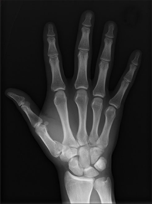
A 42-year-old, right-handed man sustained an injury to his right thumb base following a fall during a rugby match. He was reviewed in our clinic four days after the injury. Apart from bruising over the right thenar eminence, there was pain on movement of the right thumb TMCJ. Plain radiography showed a fracture-dislocation of the right thumb TMCJ (Fig. 1). Similar to Bennett’s fracture-dislocation, there was dorsal dislocation of the distal fragment. However, unlike a typical Bennett’s injury, the proximal fragment, which is usually attached to the DAOL, was found to be rotated (Fig. 2).
Fig. 1.

(a) Lateral and (b) anteroposterior radiographs of the right thumb show a fracture-dislocation of the TMCJ.
Fig. 2.

Anteroposterior radiograph of the right hand shows the distal fragment of the fracture-dislocation being pronated and dislocated dorsally.
In view of the unstable nature of the injury, surgical management was adopted. Under general anaesthesia, a Wagner approach to the patient’s right thumb metacarpal base was used for exposure, with dissection down to the TMCJ, and the capsule was opened. The proximal fragment, containing most of the articular surface, was dislocated and pronated, with the articular surface facing dorsoradially, instead of facing the trapezium proximally. The distal fragment was pronated and dislocated dorsally.
The fracture fragments were reduced (Fig. 3) and temporarily pinned with two 1.0 mm Kirschner wires. Definitive fixation was performed with the Synthes® 2.0 mm modular hand system, with restoration of the joint surface (Fig. 4). After fixation of the fracture, the patient’s TMCJ was noted to be relatively unstable compared to the contralateral thumb TMCJ; the metacarpal base was mildly subluxable dorsally. The fixation was protected with a thumb spica splint.
Fig. 3.

(a) Pre-reduction intraoperative photograph shows the distal fragment pronated and dislocated dorsally (A); the dislocated proximal fragment, with the articular surface facing dorsoradially (B); and the articular surface of the trapezium (C). (b) Post-reduction intraoperative photograph shows restoration of a congruent trapeziometacarpal joint.
Fig. 4.

Intraoperative radiographs show the fixation with the Synthes® 2.0 mm modular hand system.
Our postoperative management plan was to the keep the joint immobilised for four weeks to allow for ligamentous healing before allowing range of motion exercises at the TMCJ. However, our patient defaulted on further reviews.
DISCUSSION
The TMCJ is lax and subluxable in the resting position, but becomes more stable in opposition. The stability of the TMCJ occurs only in the final phase of opposition and is a result of the screw-home-torque mechanism.(2) This mechanism involves tight articular congruence, and occurs with a combination of tension on the dorsal ligament complex and the DAOL. In Bennett’s fracture-dislocation, the metacarpal shaft subluxates in a dorsal, proximal and radial direction due to the pull of the abductor pollicis longus, extensor pollicis longus, extensor pollicis brevis and adductor pollicis longus muscles. The proximal fragment remains in place due to an intact DAOL.(3)
In a typical Bennett’s fracture-dislocation, the volar ulnar corner of the proximal fragment of the thumb metacarpal is clearly seen on the posteroanterior views of hand radiography (Fig. 5). This is due to the fact that there is minimal, if any, rotation of the proximal fragment as the DAOL is intact. In our patient, this volar ulnar corner where the DAOL is attached is no longer clearly seen (Fig. 2). This raises the suspicion that the fragment is rotated and the injury is therefore not a typical Bennett’s fracture-dislocation.
Fig. 5.
Anteroposterior radiograph of the right hand shows a typical Bennett’s fracture-dislocation.
Narushima et al(4) reported a similar case where the characteristics of the proximal fragment were similar to that found in our patient – the fragment consisted of most of the articular surface, which faced in a radial direction. In Narushima et al’s reported case, however, the distal fragment was not dislocated8dorsally, and they hypothesised that the dorsoradial ligament was intact.(5) The pathomechanics behind the injury in our patient is similar, with an added dorsoradial ligament injury.
In our patient, apart from rotation of the proximal fragment, the distal fragment was also dislocated dorsally, suggesting a dorsoradial ligament rupture. The ligamentous injury seen in our patient was a combination of that seen in a typical Bennett’s fracture-dislocation and that seen in the case reported by Narushima et al.(4)
Being an injury that consists of both a bony and a significant ligamentous injury, management should target both components. While reduction and stabilisation of the metacarpal resolves the bony injury issue, ligamentous injury should be addressed concurrently.
Following fixation of the fracture, intraoperative assessment suggested residual instability in the TMCJ, which we temporarily immobilised to allow ligamentous healing. An alternative would have been to pin the joint temporarily instead of splinting. However, we decided against pinning the joint, as we correctly suspected that the patient may be lost to follow-up. Should our patient return with persistent TMCJ instability, our plan is to perform a TMCJ ligament reconstruction.
ACKNOWLEDGEMENTS
We would like to thank Dr Chew Yoon Chong Winston for his assistance in the preparation of this manuscript.
REFERENCES
- 1.Hove LM. Fractures of the hand. Distribution and relative incidence. Scand J Plast Reconstr Surg Hand Surg. 1993;27:317–9. [PubMed] [Google Scholar]
- 2.Edmunds JO. Traumatic dislocations and instability of the trapeziometacarpal joint of the thumb. Hand Clin. 2006;22:365–92. doi: 10.1016/j.hcl.2006.05.001. [DOI] [PubMed] [Google Scholar]
- 3.Carlsen BT, Moran SL. Thumb trauma: Bennett fractures, Rolando fractures, and ulnar collateral ligament injuries. J Hand Surg Am. 2009;34:945–52. doi: 10.1016/j.jhsa.2009.03.017. [DOI] [PubMed] [Google Scholar]
- 4.Narushima Y, Hara A, Kusunose K. An unusual fracture dislocation of the trapeziometacarpal joint of the thumb: a case report. Hand Surg. 2010;15:57–60. doi: 10.1142/S0218810410004564. [DOI] [PubMed] [Google Scholar]
- 5.Strauch RJ, Behrman MJ, Rosenwasser MP. Acute dislocation of the carpometacarpal joint of the thumb: an anatomic and cadaver study. J Hand Surg. 1994;19:93–8. doi: 10.1016/0363-5023(94)90229-1. [DOI] [PubMed] [Google Scholar]