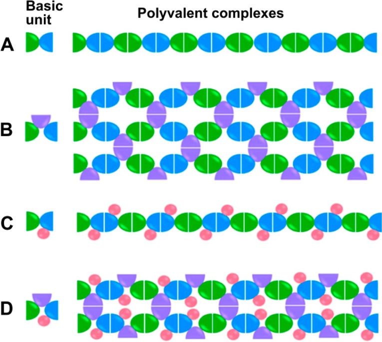
Figure 8.

Depiction of polyvalent complex formation. For each type, A–D, the basic unit (one protein fusion) is shown at the left, with each color indicating a part of the fusion that consists of a homodimer-forming protein (blue, green, and purple) or nonhomodimer-forming protein for antigen display (pink). Depending on the number of homodimer-forming proteins included in the basic fusion protein unit, the resulting self-assembled polyvalent complexes were of a linear (A,C) or network (B,D) morphology, as seen in micrographs in the original publication. Reprinted from Wang et al.,92 Copyright 2013 with permission from Elsevier.
